![]()
Section 1: THORAX
Thorax Questions
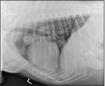
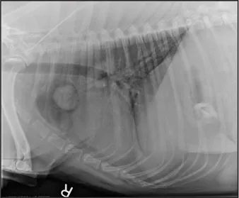
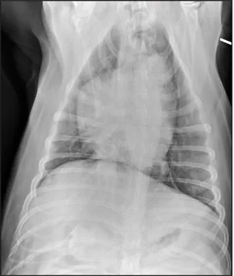
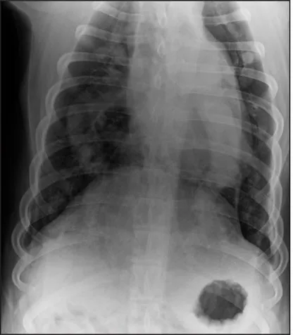
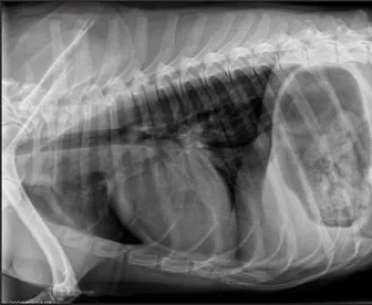
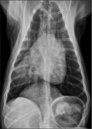
CASE 1.1 A 7-year-old neutered male Labrador Retriever who was hit by a car. You obtain these thoracic radiographs: Figs. 1.1a, b, left and right lateral projections, respectively; Figs. 1.1c, d, ventrodorsal and dorsoventral projections, respectively.
1 What are your radiographic findings?
2 What is your radiographic diagnosis?
1.1a
1.1b
1.1c
1.1d
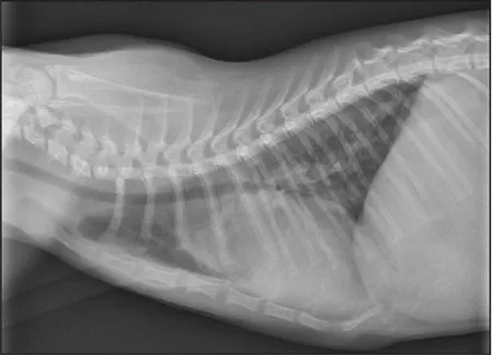
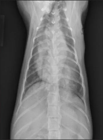
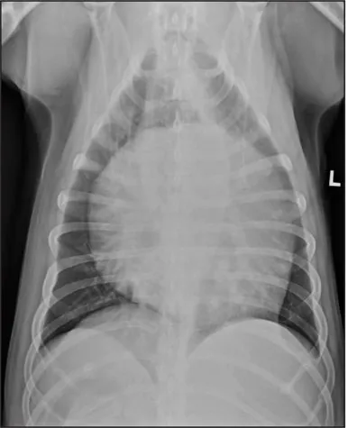
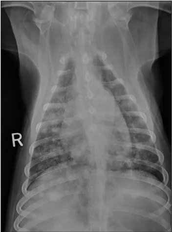
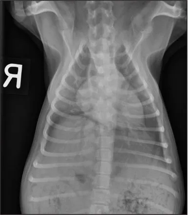
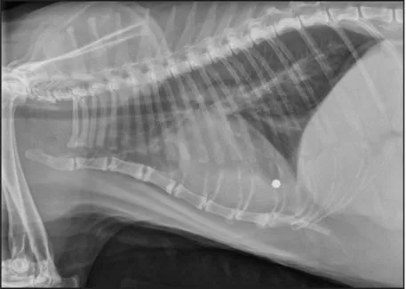
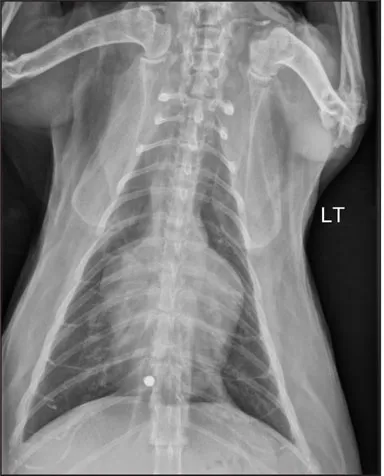
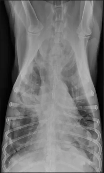
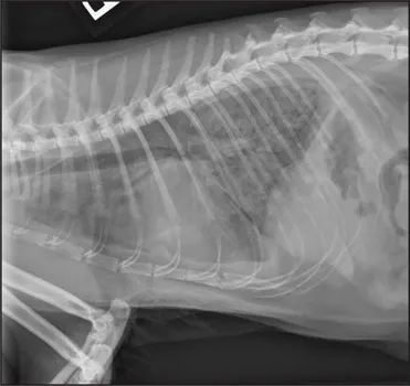
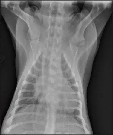
CASE 1.2 A 4-month-old female domestic shorthair cat with shallow breathing and a grade III/VI continuous murmur at the left cranial base. You obtain these thoracic radiographs: Fig. 1.2a, right lateral projection; Fig. 1.2b, dorsoventral projection.
1 What are your radiographic findings?
2 What is your radiographic diagnosis?
3 Is additional imaging needed?
1.2a
1.2b
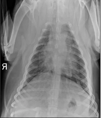
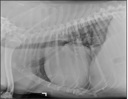
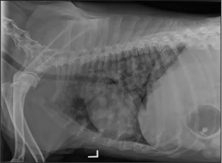
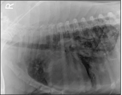
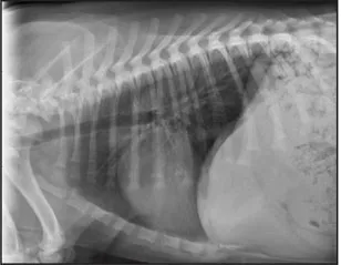
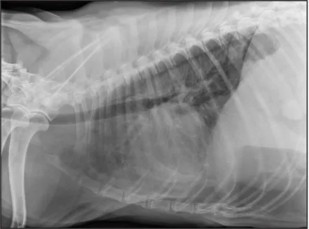
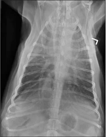
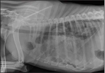
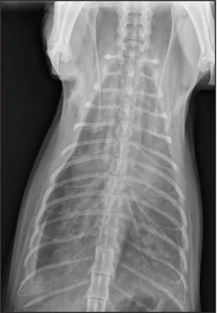
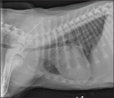
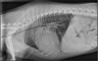
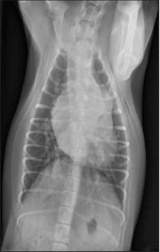
CASE 1.3 An 8-year-old male German Shepherd Dog with lethargy and muffled heart sounds. No heart murmur ausculted. You obtain these thoracic radiographs: Fig. 1.3a, left lateral projection; Fig. 1.3b, dorsoventral projection.
1 What are your radiographic findings?
2 What is your radiographic diagnosis?
3 Is additional imaging needed?
1.3a
1.3b
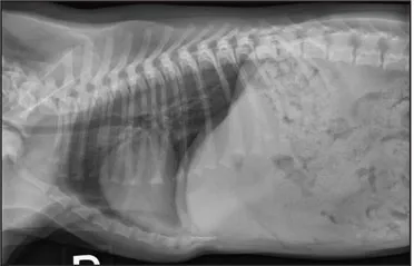
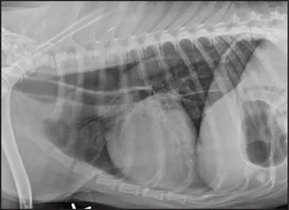
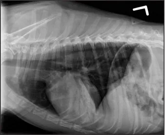
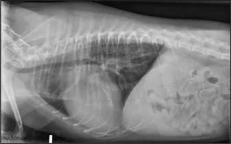
CASE 1.4 A 13-year-old spayed female mixed breed dog with a history of polyuria and polydipsia and abdominal distension. You obtain these thoracic radiographs: Fig. 1.4a, left lateral projection; Fig. 1.4b, dorsoventral projection.
1 What are your radiographic findings?
2 What is your radiographic diagnosis?
3 Are any additional radiographs needed?
1.4a
1.4b
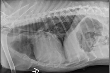
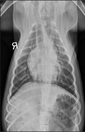
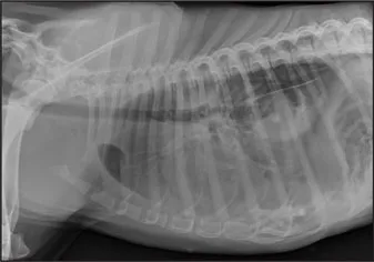
CASE 1.5 An 8-year-old spayed female English Mastiff with a 2-week history of labored breathing, anorexia, and weight loss. You obtain these thoracic radiographs: Fig. 1.5a, right lateral projection; Fig. 1.5b, dorsoventral projection.
1 What are your radiographic findings?
2 What is your radiographic diagnosis?
1.5a
1.5b
CASE 1.6 A 10-week-old male Beagle with abnormal mentation, tachypnea, and ptyalism. On physical examination, a hemorrhagic and erythematous lesion is found in the oral cavity. You obtain these thoracic radiographs: Figs. 1.6a, b, left lateral and right lateral projections, respectively; 1.6c, dorsoventral projection.
1 What are your radiographic findings?
2 What is your radiographic diagnosis?
3 Are any additional radiographic views needed?
1.6a
1.6b
1.6c
CASE 1.7 A 9-year-old spayed female Vizla with a 3-week history of stumbling and weakness following exercise and regurgitation following eating. You obtain these thoracic radiographs: Figs. 1.7a, b, left and right lateral projections, respectively; Fig. 1.7c, dorsoventral projection.
1 What are your radiographic findings?
2 What is your radiographic diagnosis?
1.7a
1.7b
1.7c
CASE 1.8 A 12-year-old neutered male Jack Russell Terrier with a productive cough. You obtain these thoracic radiographs: Figs. 1.8a, b, left and right lateral projections, respectively; Fig. 1.8c, ventrodorsal projection.
1 What are your radiographic findings?
2 What is your radiographic diagnosis?
1.8a
1.8b
1.8c
CASE 1.9 A 10-year-old neutered male Golden Retriever with labored breathing and exercise intolerance. You obtain these throacic radiographs: Fig. 1.9a, left lateral projection; Fig. 1.9b, dorsoventral projection.
1 What are your radiographic findings?
2 What is your radiographic diagnosis?
3 Are additional radiographic projections needed?
1.9a
1.9b
CASE 1.10 A 3-year-old neutered male domestic shorthair cat with acute hindlimb paralysis and tachycardia. You obtain these thoracic radiographs: Fig. 1.10a, left lateral projection; Fig. 1.10b, dorsoventral projection.
1 What are your radiographic findings?
2 What is your radiographic diagnosis?
3 Is additional imaging needed?
1.10a
1.10b
CASE 1.11 A 4-year-old female German Shorthair Pointer with a 1-week history of a cough, dyspnea, and lethargy. You obtain these thoracic radiographs: Figs. 1.11a, b, left and right lateral projections, respectively; Fig. 1.11c, ventrodorsal projection.
1 What are your radiographic findings?
2 What is your radiographic diagnosis?
1.11a
1.11b
1.11c
CASE 1.12 A 9-year-old spayed female cat with a cough and lethargy. Crackles and wheezes were auscultated on physical examination. You obtain these thoracic radiographs: Fig. 1.12a, right lateral projection; 1.12b, dorsoventral projection.
1 What are your radiographic findings?
2 What is your radiographic diagnosis?
3 Is further imaging needed?
1.12a
1.12b
CASE 1.13 A 3-month-old male Newfoundland with episodes of weakness with cyanosis and an occasional, dry cough. On auscultation, a grade V/VI holosystolic basilar murmur was identified. You obtain these thoracic radiographs: Fig. 1.13a, right lateral projection; 1.13b, dorsoventral projection.
1 What are your radiographic findings?
2 What is your radiographic diagnosis?
3 Is further imaging needed?
1.13a
1.13b
CASE 1.14 A 5-month-old female Shetland Sheepdog with exercise intolerance and shortness of breath. You obtain these thoracic radiographs: Figs. 1.14a, b, left and right lateral projections, respectively; Fig. 1.14c, dorsoventral projection.
1 What are your radiographic findings?
2 What is your radiographic diagnosis?
1.14a
1.14b
1.14c
CASE 1.15 A 2-year-old neutered male mixed breed canine with intermittent vomiting, which has recently increased to three times daily, and muffled heart sounds. You obtain these thoracic radiographs: Fig. 1.15a, left lateral projection; Fig. 1.15b, dorsoventral projection.
1 What are your radiographic findings?
2 What is your radiographic di...