Measurement in Ultrasound
  1. 414 pages
  2. English
  3. ePUB (mobile friendly)
  4. Available on iOS & Android
eBook - ePub

About this book

Measurement and interpretation of key ultrasound parameters are essential to differentiate normal anatomy from pathology. By using Measurement in Ultrasound, trainee radiologists and ultrasonographers can gain an appreciation of such measurements, while practitioners can use it as a valuable reference in the clinical setting.

The book follows a consistent format throughout for ease of reference and features useful information on preparation and positioning of the patient for ultrasound, the type of transducer and method to be used, the appearance of the resulting ultrasound images and the measurements to be derived from them.

Designed for frequent use in everyday practice, the book includes more than 150 high-quality ultrasound images annotated with key measurements and accompanied by concise explanatory text. Normal variants are provided, along with ranges for features that can change during development and in disease.

This new edition covers relevant developments in ultrasound. Where appropriate, updated ultrasound measurements that have arisen are also included and key references are provided as an aid to further study.

Frequently asked questions

Yes, you can cancel anytime from the Subscription tab in your account settings on the Perlego website. Your subscription will stay active until the end of your current billing period. Learn how to cancel your subscription.
At the moment all of our mobile-responsive ePub books are available to download via the app. Most of our PDFs are also available to download and we're working on making the final remaining ones downloadable now. Learn more here.
Perlego offers two plans: Essential and Complete
  • Essential is ideal for learners and professionals who enjoy exploring a wide range of subjects. Access the Essential Library with 800,000+ trusted titles and best-sellers across business, personal growth, and the humanities. Includes unlimited reading time and Standard Read Aloud voice.
  • Complete: Perfect for advanced learners and researchers needing full, unrestricted access. Unlock 1.4M+ books across hundreds of subjects, including academic and specialized titles. The Complete Plan also includes advanced features like Premium Read Aloud and Research Assistant.
Both plans are available with monthly, semester, or annual billing cycles.
We are an online textbook subscription service, where you can get access to an entire online library for less than the price of a single book per month. With over 1 million books across 1000+ topics, we’ve got you covered! Learn more here.
Look out for the read-aloud symbol on your next book to see if you can listen to it. The read-aloud tool reads text aloud for you, highlighting the text as it is being read. You can pause it, speed it up and slow it down. Learn more here.
Yes! You can use the Perlego app on both iOS or Android devices to read anytime, anywhere — even offline. Perfect for commutes or when you’re on the go.
Please note we cannot support devices running on iOS 13 and Android 7 or earlier. Learn more about using the app.
Yes, you can access Measurement in Ultrasound by Paul S. Sidhu, Wui K. Chong, Keshthra Satchithananda, Paul S. Sidhu,Wui K. Chong,Keshthra Satchithananda in PDF and/or ePUB format, as well as other popular books in Medicine & Anatomy. We have over one million books available in our catalogue for you to explore.

Information

Publisher
CRC Press
Year
2017
Print ISBN
9781138474048
eBook ISBN
9781315355917
Edition
2
Subtopic
Anatomy

1 Upper Abdominal

Anu E. Obaro, Venus
Hedayati, Colin R. Deane,
Keshthra Satchithananda and
Paul S. Sidhu
Liver
Liver size
Liver fibrosis assessment
Biliary tree
Gallbladder and gallbladder wall
Spleen
Pancreas
Pancreatic duct (adult)
Adrenal glands (adult)
Diaphragm
Upper Abdominal Vasculature
Portal vein
Hepatic veins
Hepatic artery
Celiac and superior mesenteric arteries
Doppler ultrasound assessment of post-prandial intestinal blood flow
Inferior mesenteric artery

Liver size

Preparation

None.

Position

Supine, right anterior oblique to demonstrate the porta hepatis.

Transducer

2.0–6.0 MHz curvilinear transducer.

Method

Longitudinal views are taken in the midclavicular and midline positions, and measurements obtained. Anteroposterior diameters are also measured at the midpoint of the longitudinal diameters. All measurements are taken on deep inspiration.

Appearance

Uniform pattern of medium-strength echoes.

Measurements

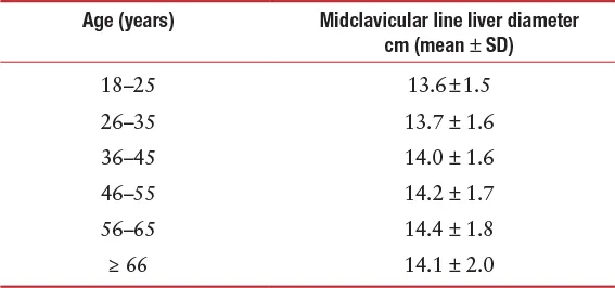
There are significant variations in liver size due to gender, age, body mass index and weight.
Diameter
Female cm (mean ± SD)
Male cm (mean ± SD)
Midclavicular line (largest craniocaudal diameter)
14.9 ± 1.6
15.1 ± 1.5
Images
In the transverse plane, the normal caudate lobe should be less than 2/3 of the size of the right lobe.
Images
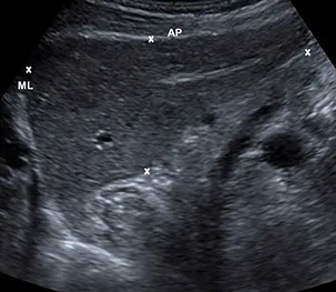
A midline longitudinal view through the left lobe of the liver, demonstrating the anteroposterior diameter (AP) and the midline longitudinal length (ML).
Images
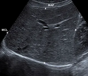
A midclavicular longitudinal view through the right lobe of the liver, with a midclavicular anteroposterior diameter (MAP) and a midclavicular longitudinal length (MCL).

Further Reading

Kratzer W., Fritz V., Mason R.A., Haenle M.M., Kaechele V. Factors affecting liver size: A sonographic survey of 2080 subjects. J Ultrasound Med.2003; 22:1155–1161.
Patzak M, Porzner M, Oeztuerk S, Mason RA, Wilhelm M, Graeter
T, Kratzer W., Haenle M.M., Akinli A.S.. Assessment of liver size by ultrasonography. J Clin Ultrasound.2014; 42:399–404.

Liver fibrosis assessment

Preparation

Patient should fast for 6–8 hours prior to the examination.

Position

Supine, right anterior oblique.

Transducer

2.0–6.0 MHz curvilinear transducer.

Method

Measurements are obtained from an intercostal view, to interrogate an area of the liver at least 2 cm deep to the liver capsule, away from major vessels. The right arm is raised above the head. The sample box is placed over the area selected, ideally in a perpendicular position, and a total of 10 measurements are obtained in brief suspended respiration, from the same area of liver. Segments 5 and 6 are normally interrogated; the left lobe of the liver should be avoided. Different ultrasound machines have different methods of obtaining readings and different display methods, and the readings are not transferable between machines. Transient elastography (TE) or “fibroscan” and acoustic radiation force impulse (ARFI) imaging are the most commonly used methods. A fibroscan does not produce an ultrasound image, and provides measurements in kilopascals (kPa).

Appearance

The measurements can be expressed in velocity of shear wave (m/sec). The level of liver fibrosis is calculated and classified according to the METAVIR (F0–F4) or ISHAK (0–6) scoring system to ascertain normality, the degree of liver fibrosis, or the presence of cirrhosis. The most common diseases for which fibrosis will be assessed are Hepatitis C virus (HCV), Hepatitis B virus (HBV), and alcoholic liver disease.

Measurements

Acoustic radiation force impulse (ARFI) imaging
(usi...

Table of contents

  1. Cover Page
  2. Half title
  3. title
  4. copy
  5. fmchapter
  6. bio
  7. Adults
  8. 1 Upper Abdominal
  9. 2 URINARY TRACT
  10. 3 Organ Transplantation
  11. 4 Genital Tract
  12. 5 Gastrointestinal Tract
  13. 6 Superficial Structures
  14. 7 Peripheral Vascular (Arterial)
  15. 8 Peripheral Vascular (Venous)
  16. 9 Musculoskeletal System
  17. 10 UPPER ABDOMEN
  18. 11 Renal Tract
  19. 12 Pediatric Uterus, Ovary, and Testis
  20. 13 NEONATAL BRAIN
  21. Part Obstetrics
  22. 14 Obstetrics
  23. Index