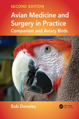
Avian Medicine and Surgery in Practice is an invaluable quick reference resource for clinicians and a useful study guide for veterinary students.
In this practical and beautifully illustrated book, early chapters cover physical examination, advice on interpreting diagnostic tests, and avian anatomy and physiology. Disorders affecting the different body regions and systems make up the majority of the book from the external—skin, feathers, eyes, legs and feet—to the internal including the gastrointestinal tract and the cardiovascular system. Further aspects of avian medicine discussed in the book include behavioural problems, incubation of eggs, paediatrics and surgery.
Written by an expert with more than 30 years of clinical experience in avian medicine, the new edition is thoroughly revised with updated diseases, new and expanded clinical techniques, and over 100 new color illustrations. It also adds four important new chapters: Husbandry, Grooming and Nutrition, Diagnostic Imaging, Endoscopy, and Oncology as well as new sections on cardiovascular anatomy and neuroanatomy.
Although veterinarians are taught avian anatomy in veterinary school, if one is unfamiliar with its clinical relevance it is always worth refreshing one’s memory. This chapter will highlight those features of avian anatomy and physiology that are relevant to the clinician. It will not seek to be a comprehensive review of the subject. The focus in this chapter is on companion birds and, as such, anatomical structures such as the phallus (found in ratites and waterfowl) will not be discussed. Without a basic understanding of anatomy and physiology, it becomes difficult to understand the pathophysiology of disease and how treatment will affect the patient as a whole.
THE SKIN, FEATHERS, NAILS AND BEAK
Avian skin is attached both to the underlying muscles and, more tightly, to the skeleton. It consists of the thin epidermis (only 10 cell layers deep) and the thicker underlying dermis. Feather follicles originate from the dermis. True glands are absent from much of the skin, although epidermal cells may secrete a lipoid sebaceous material. The external ear has glands that secrete a waxy material, but the only other gland found on the skin is the uropygial gland. This gland, absent in ratites, many pigeons, woodpeckers and some parrots (Amazona, Anodorhynchus and Cyanospitta), is a bi-lobed gland on the dorsum of the tail. It secretes a lipoid sebaceous material, spread over feathers during grooming, which may assist in waterproofing, cleaning and reducing skin infections. It is thought it may also have a role in sex identification; uropygial secretions may play a role in ultraviolet reflectivity, which in turn may be used by birds for sex differentiation.
Scales, raised areas of highly keratinised epidermis separated by folds of less keratinised skin, cover the non-feathered part of the leg, known as the podotheca. The claws, which enclose the terminal phalanx of each digit, are made up of two plates: the strongly keratinised dorsal plate enclosing dorsal and lateral aspects of the phalanx, and the softer ventral plate forming the sole of the claw. The dorsal plate grows faster than the ventral plate, therefore the nails curve downwards.
The bones of the upper and lower jaw are covered in horny keratin, called rhamphotheca (Fig. 1.1); the mandibular rhamphotheca is known as the gna-thotheca and the maxillary rhamphotheca is called the rhinotheca. The dorsal midline of the rhinotheca is the culmen and the ventral midline of the gnathotheca is the gonys. The cutting edge of both the upper and lower beak is the tomia, while the soft tissue between the mandibular rami is the inter-ramular region (Fig. 1.2). Histologically the rhamphotheca resembles skin, with the dermis attached to the periosteum of the underlying bone. The epidermis is modified, in that the stratum corneum is thickened and hardened, as the cells contain free calcium phosphate and crystals of hydroxyapatite. The neurological innervation of the upper beak is the ophthalmic and maxillary divisions of the trigeminal nerve, while the mandibular division of the trigeminal nerve innervates the lower beak. The cere, the fleshy area around the nares, is found only in owls, parrots and pigeons.
The unique structure of avian skin is the feathers. These arise from feather follicles, arranged in tracts around the body known as pterylae. The featherless skin between these tracts is called apterylae. Each follicle is a cylindrical pit in the skin, lined with epidermis and dermis. At the base of the follicle is the dermal papilla, a small mound of dermis that enters the proximal shaft of the feather (the calamus) through a small hole known as the inferior umbilicus. The epidermis covering the dermal papilla is continuous with the calamus and with a thin layer of epidermis lining the follicle (Fig. 1.3).
Figure 1.1 A red-tailed black cockatoo. Note that the keratin covering its beak (the rhamphotheca) is histologically similar to skin.
Figure 1.2 Labelled diagram of a bird beak with key parts and their locations.
Each feather shaft consists of the calamus, embedded in follicle, and the rachis, the main shaft beyond the calamus. They are distinguished by the distal (superior) umbilicus, a small opening into the shaft found at the junction of the rachis and calamus. Occasionally there is an after feather (the hypopenna), a small extra feather on the rim of the distal umbilicus (Figs 1.4, 1.5).
Figure 1.3 Diagram of the internal structures of the feather follicle.
Coming off the rachis are the barbs and coming off the barbs are the barbules, filaments that interlock to form the vane (Fig. 1.6). This is the pen-naceous region. The vanes are asymmetrical, with the external vane narrower than the internal vane. On the dorsal wing the external vane of one feather overlaps the internal vane of the next. Just below the vane is the plumaceous region, where a few downy barbs fail to interlock.
Within the calamus of an immature feather is the pulp, a loose reticulum of mesoderm with an axial artery and vein. This pulp retracts as the feather matures, leaving pulp caps (empty chambers within the calamus).
There are seven types of feathers (Fig. 1.7): contour, semiplume, down, powder down, hypopenna, filoplume and bristle.
• Contour feathers include the flight feathers and the body feathers. The flight feathers on the tail are called the retrices. The flight feathers on the wings are known as the remiges: the primaries (9–11) in total arise from the periosteum of the metacarpus; the secondaries (6–32) in total arise from the periosteum of the ulna; and the tertiaries arise from the humeral area. Overlying them are the coverts (Fig. 1.8).
• Semi-plume feathers have a wholly fluffy vane, with the rachis longer than barb. They lie along pterylae margins, acting as insulation.
• Down feathers are also wholly fluffy, but the rachis is either absent or shorter than the longest barb. Distribution varies between species.
• Powder down feathers are structured like down feathers, although some are semi-plumes or contour feathers. They shed a fine waxy powder, which is actually keratin flakes. This powder forms a waterproofing coat over the contour feathers and may play a role in keeping the bird clean. Powder down feathers are usually grouped in patches (e.g. on the thigh), although some species have them widely distributed. They are found in herons, parrots, toucans, pigeons and bowerbirds.
• Hypopennae (1–5) in total are small feathers projecting from the distal umbilicus of penna-ceous and plumaceous feathers (after feathers). They are usually not associated with retrices or longer remiges.
• Filoplumes have a long fine shaft with a tuft of short barbs/barbules at the end. They possibly have a sensory/proprioceptive role and are found close to the follicles of contour feathers.
• Bristles have a stiff rachis, with either a few barbs at the proximal end or no barbs at all. They are found around the mouth, nares and eyes, and possibly have a tactile function.
Figure 1.4 Diagram of the external parts of a feather.
Figure 1.5 Closer view of the calamus and rachis parts of a feather.
Figure 1.6 Closer view of the pennaceous portion of a feather.
Figure 1.7 Illustration of the range and shapes of different types of feather.
Figure 1.8 Schematic diagram of the location and types of feathers that make up a parrot wing.
The colour of feathers is the result of the combination of pigments and feather structure. Carotenoids or psittacins (yellow pigments absorbed from the diet, including reds, oranges and pinks) create the foreground colour. Melanins are the grey pigments (including black, grey and brown) that create the background and also the foreground colour. Each feather barb has a cortex (an outer layer) containing either carotenoid pigments (psittacins) or melanin pigments. If melanin is in the cortex, it is known as foreground colour and produces black, greys, dark browns and chestnut reds. This is the marking seen in many birds. The barb also has a medulla, which only ever contains melanin (background melanin). All of these pigments are distributed in different layers and, when combined with special features of barb structure that affect the passage of light, produce the spectrum of colours seen.
Moulting, the shedding of old, worn feathers and the renewal of plumage, is a regular event. It is controlled by a wide range of factors including thyroid activity, reproductive hormones, photoperiod, body condition, age and diet. After a series of juvenile moults to attain adult plumage, most birds go on to moult one to two times annually. These moults, often referred to as the prenuptial and postnuptial moults, occur in spring and autumn respectively. The pattern of moulting is orderly and in the following progression (with some overlap): the inner primaries; the outer primaries; the secondaries and tail feathers; and finally the body contour feathers. It is usually bilaterally symmetrical and is paced so as to avoid loss of flight capacity at any time.
When it is time to moult an old feather, a proliferation of epidermal cells at the base of the follicle (the epidermal collar) separates the old feather from the dermal papilla and allows it to shed. These epidermal cells then start to group themselves into two series of spiral barb ridges. The tips of these ridges end along a longitudinal line on the ventral aspect of the feather (the seam). On the dorsal side of feather the epidermis thickens to form the rachis. Within this structure is the dermal core, consisting of the axial blood vessels, with mesoderm around them. As it grows the feather emerges from the follicle as a pointed projection with a dermal core and an epidermal cover (sheath). This sheath then progressively ruptures, freeing the barbs that have separated along the seam and allowing the feather to open. Much of the increased grooming activity seen in birds at this time is to remove this sheath.
Because birds lack sweat glands, they rely on evaporative heat loss from the respiratory tract and heat transfer through apterylae (the featherless tracts of skin) to cool their bodies. To do this, many birds hold their feathers close to the body and may extend their wings, exposing the apterylae. Conversely, to retain body heat when ill or cold, they fluff their feathers up to trap body heat against the skin. (Ostriches do the reverse, i.e. they raise their feathers to promote heat loss and hold them close to conserve body heat.)
THE SKELETON
General
Bones serve two major functions: they provide structural support for the muscular system and they act as a reservoir for calcium and phosphorus (Fig. 1.9). Although the structural make-up of bone is similar across all animal species, there are some specific differences between mammalian and avian bones. The requirement for flight means that birds have evolved with bones that are lightweight, but aerodynamically strong. They have thin brittle cortices and wide medullas that may, in some species and some bones, be pneumatic. Under the influence of oestrogen during the breeding season, many hens will lay down medullary bone (extra bone in the medullary cavities of the long bones) to form a calcium reservoir for egg production.
Figure 1.9 Labelled diagram of the skeleton of a bird.
The blood supply to bones arises from periosteal, medullary, metaphyseal and epiphyseal vessels. The periosteal blood supply is the predominant source of blood to the bone and its disruption, either by trauma or surgical repair of a fracture, may result in delayed healing or even complete failure to heal (a non-union).
Skull
The upper jaw of psittacine birds consists of three bones: the premaxilla, the nasal bone and the maxilla (Fig. 1.10). Together they form a rigid block hinged to the braincase by the prokinetic craniofacial joint (Fig. 1.11). The elastic zone of this joint allows movement of the upper jaw. The palate, made up of the palatal processes of the premaxillary and maxillary bones, the palatine...
Table of contents
Cover
Title Page
Copyright Page
Table of Contents
Preface
Abbreviations
Us and international unit (blood values) conversion
Chapter 1 Clinical Anatomy and Physiology
Chapter 2 Husbandry, Grooming and Nutrition
Chapter 3 The physical Examination
Chapter 4 Clinical Techniques
Chapter 5 Diagnostic Imaging
Chapter 6 Endoscopy
Chapter 7 Interpreting diagnostic Tests
Chapter 8 Supportive Therapy
Chapter 9 Differential Diagnoses
Chapter 10 Diseases of the Skin and Feathers
Chapter 11 Disorders of the Beak and Cere
Chapter 12 Disorders of the Eye
Chapter 13 Disorders of the Ear
Chapter 14 Diseases of the LEGS, Feet and Toes
Chapter 15 Disorders of the Musculoskeletal System
Chapter 16 Diseases of the Gastrointestinal Tract
Chapter 17 Disorders of the Liver
Chapter 18 Disorders of the Pancreas
Chapter 19 Diseases of the Respiratory System
Chapter 20 Disorders of the Cardiovascular System
Chapter 21 Disorders of the Lymphatic and haematopoietic Systems
Chapter 22 Disorders of the Nervous System
Chapter 23 Disorders of the Reproductive Tract
Chapter 24 Disorders of the Urinary System
Chapter 25 Behavioural Problems
Chapter 26 Incubation of Eggs
Chapter 27 Paediatrics
Chapter 28 Analgesia and Anaesthesia
Chapter 29 Surgery
Chapter 30 Oncology
Appendix 1: Formulary
Appendix 2: Reference Intervals for Commonly Kept Companion Birds
Appendix 3: Biological Values for Some Common Companion Bird Species
Index
Trusted by 375,005 students
Access to over 1.5 million titles for a fair monthly price.
Study more efficiently using our study tools.
Frequently asked questions
Yes, you can cancel anytime from the Subscription tab in your account settings on the Perlego website. Your subscription will stay active until the end of your current billing period. Learn how to cancel your subscription
No, books cannot be downloaded as external files, such as PDFs, for use outside of Perlego. However, you can download books within the Perlego app for offline reading on mobile or tablet. Learn how to download books offline
Perlego offers two plans: Essential and Complete
Essential is ideal for learners and professionals who enjoy exploring a wide range of subjects. Access the Essential Library with 800,000+ trusted titles and best-sellers across business, personal growth, and the humanities. Includes unlimited reading time and Standard Read Aloud voice.
Complete: Perfect for advanced learners and researchers needing full, unrestricted access. Unlock 1.5M+ books across hundreds of subjects, including academic and specialized titles. The Complete Plan also includes advanced features like Premium Read Aloud and Research Assistant.
Both plans are available with monthly, semester, or annual billing cycles.
We are an online textbook subscription service, where you can get access to an entire online library for less than the price of a single book per month. With over 1.5 million books across 990+ topics, we’ve got you covered! Learn about our mission
Look out for the read-aloud symbol on your next book to see if you can listen to it. The read-aloud tool reads text aloud for you, highlighting the text as it is being read. You can pause it, speed it up and slow it down. Learn more about Read Aloud
Yes! You can use the Perlego app on both iOS and Android devices to read anytime, anywhere — even offline. Perfect for commutes or when you’re on the go. Please note we cannot support devices running on iOS 13 and Android 7 or earlier. Learn more about using the app
Yes, you can access Avian Medicine and Surgery in Practice by Bob Doneley in PDF and/or ePUB format, as well as other popular books in Medicine & Veterinary Medicine. We have over 1.5 million books available in our catalogue for you to explore.