- 192 pages
- English
- ePUB (mobile friendly)
- Available on iOS & Android
eBook - ePub
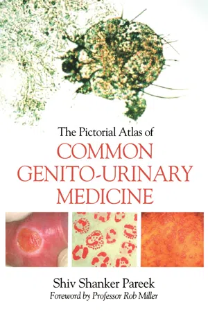
The Pictorial Atlas of Common Genito-Urinary Medicine
About this book
Sexually transmitted infections remain a global health concern with the World Health Organization reporting over 340 million new cases of bacterial and protozoal STI every year, worldwide. They are frequently associated with a significant degree of medical and psychological morbidity. This full-colour pictorial guide is a unique, single volume compilation of common diseases in genito-urinary medicine. It covers a wide range of ailments from sexually transmitted infections to dermatological conditions affecting the genito-urinary system. Each high-quality image is accompanied by a clear, concise description to aid diagnosis. As well as providing a diagnostic tutorial for trainees in genito-urinary medicine, this book is a reference for any healthcare professional encountering these diseases. Candidates for the Diploma of Genito-urianry Medicine and the Knowledge Based Assessment in Genito-Urinary Medicine will find this visual compendium invaluable.
Information
Chapter 1: Genitalia
1.1: Male internal and external genitalia (Fig. 1.1, next page)
- Urinary bladder – collects urine prior to its excretion from the body. It has a capacity of approximately 500 mL. The daily volume of urine excreted depends on the level of hydration. The bladder comprises four layers: serous, muscular, submucous, and mucous. Inside the bladder, the trigone and internal orifices of the urethra and the two ureters are noticeable.
- Ureter – each kidney has a ureter carrying urine from the kidney to the bladder. The ureters are each about 250 mm in length.
- Seminal vesicles – these are paired glands located on the posteroinferior aspect of the urinary bladder. A major part of the fluid in semen is secreted by these glands. The secretion is alkaline in nature and contains fructose.
- Rugae – these are multiple mucosal folds inside an empty bladder.
- Urethra – this starts from the internal urethral orifice in the urinary bladder and runs to the external urethral opening at the glans penis. It is divided into three parts: the prostatic urethra, the membranous urethra, and the spongy urethra (also called the penile urethra). The total length of the male urethra is 175 mm to 200 mm. The urethra has two main functions: to allow urine to empty from the bladder and to allow the flow of semen during ejaculation.
- Prostatic urethra – this is the part of the urethra passing through the prostate. It is about 24 mm in length.
- Membranous urethra – this is a short section of the urethra between the prostatic urethra and the spongy urethra. The length of the membranous urethra is 10 mm to 15 mm.
- Spongy (penile) urethra – this is the longest portion of the urethra, about 150 mm to 160 mm in length, and is located inside the corpus spongiosum penis. It runs along the ventral surface of the penis.
- Cowper’s glands (also called bulbourethral glands) – these are a pair of small glands situated posterior to, and lateral to, the membranous urethra. They secrete clear fluid which is added to semen.
- Epididymis – this structure is present on the posterior side of each testis and is a coiled part of the spermatic duct which connects to the vas deferens. The epididymis accumulates and matures sperm cells and carries them to the vas deferens prior to ejaculation. The epididymis has three parts: head, body, and tail.
 Figure 1.1 Male genital organs.
 - Vas deferens – this is a muscular tube which extends from the epididymis to the prostate, via the seminal vesicle, where it connects to the urethra. It carries mature sperm to the urethra for ejaculation.
- Testes – there are two oval-shaped testes (singular: testis) within the scrotum. The spermatic cord starts from the top of each testis. The testes produce testosterone (the primary male sex hormone) and sperm cells. Testes contain seminiferous tubules in which spermatogenesis occurs.
- Tyson’s glands (also called pearly penile papules) – these are similar to sebaceous glands and are situated around the corona and inner surface of the prepuce of the penis. They are not always present and are not thought to be pathological. The glands may be analogous to preputial glands found in other mammals.
- Coronal ridge – this separates the shaft of the penis from the glans.
- Trigone – this is the internal triangular area of the urinary bladder between the two ureter orifices and the internal urethral opening.
- Prostate gland – it contains two prostatic ducts which connect to the prostatic urethra. The gland produces fluid which is added to the ejaculate.
- Gians penis – this is the head of the penis. It is reddish in colour, smooth and shiny. When the penis is erect, the glans becomes moist and very sensitive.
- Prepuce – this is the outer retractable foreskin of the penis. Its function is to cover and protect the glans penis.
- Littre’s glands (also called periurethral glands) – these are situated in the urethra and secrete mucus which is mixed with semen during ejaculation.
1.2: Female internal and external genitalia (Fig. 1.2, next page)
- Vagina – this is a muscular canal which extends from the cervix to the surface of the body. It is normally 150 mm to 180 mm in length and is lined by mucous membrane. The vagina has a few tiny secretory glands which lubricate it.
- Bartholin’s glands – these were documented in the literature in 1677. They are paired glands situated on either side of the lower ends of the labia minora near the vaginal opening. Each measures 10 mm to 30 mm in diameter and produces mucus via Bartholin’s ducts when the woman is sexually aroused. An infected Bartholin’s gland develops a Bartholin’s abscess.
- Skene’s glands – these glands are also reported as paraurethral glands. They are present on the upper wall of the vagina near the urethra. They are close to the area described as ‘the G-spot’ and are analogous to the male prostate gland.
- Vestibule – this is defined as the space between the labia minora and the opening of the vaginal orifice.
 Figure 1.2 Female genital organs.
 - Clitoris – this is a very sensitive organ which is actually a bundle of nerves. It is the size of a pea and located at the top of the labia minora, 20 mm to 30 mm above the urethral opening.
- Cervix – this is the narrow portion of the uterus which opens into the vagina. The segment of cervix opening in the vagina is called the external os. The segment between the external os and uterus is called the endocervical canal; this ends at the internal os.
- Labia majora – these are two large folds which cover the labia minora. They are composed of fatty tissue and vary from woman to woman. They are often called the ‘outer lips’.
- Labia minora – these are a smaller pair of skin folds, often called the ‘inner lips’. They vary from woman to woman.
- Uterus – the uterus is a pear-shaped organ located in the lower abdomen between the bladder and rectum. The portion above the cervix is termed the corpus; it has two layers, a thin inner layer, the endometrium, and a thick, outer muscle layer, the myometrium.
- Ovary – there are two ovaries, one on each side of the uterus, They produce eggs and female hormones. They are each about the size of an almond.
- Fallopian tube or oviduct – each ovary is connected to the uterus by a fallopian tube (oviduct) which transports eggs from the ovary to the uterus.
- Urethra – the female urethra is shorter than the male urethra and measures about 40 mm. It is situated above the front wall of the vagina and consists of three layers: an outer muscular layer, spongy erectile tissue, and an inner mucous membrane.
Chapter 2: Syphilis
History
Syphilis is a disease with a long history; it was present in the New World prior to 1492 and possibly introduced into Europe at about this time. There is some documented evidence of syphilis among sailors who returned to Europe from the New World in 1493, perhaps even by Columbus. Girolamo Fracastoro (1478–1553) is best known for giving the disease the name ‘syphilis’; it was taken from an epic poem he wrote about the disease in 1530, entitled ‘Syphilis sive morbus Gallicus’, which translates as ‘Syphilis or the French dise...
Table of contents
- Cover
- Title Page
- Copyright Page
- Contents
- Foreword
- Preface
- Acknowledgements
- Dedication
- Sources of Information
- 1: Genitalia
- 2: Syphilis
- 3: Gonorrhoea
- 4: Urethritis due to Chlamydia Trachomatis
- 5: Non-specific Urethritis
- 6: Pelvic Inflammatory Disease
- 7: Prostatitis
- 8: Epididymitis and Epididymo-orchitis
- 9: Reiter’s Syndrome
- 10: Bacterial Vaginosis
- 11: Trichomoniasis
- 12: Genital Candidiasis
- 13: Tropical Genital and Sexually Acquired Infections
- 14: Non-venereal Endemic Treponematoses
- 15: Urinary Tract Infection
- 16: Genital Herpes
- 17: Human Papillomavirus
- 18: Molluscum Contagiosum
- 19: Sexually Acquired Viral Hepatitis B and C
- 20: Scabies
- 21: Pediculosis Pubis
- 22: Dermatoses Affecting the Genital Area
- 23: Malignant and Premalignant Conditions Affecting the Genital Area
- 24: Miscellaneous Conditions Affecting the Genitalia
- Appendix: SHHAPT Codes
- Index
Trusted by 375,005 students
Access to over 1.5 million titles for a fair monthly price.
Study more efficiently using our study tools.
Frequently asked questions
Yes, you can cancel anytime from the Subscription tab in your account settings on the Perlego website. Your subscription will stay active until the end of your current billing period. Learn how to cancel your subscription
No, books cannot be downloaded as external files, such as PDFs, for use outside of Perlego. However, you can download books within the Perlego app for offline reading on mobile or tablet. Learn how to download books offline
Perlego offers two plans: Essential and Complete
- Essential is ideal for learners and professionals who enjoy exploring a wide range of subjects. Access the Essential Library with 800,000+ trusted titles and best-sellers across business, personal growth, and the humanities. Includes unlimited reading time and Standard Read Aloud voice.
- Complete: Perfect for advanced learners and researchers needing full, unrestricted access. Unlock 1.5M+ books across hundreds of subjects, including academic and specialized titles. The Complete Plan also includes advanced features like Premium Read Aloud and Research Assistant.
We are an online textbook subscription service, where you can get access to an entire online library for less than the price of a single book per month. With over 1.5 million books across 990+ topics, we’ve got you covered! Learn about our mission
Look out for the read-aloud symbol on your next book to see if you can listen to it. The read-aloud tool reads text aloud for you, highlighting the text as it is being read. You can pause it, speed it up and slow it down. Learn more about Read Aloud
Yes! You can use the Perlego app on both iOS and Android devices to read anytime, anywhere — even offline. Perfect for commutes or when you’re on the go.
Please note we cannot support devices running on iOS 13 and Android 7 or earlier. Learn more about using the app
Please note we cannot support devices running on iOS 13 and Android 7 or earlier. Learn more about using the app
Yes, you can access The Pictorial Atlas of Common Genito-Urinary Medicine by Shiva Pareek in PDF and/or ePUB format, as well as other popular books in Medicine & Dermatology. We have over 1.5 million books available in our catalogue for you to explore.