eBook - ePub
Dermoscopy in General Dermatology
- 354 pages
- English
- ePUB (mobile friendly)
- Available on iOS & Android
eBook - ePub
Dermoscopy in General Dermatology
About this book
This lavishly illustrated guide from experts will enable practitioners to get the most out of dermoscopy for investigations and treatments in general dermatology.
Trusted by 375,005 students
Access to over 1.5 million titles for a fair monthly price.
Study more efficiently using our study tools.
Information
Part IInflammatory Diseases
1Papulosquamous disorders
Aimilios Lallas and Enzo Errichetti
1.1Psoriasis
1.1.1Introduction
Psoriasis is a common, chronic, and recurrent inflammatory disease characterized by heritability, phenotypic variability, and possible association to psoriatic arthritis and metabolic syndrome. Psoriasis is considered as a hyperproliferative disorder, but this increased proliferation of keratinocytes is the result of a cascade of immunologic reactions driven by inflammatory mediator cells and cytokines.1–3
1.1.2Clinical presentation
Psoriasis is typified by the presence of well-demarcated, scaly erythematous plaques of various sizes, typically covered by adherent silvery scales (Figure 1.1). The most frequent sites of involvement are the scalp, elbows, and knees, followed by lower back, buttocks, nails, umbilical region, trunk, palms, and soles. However, psoriatic lesions might develop on any body site. The severity of manifestations varies from very few small plaques to involvement of the largest part of the skin (erythroderma).1–3 The main clinical types of psoriasis are the following:
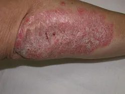
Figure 1.1The typical psoriatic lesion: demarcated erythematous plaque with stuck-on white-silvery scales.
1.1.2.1Plaque Psoriasis
Plaque psoriasis, also known as psoriasis vulgaris, is the most frequent clinical variant of the disease, typified by lesions as described earlier. Initially, the psoriatic lesions appear as red scaling papules that grow and coalesce to form round-oval plaques covered by thick silvery scales (Figure 1.2). The intensity of hyperkeratosis depends on the anatomic body site, being heavy on the scalp or palms and soles and absent in intertriginous areas (Figure 1.3). The scales are typically adherent in the center and looser at the periphery. When removed, small bleeding points appear (Auspitz sign). The most frequent anatomic sites of involvement have been mentioned previously. Psoriatic lesions may also develop on sites of physical trauma (Koebner’s phenomenon). In general, the disease is asymptomatic. However, pruritus might be present in a considerable proportion of patients.1–3
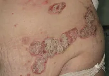
Figure 1.2Psoriatic lesions often coalesce to form larger plaques.
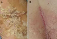
Figure 1.3Palmar psoriatic lesion characterized by intense hyperkeratosis (A). Psoriatic lesion on the intergluteal fold lacks hyperkeratosis (B).
1.1.2.2Guttate Psoriasis
Guttate psoriasis is characterized by the acute onset of multiple small, red, scaly papules, often following an acute infection, such as streptococcal pharyngitis (Figure 1.4). Guttate psoriasis might represent the first manifestation of psoriasis or may occur as an acute exacerbation of preexisting plaque psoriasis.1–3
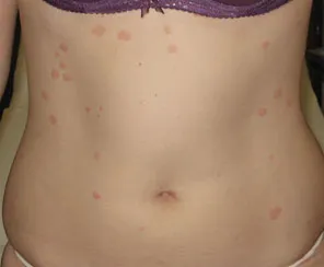
Figure 1.4Guttate psoriasis is typified by small papules/plaques of recent onset located mainly on the trunk.
1.1.2.3Inverse Psoriasis...
Table of contents
- Cover
- Half Title Page
- Dedication
- Title Page
- Copyright Page
- Contents
- Preface
- Contributors
- Introduction
- Part IInflammatory Diseases
- Part IIInfiltrative Diseases
- Part IIIInfectious Diseases
- Part IVHair and Nail Diseases
- Part VSkin of Color
- Appendix I: Differential diagnosis of erythematosquamous macules/papules on the trunk and/or extremities
- Appendix II: Differential diagnosis of erythematous macules/plaques on the face
- Appendix III: Differential diagnosis of palmar/plantar keratoderma
- Appendix IV: Differential diagnosis of sclero- atrophic patches on the trunk and/or extremities
- Appendix V: Differential diagnosis of hyperpigmented macules/papules on the trunk and/or extremities
- Appendix VI: Differential diagnosis of hypopigmented macules on the trunk and/or extremities
- Appendix VII: Differential diagnosis of itchy papules/nodules on the trunk and/or extremities
- Appendix VIII: Differential diagnosis of inflammatory papules along Blaschko’s lines
- Appendix IX: Differential diagnosis of purpuric macules/patches
- Appendix X: Differential diagnosis of nonscarring alopecia
- Appendix XI: Differential diagnosis of scarring alopecia
- Appendix XII: Differential diagnosis of hair casts
- Appendix XIII: Differential diagnosis of onycholysis
- Appendix XIV: Differential diagnosis of pitting of the nail plate
- Index
Frequently asked questions
Yes, you can cancel anytime from the Subscription tab in your account settings on the Perlego website. Your subscription will stay active until the end of your current billing period. Learn how to cancel your subscription
No, books cannot be downloaded as external files, such as PDFs, for use outside of Perlego. However, you can download books within the Perlego app for offline reading on mobile or tablet. Learn how to download books offline
Perlego offers two plans: Essential and Complete
- Essential is ideal for learners and professionals who enjoy exploring a wide range of subjects. Access the Essential Library with 800,000+ trusted titles and best-sellers across business, personal growth, and the humanities. Includes unlimited reading time and Standard Read Aloud voice.
- Complete: Perfect for advanced learners and researchers needing full, unrestricted access. Unlock 1.5M+ books across hundreds of subjects, including academic and specialized titles. The Complete Plan also includes advanced features like Premium Read Aloud and Research Assistant.
We are an online textbook subscription service, where you can get access to an entire online library for less than the price of a single book per month. With over 1.5 million books across 990+ topics, we’ve got you covered! Learn about our mission
Look out for the read-aloud symbol on your next book to see if you can listen to it. The read-aloud tool reads text aloud for you, highlighting the text as it is being read. You can pause it, speed it up and slow it down. Learn more about Read Aloud
Yes! You can use the Perlego app on both iOS and Android devices to read anytime, anywhere — even offline. Perfect for commutes or when you’re on the go.
Please note we cannot support devices running on iOS 13 and Android 7 or earlier. Learn more about using the app
Please note we cannot support devices running on iOS 13 and Android 7 or earlier. Learn more about using the app
Yes, you can access Dermoscopy in General Dermatology by Aimilios Lallas, Enzo Errichetti, Dimitrios Ioannides, Aimilios Lallas,Enzo Errichetti,Dimitrios Ioannides in PDF and/or ePUB format, as well as other popular books in Medicine & Dermatology. We have over 1.5 million books available in our catalogue for you to explore.