Basic knowledge of radiology is essential for medical students regardless of the specialty they plan to enter. Hospital patients increasingly undergo some form of imaging, ranging from plain film through to CT and MRI. As technologies and techniques advance and radiology grows in scope, medical school curricula are reflecting its increased importance. This book provides a mixture of case-based teaching, structured questions, and self-assessment techniques relevant to the evolving modern curriculum. It covers critical areas including knowledge of when to investigate a patient, which modality best answers a specific clinical question and how to interpret chest and abdominal x-rays. Along with final year medical students, this book will also benefit postgraduate FY1 and FY2 junior doctors and those in the earlier clinical years who wish to expland their radiology knowledge. It also provides a useful basic radiology primer for the early MRCP and MRCS examinations. 'It is a great honour to be asked to provide a foreword for this excellent and unusual text. There is an eminently practical range of topics covered in this book and this reflects the commonsense approach by the authors. The images are good and the explanatory text educationally valuable and very much to the point.' - From the Foreword by Professor Adrian K. Dixon
eBook - ePub
Radiology for Undergraduate Finals and Foundation Years
Key Topics and Question Types
- 224 pages
- English
- ePUB (mobile friendly)
- Available on iOS & Android
eBook - ePub
Radiology for Undergraduate Finals and Foundation Years
Key Topics and Question Types
About this book
Trusted by 375,005 students
Access to over 1.5 million titles for a fair monthly price.
Study more efficiently using our study tools.
Information
Topic
MedicineChapter 1
Introduction
RADIOLOGY: A BRIEF HISTORY
The ‘birth’ of radiology can be considered as being in 1895 with the discovery of X-rays, by German physicist Wilhelm Conrad Röntgen. The ‘X-rays’ he produced are still referred to by some as ‘Röntgen rays’, or the English language version ‘Roentgen rays’, in his honour. Soon after their discovery, X-rays were being used for various applications including fitting shoes, and diagnostic medical imaging. Initially, a variety of hospital personnel conducted radiography including physicists, photographers, doctors, nurses, and engineers. The medical specialty of radiology grew up over many years around the new technology. The origins of the British Institute of Radiology (BIR) can be traced back to a first meeting held on 2 April 1897 to form ‘The X-ray Society’. The first general meeting of the new society renamed ‘The Röntgen Society’ in honour of Wilhelm Röntgen, was held on 3 June 1897. Eventually, as the technique evolved, the Society of Radiographers was formed in 1920, and from the 1930s doctors were appointed with a specific interest in the use of X-rays for diagnosis or therapy, thus the specialty of radiology was formed.
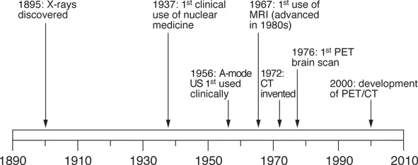
Figure 1.1 Radiology timeline.
AN EVOLVING SPECIALTY
The practice of medicine and surgery has changed immeasurably in recent years with an increasing reliance on diagnostic tests, whether they are biochemical, haematological, or radiological. Patients are better informed and have greater expectations than ever before. Consequently, it is crucial that clinicians have a basic understanding of radiology in order that they can optimise investigations, understand the risks and benefits of different studies, interpret basic investigations and be able to communicate effectively with their patients about these in the clinic or the ward situation.
In response to these developments, radiology has been increasingly incorporated into the medical school curriculum through lectures and film-based teaching. Radiology lends itself very well to examinations and these can take many forms, from multiple choice questions through to the objective structured clinical examination (OSCE) and the viva voce.
The aim of the first sections of this book is to provide medical students with a basic knowledge of the radiology department. What do the tests involve? What issues do I need to consider? What is the radiation dose? How should I present the radiological image? The later chapters aim to educate the reader through the various types of questions that may be encountered: OSCE questions, multiple choice questions, single best answer questions, short answer questions, extended matching items, and viva topics. Each question has a full answer, and the book covers a broad range of radiological topics that one might encounter at this stage of medical education.
Chapter 2
The radiology imaging modalities
X-RAYS/FLUOROSCOPY
X-rays are high energy electromagnetic waves. In diagnostic radiology, they are emitted from a point source, directed through the relevant part of the body and onto a radiographic plate (previously a film). As they pass through the body, X-rays are absorbed to different degrees by different tissues, which is dependent upon their density and thickness, i.e. bone absorbs more than soft tissue, which in turn absorbs more than air. This gives the appearance of white for bone, black for air and grey for the soft tissues when viewed. Fluoroscopy uses continuous low dose X-rays to give a moving image and can be used to give functional information, for example in the evaluation of the GI tract.
Main uses:
• Chest X-ray.
• Abdominal X-ray.
• Extremity films: fractures, arthritis.
• Barium fluoroscopy studies.
• Interventional fluoroscopy (see below).
• Mammography.
Advantages | Disadvantages |
Relatively low radiation dose | Poor soft tissue differentiation |
Quick | Generally poor sensitivity and specificity |
Cheap | |
Available |
COMPUTED TOMOGRAPHY
Computed tomography (CT) comprises a gantry housing an X-ray tube, a bank of detectors, and a table on which the patient lies. The patient moves through the gantry as the X-ray tube and detectors rotate around them at high speed. This creates a threedimensional volume of data within a matter of seconds that can be reconstructed to provide images in any plane. CT uses high doses of ionising radiation to achieve these images, typically up to 10 milli-Sieverts (mSv), the equivalent of around three years of background radiation. Therefore, it is crucial to weigh the risks and benefits of the procedure in each case. CT is widely used to image the brain, thorax, abdomen and pelvis, enabling rapid imaging of even the sickest patient.
Main uses:
• Head injury.
• Abdominal / chest trauma.
• Detection and staging of malignancy.
• Investigation of the acute abdomen.
Advantages | Disadvantages |
Excellent spatial resolution | Ionising radiation |
Available | Nephrotoxicity of iodinated contrast media |
Linear relationship of contrast dose allows | High volume of images for the radiologist to |
characterisation of lesion content | interpret |
High sensitivity and specificity | Cost |
Multi-planar reconstruction | |
Quick |
ULTRASOUND
Ultrasound (US) uses sound waves of 2–15 MHz (above the range of human hearing) to produce images. The ultrasound probe acts as both emitter and receiver of the ultrasound waves in order to create an image. Ultrasound does not utilise any ionising radiation and is considered safe in pregnancy and for use in children, although there is a theoretical risk from the heating of tissues. Ultrasound has many applications and may be used to image the brain (neonates), soft tissues, peripheral vascular system, abdomen and pelvis, as well as the developing foetus. The major limitations of ultrasound are its inability to pass through air (lung, colon) or bone. In obese patients, abdominal or pelvic US may be limited by the depth of penetration of the sound waves.
Main uses:
• Renal tract imaging.
• Biliary imaging / cholecystitis.
• Liver imaging / lesion characterisation.
• Deep vein thrombosis.
• Doppler imaging: carotid stenoses; AAA; post liver / renal transplant vascularity.
• Gynaecological imaging.
• Musculoskeletal imaging.
• Others: breast, thyroid, testes.
Advantages | Disadvantages |
Available | Operator dependent |
Portable | Limited depth penetration |
Non-invasive | Limited anatomical access: US waves cannot penetrate through bone or air |
No ionising radiation | Patient factors: e.g. liver high under the rib cage; limited in obesity |
Real-time imaging(e.g. biopsies) |
MAGNETIC RESONANCE IMAGING
Magnetic resonance imaging (MRI) involves placing a patient on a table in a relatively confined space (often described by patients as a tunnel) within a strong magnetic field. This causes some alignment of the hydrogen atoms within the body along the magnetic field. A series of radiofrequency waves are then used to displace these atoms and, as they return to their original positions, they emit a small radiofrequency signal. An image is constructed from these signals – essentially a map of hydrogen atoms. However, th...
Table of contents
- Cover
- Half Title
- Title Page
- Copyright Page
- Table of Contents
- Foreword
- Preface
- About the editors
- List of abbreviations
- 1 Introduction
- 2 The radiology imaging modalities
- 3 Key topics in radiology: contrast media, radiation protection and the future of radiology
- 4 Objective structured clinical examination (OSCE)
- 5 Multiple choice questions (MCQs)
- 6 Extended matching item (EMI)
- 7 Single best answer (SBA)
- 8 Short answer questions (SAQs)
- 9 Viva voce
- Bibliography
- Index of answers
- Index
Frequently asked questions
Yes, you can cancel anytime from the Subscription tab in your account settings on the Perlego website. Your subscription will stay active until the end of your current billing period. Learn how to cancel your subscription
No, books cannot be downloaded as external files, such as PDFs, for use outside of Perlego. However, you can download books within the Perlego app for offline reading on mobile or tablet. Learn how to download books offline
Perlego offers two plans: Essential and Complete
- Essential is ideal for learners and professionals who enjoy exploring a wide range of subjects. Access the Essential Library with 800,000+ trusted titles and best-sellers across business, personal growth, and the humanities. Includes unlimited reading time and Standard Read Aloud voice.
- Complete: Perfect for advanced learners and researchers needing full, unrestricted access. Unlock 1.5M+ books across hundreds of subjects, including academic and specialized titles. The Complete Plan also includes advanced features like Premium Read Aloud and Research Assistant.
We are an online textbook subscription service, where you can get access to an entire online library for less than the price of a single book per month. With over 1.5 million books across 990+ topics, we’ve got you covered! Learn about our mission
Look out for the read-aloud symbol on your next book to see if you can listen to it. The read-aloud tool reads text aloud for you, highlighting the text as it is being read. You can pause it, speed it up and slow it down. Learn more about Read Aloud
Yes! You can use the Perlego app on both iOS and Android devices to read anytime, anywhere — even offline. Perfect for commutes or when you’re on the go.
Please note we cannot support devices running on iOS 13 and Android 7 or earlier. Learn more about using the app
Please note we cannot support devices running on iOS 13 and Android 7 or earlier. Learn more about using the app
Yes, you can access Radiology for Undergraduate Finals and Foundation Years by Tristan Barrett,Nadeem Shaida,Ashley Shaw in PDF and/or ePUB format, as well as other popular books in Medicine & Medical Theory, Practice & Reference. We have over 1.5 million books available in our catalogue for you to explore.