- 352 pages
- English
- ePUB (mobile friendly)
- Available on iOS & Android
eBook - ePub
Tooth Whitening Techniques
About this book
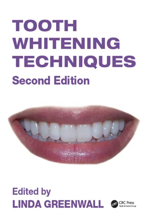
The field of tooth whitening has continued to develop as more and more dental practitioners have turned to cosmetic dentistry and associated aesthetic facial procedures. This new edition of an acclaimed text covers recent technical innovations, but also looks at the latest innovations in practice to treat the single tooth or lesions and white spots. The editor is extremely well placed to give expert advice on how to incorporate whitening into a full aesthetic facial practice.
Tools to learn more effectively

Saving Books

Keyword Search

Annotating Text

Listen to it instead
1 | DISCOLORATION OF TEETH |
INTRODUCTION
Tooth discoloration is a common problem leading patients to seek treatment to have the discoloration removed. People of various ages may be affected, and it can occur in both primary and secondary teeth. The etiology of dental discoloration is multifactorial, and different parts of the tooth can take up different stains. This effect is a result of the anatomy of the tooth. Intrinsic discoloration increases with increasing age and is more common in men (Eriksen and Nordbo 1978). It may affect 31% of men and 21% of women (Ness et al. 1977). The result is a complex of physical and chemical interactions with the tooth surface. The aim of this chapter is to assess the etiology of tooth discoloration and the mechanisms by which teeth stain. It is the intention of this chapter to explain the complexity of tooth discoloration.
COLOR OF NATURAL HEALTHY TEETH
Teeth are polychromatic (Louka 1989). The color varies among the gingival, incisal, and cervical areas according to the thickness, reflectance of different colors, and translucency in enamel and dentin (see Figure 1.2). The color of healthy teeth is primarily determined by the dentin and is modified by the following:
• The color of the enamel covering the crown.
• The translucency of the enamel, which varies with different degrees of calcification.
• The thickness of the enamel, which is greater at the occlusal or incisal edge of the tooth and thinner at the cervical third (Dayan et al. 1983).
• The intensity, thickness, structure of the dentin.
• Presence of secondary or tertiary dentin trauma.
• Existing restorations.
CLASSIFICATION OF DISCOLORATION
Many researchers classify staining as either extrinsic or intrinsic (Dayan et al. 1983, Hayes et al. 1986, Teo 1989). There is confusion concerning the exact definitions of these terms. Feinman et al. (1987) described extrinsic discoloration as that occurring when an agent stains or damages the enamel surface of the teeth, and intrinsic staining as occurring when internal tooth structure is penetrated by a discoloring agent. According to these definitions, the terms staining and discoloration are used synonymously. However, extrinsic staining will be defined here as staining that can be easily removed by a normal prophylactic cleaning (Dayan et al. 1983). Intrinsic staining is defined here as endogenous staining that has been incorporated into the tooth matrix and thus cannot be removed by prophylaxis.
Some discoloration is a combination of both types of staining and may be multifactorial. For example, nicotine staining on teeth is extrinsic staining that becomes intrinsic staining. The modified classification of Dzierzak (1991), Hayes et al. (1986), and Nathoo (1997) will be used as a guide. See Table 1.1.
STAINS DURING ODONTOGENESIS (PRE-ERUPTIVE)
These alter the development and appearance of the enamel and dentin on permanent teeth.
Developmentally defective enamel and dentin
Defects of enamel development (Figures 1.6A and 1.6B) can be caused by, for example, amelogenesis imperfecta (Figure 1.5), dentinogenesis imperfecta, and enamel hypoplasia. The defects in enamel are either hypocalcific or hypoplastic (Rotstein 1998). Enamel hypocalcification is a distinct brownish or whitish area found on the buccal aspects of teeth (see Figure 1.5). The enamel is well formed and the surface is intact. Many of these white and brown discolorations can be removed with whitening in combination with microabrasion (see Chapter 10). Enamel hypoplasia is developmental defective enamel. The surface of the tooth is defective and porous and may be readily discolored by materials in the oral cavity. Depending on the severity and extent of the dysplasia, the enamel surface may be whitened with varying degrees of success. Some enamel white hypoplastic lesions are due to exposure of chemicals (such as bisphenol a) peri- or postnatally (Jedeon et al. 2013).
Fluorosis
This staining is caused by excessive fluoride uptake with the developing enamel layers. The fluoride source can be from the ingestion of excessive fluoride in the drinking water or from overuse of fluoride supplements (Ismail and Hasson 2008) or fluoride toothpastes (Shannon 1978). It occurs within the superficial enamel and appears as white or brown patches of irregular shape and form (Figure 1.7A). The acquisition of stain, however, is posteruptive. The teeth are not discolored on eruption, but because the surface is porous they gradually absorb the colored chemicals present in the oral cavity (Rotstein 1998). Staining caused by fluorosis manifests in three different ways: as simple fluorosis, opaque fluorosis, or fluorosis with pitting (Nathoo and Gaffar 1995). Simple fluorosis appears as brown pigmentation on a smooth enamel surface, whereas opaque fluorosis appears as gray or white flecks on the tooth surface (Figure 1.7B). Fluorosis with pitting occurs as defects in the enamel surface, and the color appears to be darker.
Table 1.1 Etiology of tooth discoloration
Extrinsic stains • Plaque (Figure 1.26), chromogenic bacteria, surface protein denaturation • Mouthwashes (e.g., chlorhexidine) • Beverages (tea [Figure 1.28], coffee [Figures 1.27 and 1.31], red wine, cola) • Foods (curry, cooking oils and fried foods [Figure 1.33], foods with colorings, berries, beetroot) • Illness • Antibiotics (erythromycin [Figure 1.29], amoxicillins [Figures 1.6A and 1.30]) • Iron supplements (Figures 1.34) |
Intrinsic stains |
Pre-eruptive |
Disease • Hematologic diseases (Figure 1.15) • Liver diseases • Diseases of enamel and dentin (Figure 1.5) |
Medication • Tetracycline stains (Figures 1.8–1.10) • Other antibiotics • Fluorosis stains (Figure 1.7) |
Posteruptive • Trauma (Figure 1.4), intrapulpal hemorrhage (Figure 1.14A), pulp necrosis (Figure 1.15B) • Primary and secondary caries (Figure 1.27) and erosion, buccal (Figure 1.36) and palatal (Figures 1.22–1.24, 1.38) • Dental restorative materials (Figures 1.17 and 1.18), endodontic materials (Figure 1.4, lower right incisor tooth) • Aging (Figure 1.25) • Smoking (Figures 1.33 and 1.35) • Chemicals • Som... |
Table of contents
- Cover
- Half Title
- Title Page
- Copyright Page
- Table of Contents
- Foreword
- Preface to Second Edition
- Acknowledgments
- Contributors
- 1 Discoloration of Teeth
- 2 The Science of Tooth Whitening
- 3 Tooth Whitening Materials
- 4 Treatment Planning for Successful Whitening
- 5 The Home Whitening Technique
- 6 Home Whitening Trays: How to Make Them
- 7 In-Office Power Bleaching
- 8 Intracoronal Bleaching of Nonvital Teeth
- 9 Single Tooth Whitening of Vital Teeth
- 10 Tooth Whitening, the Microabrasion Technique, and White Spot Eradication
- 11 Molar Incisor Hypoplasia
- 12 Management of Tetracycline Discoloration
- 13 Whitening Treatments for Tetracycline Discoloration
- 14 Over-the-Counter Whitening Strips
- 15 Combining Whitening Techniques and Minimally Invasive Treatments
- 16 The Effect of Whitening on Restorative Materials
- 17 A Guide to Esthetic Treatment after Whitening
- 18 Comparison of Tooth Bleaching Results
- 19 Nightguard Vital Bleaching: Post-Treatment Effects, Longevity, and Long-Term Results
- 20 Tooth Sensitivity Associated with Tooth Whitening
- 21 Safety and Toxicologic Considerations for Tooth Bleaching
- 22 Whitening for Patients Younger than 18: Index of Treatment Need
- 23 Managing and Developing a Successful Whitening Practice
- 24 Whitening, Therapeutic Esthetics, and Oral Health Improvement: The Future
- Index
Frequently asked questions
Yes, you can cancel anytime from the Subscription tab in your account settings on the Perlego website. Your subscription will stay active until the end of your current billing period. Learn how to cancel your subscription
No, books cannot be downloaded as external files, such as PDFs, for use outside of Perlego. However, you can download books within the Perlego app for offline reading on mobile or tablet. Learn how to download books offline
Perlego offers two plans: Essential and Complete
- Essential is ideal for learners and professionals who enjoy exploring a wide range of subjects. Access the Essential Library with 800,000+ trusted titles and best-sellers across business, personal growth, and the humanities. Includes unlimited reading time and Standard Read Aloud voice.
- Complete: Perfect for advanced learners and researchers needing full, unrestricted access. Unlock 1.4M+ books across hundreds of subjects, including academic and specialized titles. The Complete Plan also includes advanced features like Premium Read Aloud and Research Assistant.
We are an online textbook subscription service, where you can get access to an entire online library for less than the price of a single book per month. With over 1 million books across 990+ topics, we’ve got you covered! Learn about our mission
Look out for the read-aloud symbol on your next book to see if you can listen to it. The read-aloud tool reads text aloud for you, highlighting the text as it is being read. You can pause it, speed it up and slow it down. Learn more about Read Aloud
Yes! You can use the Perlego app on both iOS and Android devices to read anytime, anywhere — even offline. Perfect for commutes or when you’re on the go.
Please note we cannot support devices running on iOS 13 and Android 7 or earlier. Learn more about using the app
Please note we cannot support devices running on iOS 13 and Android 7 or earlier. Learn more about using the app
Yes, you can access Tooth Whitening Techniques by Linda Greenwall in PDF and/or ePUB format, as well as other popular books in Medicine & Dentistry. We have over one million books available in our catalogue for you to explore.