There is a growing need to understand and combat potential radiation damage problems in semiconductor devices and circuits. Assessing the billion-dollar market for detection equipment in the context of medical imaging using ionizing radiation, Electronics for Radiation Detection presents valuable information that will help integrated circuit (IC) designers and other electronics professionals take full advantage of the tremendous developments and opportunities associated with this burgeoning field.
Assembling contributions from industrial and academic experts, this book—
Addresses the state of the art in the design of semiconductor detectors, integrated circuits, and other electronics used in radiation detection
Analyzes the main effects of radiation in semiconductor devices and circuits, paying special attention to degradation observed in MOS devices and circuits when they are irradiated
Explains how circuits are built to deal with radiation, focusing on practical information about how they are being used, rather than mathematical details
Radiation detection is critical in space applications, nuclear physics, semiconductor processing, and medical imaging, as well as security, drug development, and modern silicon processing techniques. The authors discuss new opportunities in these fields and address emerging detector technologies, circuit design techniques, new materials, and innovative system approaches.
Aimed at postgraduate researchers and practicing engineers, this book is a must for those serious about improving their understanding of electronics used in radiation detection. The information presented here can help you make optimal use of electronic detection equipment and stimulate further interest in its development, use, and benefits.
Trusted by 375,005 students
Access to over 1 million titles for a fair monthly price.
1 The Future of Medical Imaging Understanding Our True Limitations
Mark Nadeski and Gene Frantz
CONTENTS
1.1Introduction
1.2Where Are We Going?
1.2.1The EyeCam
1.3Making Health Care More Personal
1.3.1Advances in Digital and Medical Imaging
1.3.2How Telecommunications Complements Medical Imaging
1.3.3Automated Monitoring
1.4The Future of Technology
1.4.1Remembering Our Focus
1.4.2What We Can Expect from Technology
1.4.3Development Cost
1.4.4Performance
1.4.5Multiprocessor Complexity
1.4.5.1Multiprocessing Elements
1.4.6Power Dissipation
1.4.6.1Lower Power into the Future
1.4.6.2Perpetual Devices
1.4.7Integration through SoC and SiP
1.5Defining the Future
References
1.1 INTRODUCTION
There are those who fear that technology is nearly at the physical limitations of our understanding of nature, so where can we possibly go from here? However, technology is not where our limits lie. Integrated circuits (ICs) have always exceeded our ability to fully utilize the capacity they make available to us, and the future will be no exception. Indeed, technology does not drive innovation: Innovation and human imagination are the forces that drive technology.
1.2 WHERE ARE WE GOING?
The broad field of medical imaging has seen some truly spectacular advances in the last half-century that most of us take for granted. Once marvels only in the laboratory, advances such as real-time and Doppler ultrasonography, functional nuclear medicine, computed tomography, magnetic resonance imaging, and interventional angiography have all become available in clinical settings.
It’s easy to sit back in wonder at how far the field of medical imaging has come. However, in this chapter we will glimpse the future. Some of this future is quickly taking shape today, though some of it will not arrive for years, if not decades.
Specifically, we’ll look at how advances in medical imaging are based on existing technology; how these technologies will provide more capacity and capabilities than we can conceivably exploit; and how they finally lead to the conclusion that the future of medicine is not limited by what we know, but rather by what we can imagine.
Let’s begin by looking at the edge of what is real—that wonderful place where ideas are transformed into reality.
1.2.1THE EYECAM
For centuries, humanity has dreamed of being able to make the blind see. And, for as long, restoring a person’s eyesight has been considered a feat commonly categorized as “a miracle.”
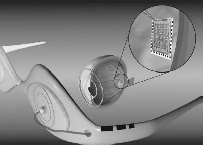
FIGURE 1.1 Example of the EyeCam, created and tested at the University of Southern California.
About 10 years ago, Texas Instruments (TI) began collaborating with a medical team at Johns Hopkins, well known for its ability to make miracles happen. The team’s goal was to develop a way to take the signal from a camera and turn it into electrical impulses that could then be used to excite the retina, as shown in Figure 1.1. If successful, they could return some level of vision back to individuals who had lost their eyesight due to retinitis pigmentosa, a disease that affects more than 100,000 people in the United States alone.
Now at the University of Southern California, this team continues to make significant progress. The project has evolved considerably over the years. Its initial conception consisted of mounting a camera on a pair of glasses that would require patients to rotate their heads in order to look around. Today the team is working to actually implant a camera module within the eye, since it is much more natural to let the eye do the moving to point the camera in the right direction. However, it’s one thing to say implanting a camera in a person’s eye is more practical than mounting it on glasses and quite another to achieve it. A number of challenges come to mind:
Size: The complete camera module has to be significantly smaller than an eyeball in order to fit.
Power: The camera must have exceptionally low power consumption. At the very least, the energy needs to be scavenged from body heat, the surrounding environment, or a yet-to-be-invented wireless power circuit.
Heat: Initial cameras may rely upon a connected power source. Even so, it is critical that the camera not produce much heat. To be practical, the camera must be able to dissipate enough power so as not to heat the eye to the point of discomfort.
Durability: The camera must be packaged in such a way as to be protected from the fluids in the eye.
Currently working with Georgia Tech University and experts at TI, the team at USC is busy making all this happen. Is such an ambitious project even possible? Although success has yet to be seen, the team envisions a successful completion of the project. And they have good reason to be confident, for they are only pressing at the edges of possibility.
Much of what lies ahead of us in medicine is the identification of technologies and devices from other parts of our world that we can apply to medical electronics. For example, Prof. Armand R. Tanguay Jr., principal investigator on the EyeCam project, acknowledges that they have many ideas about where else in the body they could implant a camera.*
Here a camera, there a camera,
In the eye a little camera.
Old Doc Donald had a patient.
E, I, E, I, O.
Certainly there is more than one verse to this song. The question we might ask ourselves is, “What do we imagine we need next?”
1.3 MAKING HEALTH CARE MORE PERSONAL
A device that can help the blind to see is a life-changing application of medical technology. Not all medical devices will have such a dramatic effect on the way we live. Most of the changes in medical care will have a much lower profile, for they will be incorporated into our daily lives. However, while their application may be more subtle, the end result will certainly be profound.
The future of medicine is based upon a firm foundation of existing technologies. What is new, in many cases, is not the technology itself, but rather how the technology is applied in new ways. Consider these key technologies:
•Digital imaging
•Telecommunications
•Automated monitoring
Each of these technologies is already firmly established in a number of disparate industries. Specifically applying them to medicine will still require creativity and hard work, but will enable entirely new applications. Perhaps most importantly, for health care providers and their patients, the resulting advances will help shift health care into becoming a more routine part of daily life, creating a future where medical devices help us to:
1.Manage our chronic conditions
2.Predict our catastrophic diseases
3.Live out our final years in the comfort of our homes
1.3.1ADVANCES IN DIGITAL AND MEDICAL IMAGING
Improving health care is the ultimate goal behind advances in medicine. As medical imaging advances, it will allow patients to have more personalized and targeted health care. Imaging, diagnosis, and treatment plans will continue to become more specialized and customized to a patient’s particular needs and anatomy. We may even see therapies that are tailored to genetics. Look at how far we’ve come already:
Migration to digital files: Photographic plates were once used to “catch” X-ray images. These plates gave way to film, which in turn is now giving way to digital radiography. Through the use of advanced digital signal processing, X-ray signals now can be converted to digital images at the point of acquisition while imposing no loss in image clarity. Digital files have a variety of benefits, including eliminating the time and cost of processing film, as well as being a more reliable storage medium that can be transferred near-instantaneously across the world.
Real-time processing: The ability to render digital images in real time expands our ability to monitor the body. Using digital X-ray machines during surgical procedures, doctors can view a precise image at the exact time of surgery. Real-time processing also increases what can be done noninvasively. For example, the Israeli company CNOGA* uses video cameras to noninvasively measure vital signs such as blood pressure, pulse rate, blood oxygen level, and carbon dioxide level simply by focusing on the person’s skin. Future applications of this technology may lead to identifying biomarkers for diseases such as cancer and chronic obstructive pulmonary disease (COPD).
Evolution from slow and fuzzy to fast and highly detailed: Today’s magnetic resonance imagers (MRIs) can provide higher quality images in a fraction of the time it took state-of-the-art machines just a few years ago. These digital MRIs are also highly flexible, with the ability to image, for example, the spine while it is in a natural, weight-bearing, standing position. With diffusion MRIs, researchers can use a procedure known as tractography to create brain maps that aid in studying the relationships between disparate brain regions. Functional MRIs, for their part, can rapidly scan the brain to measure signal changes due to changing neural activity. These highly detailed images provide deeper insights into how the brain works—insights that will be used to improve treatment and guide future imaging equipment.
Moving from diagnostic to therapeutic: High-intensity focused ultrasound (HIFU) is part of a trend in health care toward reducing the impact of procedures in terms of incision size, recovery time, hospital stays, and infection risk. But unlike many other parts of this trend, such as robot-assisted surgery, HIFU goes a step further to enable procedures currently done invasively to be done noninvasively. Transrectal ultrasound,† for example, destroys prostate cancer cells without damaging healthy, surrounding tissue. HIFU can also be used to cauterize bleeding, making HIFU immensely valuable at disaster sites, accident scenes, and on the battlefield. Focused ultrasound even has a potential role in a wide variety of cosmetic procedures, from melting fat to promoting formation of secondary collagen to eradicate pimples.
The portability of ultrasound: Ultrasound equipment continues to become more compact. Cart-b...
Table of contents
Cover
Half Title
Title Page
Copyright Page
Table of Contents
Preface
About the Editor
Contributors
Chapter 1 The Future of Medical Imaging: Understanding Our True Limitations
Chapter 2 Detector Front-End Systems in X-Ray CT: From Current-Mode Readout to Photon Counting
Chapter 3 Photon-Counting Energy-Dispersive Detector Arrays for X-Ray Imaging
Chapter 4 Planar and PET Systems for Drug Development
Chapter 5 PET Front-End Electronics
Chapter 6 Design Considerations for Positron Emission Tomography (PET) Scanners Dedicated to Small-Animal Imaging
Chapter 7 Geiger-Mode Avalanche Photodiodes for PET/MRI
Chapter 8 Current-Mode Front-End Electronics for Silicon Photomultipliers
Chapter 9 Integrated Charge-Measuring Systems for Radiation Detectors in CMOS Technologies
Chapter 10 Current- and Charge-Sensitive Signal Conditioning for Position Determination
Chapter 11 Analog-to-Digital Converters for Radiation Detection Electronics
Chapter 12 Low-Power Integrated Front-End for Timing Applications with Semiconductor Radiation Detectors
Chapter 13 Time-to-Digital Converter Circuits in Radiation Detection Systems
Index
Frequently asked questions
Yes, you can cancel anytime from the Subscription tab in your account settings on the Perlego website. Your subscription will stay active until the end of your current billing period. Learn how to cancel your subscription
No, books cannot be downloaded as external files, such as PDFs, for use outside of Perlego. However, you can download books within the Perlego app for offline reading on mobile or tablet. Learn how to download books offline
Perlego offers two plans: Essential and Complete
Essential is ideal for learners and professionals who enjoy exploring a wide range of subjects. Access the Essential Library with 800,000+ trusted titles and best-sellers across business, personal growth, and the humanities. Includes unlimited reading time and Standard Read Aloud voice.
Complete: Perfect for advanced learners and researchers needing full, unrestricted access. Unlock 1.4M+ books across hundreds of subjects, including academic and specialized titles. The Complete Plan also includes advanced features like Premium Read Aloud and Research Assistant.
Both plans are available with monthly, semester, or annual billing cycles.
We are an online textbook subscription service, where you can get access to an entire online library for less than the price of a single book per month. With over 1 million books across 990+ topics, we’ve got you covered! Learn about our mission
Look out for the read-aloud symbol on your next book to see if you can listen to it. The read-aloud tool reads text aloud for you, highlighting the text as it is being read. You can pause it, speed it up and slow it down. Learn more about Read Aloud
Yes! You can use the Perlego app on both iOS and Android devices to read anytime, anywhere — even offline. Perfect for commutes or when you’re on the go. Please note we cannot support devices running on iOS 13 and Android 7 or earlier. Learn more about using the app
Yes, you can access Electronics for Radiation Detection by Krzysztof Iniewski in PDF and/or ePUB format, as well as other popular books in Technology & Engineering & Biotechnology in Medicine. We have over one million books available in our catalogue for you to explore.