![]()
1
Mummies
Definition and Mechanisms
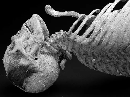
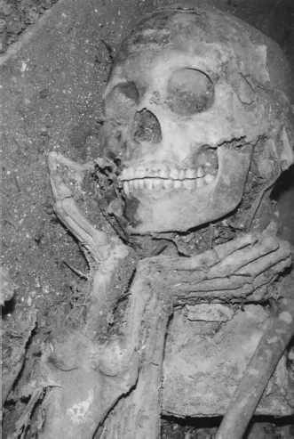
When contemplating mummies, many folks may automatically conjure up images of a linen-wrapped, well-preserved, and life-like body: there is, however, quite a bit of variability in what may be considered ‘mummified.’ Mummified remains exhibit some degree of soft tissue preservation, though the boundary between ‘mummy’ and ‘skeleton’ is somewhat ill-defined and fuzzy.1 Lynnerup (2007: 441) writes that a body is a mummy when the “soft tissue preservation is so pronounced that body parts, or the whole body, have somewhat intact skin and some preserved internal structures such as muscle fasciae, ligaments and maybe even tissue of internal organs and muscle.” Aufderheide (2003: 41) defines a mummy as “a physically preserved corpse or tissue that resembles its living morphology but resists further decay for a prolonged postmortem interval.” These definitions permit some flexibility regarding what may be classified as a ‘mummy.’ While no one would question the ‘mummy’ status of remains such as Ötzi the Tyrolean Iceman (see Figure 4.9) or the Ice Maiden from Peru (see Figure 1.2), it becomes less clear when discussing bodies that are not as well preserved. For instance, the remains described by Gaudio and colleagues (2014) and Alt et al. (2003) appear to be mostly skeletonized (Figure 1.3), with only remnants of skin and connective tissue preserved. To take this even further, while some of the prepared Chinchorro bodies have been completely stripped of flesh, they are generally considered and discussed as ‘mummies.’ Thus, while Lynnerup’s definition emphasizes soft tissue preservation, Aufderheide’s definition focuses on the final appearance of the remains and perhaps permits a bit more latitude regarding what may be considered ‘mummified remains.’ Because there are no minimum standards for being a ‘mummy,’ the decision to describe remains as mummified is effectively left up to the investigator. As will be discussed later, however, there have been recent attempts to create more systematic methods for assessing and describing the degree and amount of soft tissue preservation.
Regardless of the extent of soft tissue preservation, mummies are categorized in two ways: the physical and/or chemical processes by which soft tissue is preserved, and the degree to which these processes result from either human action or environmental factors. Categories defined by the mechanisms of mummification are based on (1) taphonomy and (2) the manner and degree to which the natural sequence and timing of tissue decomposition has been altered. Research on the taphonomy of soft tissue preservation draws heavily from the natural and biomedical sciences (e.g., Mayer et al. 1997). Research in the latter category hinges upon the reconstruction of behavior based on the presence or absence of evidence for intentional human intervention. Here the primary distinctions made are between anthropogenic and spontaneous mummification and draws more explicitly from anthropology and archaeology speaking to the broader social and environmental context in which mummification occurred.
Mechanisms of Mummification
Mummification results from the interruption of decomposition. Decomposition encompasses three different processes: autolysis, putrefaction, and decay (Carter et al. 2007; Tibbet and Carter 2008). Autolysis (‘self-digestion’) can begin almost immediately after death (Vass et al. 1992) and is due to the release of hydrolytic enzymes that begin to digest the body’s tissues. This leads to putrefaction—the breakdown of lipids, carbohydrates, and proteins and the accumulation of organic acids and gases (Tibbet and Carter 2008). Autolysis and putrefaction characterize the ‘fresh’ and ‘bloated’ phases of decomposition, respectively; rupturing of the skin marks the onset of active decay (which in turn can be subdivided into different stages, Tibbet and Carter 2008). During this phase, most of the mass of the corpse is lost through purging of cadaveric fluid. In forensic contexts, establishing the postmortem interval is dependent upon reconstructing the sequence and timing of these stages and the potential influence of environmental and anthropogenic variables.
A host of environmental factors can influence the progression of decomposition and subsequently increase the likelihood of mummification. These include the presence/absence of an aqueous medium, acidity, temperature, substrate specificity, and the presence of inhibitors such as heavy metal ions. Aufderheide (2003: 43) has identified seven primary, though not necessarily mutually exclusive, mechanisms of mummification: desiccation, thermal effects, chemical effects, anaerobiasis, excarnation, miscellaneous, and indeterminate.2 I will provide a short summary of each of these mechanisms and refer the reader to Aufderheide (2003) for a fuller treatment.
Desiccation
The most important environmental condition that influences whether soft tissue is preserved is the speed at which water is removed from the body. Desiccation is the most common mummification mechanism and involves the removal of water from the body via evaporation, osmosis, or the application of heat (Aufderheide 2003) and may be encouraged through both human/cultural intervention and environmental factors.
Human intervention in the decomposition process, including evisceration, excerebration, chemical treatments (e.g., immersion in natron), and exposure to heat, fire, or smoke, increases the likelihood of mummification by reducing the amount of water in the body or accelerating water loss. A more unusual example of intentional desiccation is the ‘self-mummification’ documented in some Buddhist priests (Hori 1962; Ritzinger and Bingenheimer 2006). It has been suggested that a gradual reduction in food and water intake resulted in dehydration and loss of body fat, which, upon death, would have encouraged mummification. In most instances, these antemortem steps were accompanied by additional postmortem measures to ensure preservation (Cuong 1982–84; Gildow and Bingenheimer 2002; Sakurai et al. 1998).
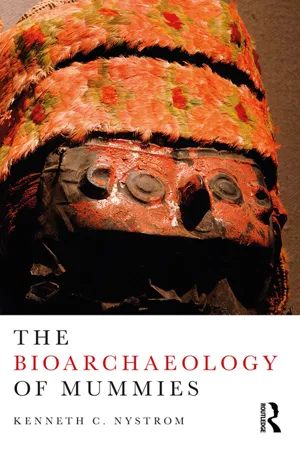
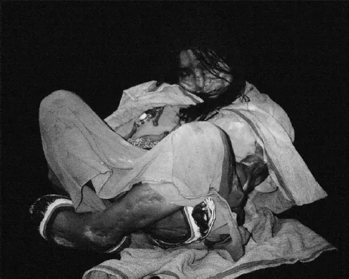
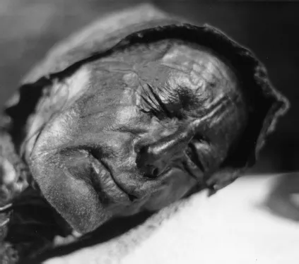
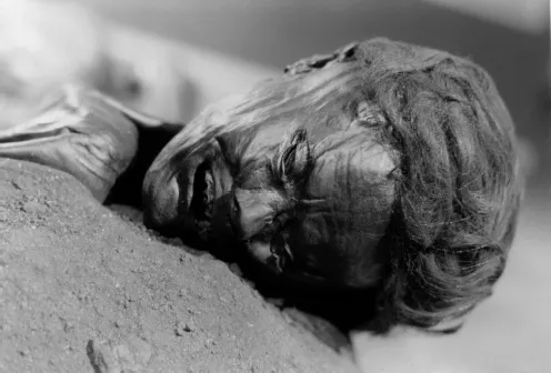
Spontaneously desiccated mummies have been described from a wide range of arid environments, from hot deserts to cold mountain-top sites. Examples of spontaneously desiccated mummies include some of the remains from the Romano-Byzantine period cemetery site of Kellis-1 in Egypt (Aufderheide et al. 2004a), the remains from the Hets Mountain cave site in Mongolia (Figure 1.1; Turner et al. 2012), the Late Horizon Volcano Llullaillaco mummies from Argentina (Figure 1.2; Ceruti 2015), and remains from Muktinath Valley in the Himalayas, Western Nepal (Figure 1.3; Alt et al. 2003).
Figure 1.1 Spontaneously desiccated mummified remains recovered from the Hets Mountain Cave site in Mongolia (Turner et al. 2012).
Figure 1.2 The Llullaillaco Maiden. Photo by Maria Constanza Ceruti 2015.
Thermal Effects
Enzymatic and bacterial activity are highly dependent upon temperature, with lower temperatures reducing, and ultimately halting, bacterial activity. Freezing environmental conditions can result in frozen ‘wet’ mummies, such as Ötzi the Tyrolean Iceman and members of the Franklin Expedition, where tissues remain hydrated (Janko et al. 2012). Alternatively, cold temperatures can also result in freeze-drying through sublimation of water, such as is observed in the Inuit mummies from Greenland (Hart Hansen and Nordqvist 1996). Exposure to heat can inhibit bacterial activity, but this may be secondary relative to the acceleration of water loss (Aufderheide 2003). Exposure to the sun or heat from a fire has been suggested to be involved in the production of the Guanche mummies (Rodríguez-Martin 1996).
Chemical Effects
Several different chemical processes can result in soft tissue preservation including the presence of heavy metals, chelation, smoking, and adipocere formation. In many cases, these chemical effects are likely secondary relative to other mummification mechanisms such as desiccation.
Figure 1.3 An example of a spontaneous mummy from the Muktinath Valley in the Himalayas, Western Nepal (Alt et al. 2003).
Heavy Metals
The presence of heavy metal ions such as arsenic, mercury, and copper reduces an enzyme’s ability to bind to a substrate that inhibits decomposition (Aufderheide 2003). The presence of mercury has been suggested to be responsible for tissue preservation observed in a 320-year-old mummy from Japan (Yamada et al. 1990) and may have been used during the Han Dynasty in China as a preservative (Werning 2010). Preservation of the “Copper Man” from Chile (Bird 1979) and a corpse from British Columbia (Schulting 1995) may be due to the presence of copper, but desiccation is likely the primary mummification mechanism in both instances (Aufderheide 2003). Recently, the presence of copper plates has been implicated in the preservation of soft tissue observed in a young child from the Zeleny Yar necropolis in Russia.3 Although results have not been published, news articles on these remains indicate that the body was wrapped in fur and then covered with copper or bronze plates. The investigators suggest that soft tissue preservation was accidental and due to burial in permafrost and the presence of copper plates. Based on the photos and descriptions provided, however, the copper plates do not appear to be in direct contact with the soft tissue and thus their contribution to the mummification process would seem to be limited.
Chelation
The preservation of soft tissue observed in bog bodies (Figures 1.4 and 1.5) results from the action of sphagnan, a pectin-like carbohydrate polymer produced by sphagnum moss. The sphagnan binds calcium ions, which not only results in the characteristic decalcification of the bones in bog bodies but also makes calcium unavailable for bacterial metabolism. Sphagnan also reacts with free amino groups and reducing sugars in what is called a Maillard reaction. This reaction results in the cross-linking of collagen fibers and in a preservation process akin to tanning (Aufderheide 2003).
Figure 1.4 The face of Tollund Man, a bog body from Denmark, discovered in 1950. Photo by Sven Rosborn.
Figure 1.5 The bog body known as Grauballe Man. Photo by Sven Rosborn.
Smoking
Mummification among the modern Anga of Koke Village in Papua New Guinea involves keeping the body in a hut with a smoky fire for approximately thirty days (Beckett et al. 2011a, b). The increase in temperature encourages desiccation, but the formaldehyde in smoke creates cross-linkages between collagen fibers that can also promote soft tissue preservation. Other mummification traditions in which smoking may have played a role are the mud-coated Chinchorro mummies (Arriaza 1995a) and the protohistoric Maori (Orchiston 1971).
Adipocere
Adipocere, more commonly known as grave wax, results from the conversion of body fat by microbial activity (species of the Clostridium genus) into saturated free fatty acids with even-numbered carbon atoms: predominantly myristic acid, palmitic acid, stearic acid, and 10-hydroxystearic acid (Bereuter et al. 1997; Mayer et al. 1997). The environmental conditions conducive for adipocere formation can be quite variable, though typically it is associated with wet or damp environments. Forensic research has demonstrated that key factors in adipocere formation include mildly alkaline reducing anaerobic conditions, warm temperatures, and some source of moisture (Fiedler and Graw 2003; Forbes et al. 2005a, b). Adipocere has been demonstrated to develop in cold, acidic environments as well, though its formation may be slower (Forbes et al. 2005a).
Aufderheide (2003: 53) considers adipocere as a form of mummification because it can be quite stable and can “resist subsequent chemical change and thus tends to preserve the tissue’s gross morphology,” though there is some disagreement (see Ubelaker and Zarenko 2011 for a brief summary). Adipocere has been observed in bodies in which preservation of soft tissue is quite good (e.g., Bereuter et al. 1997; Dickson et al. 2004; Murphy et al. 2003) as well as in instances in which remains are mostly skeletonized.
Fiedler et al. (2009: 1328) describe a substance recovered with the skeleton of a child from the Late Roman period in the city of Mainz, Germany (Figure 1.6). Most of the body was covered in a thick layer of adipocere, with only the skull and distal extremities being skeletonized. The remains were contained in a stone sarcophagus, buried in moist clay soil adjacent to the Rhine. The authors suggest that periodic flooding may have filled the sarcophagus with water, creating an anaerobic environment that led to the formation of the adipocere. Chemical analysis demonstrated that the substance in question had only traces of the fatty acid methyl esters typically found in adipocere, but the ratios in which they were found is similar to that of modern adipocere (Fiedler et al. 2009: 1332).
Surprisingly, given the high lipid content of the brain and the remarkable number of brains that have been recovered from archaeological contexts (Kim et al. 2008; Papageorgopoulou et al. 2010; Prats-Muñoz et al. 2012; see O’Connor et al. 2011 for a complete list), only two instances have occurred wherein preservation has been explained as resulting from adipocere formation. Tkocz et al. (1979) note that frequent flooding and an alkaline anaerobic clay soil at the medieval churchyard in Svendborg, Denmark, likely facilitated adipocere formation and the brain preservation. Papageorgopoulou et al. (2010) describe the brain recovered from the skull of an eighteen-month-old infant from a thirteenth-century site in Quimper-Bretagne, France (Figure 1.7). This individual was wrapped in a “leather envelope and deposited into a wooden coffin” in an acidic...