This book aims to bring to the reader an overview of different applications of brain-computer interfaces (BCIs) based on more than 20 years of experience working on these interfaces. The author provides a review of the human brain and EEG signals, describing the human brain, anatomically and physiologically, with the objective of showing some of the patterns of EEG (electroencephalogram) signals used to control BCIs. It then introduces BCIs and different applications, such as a BCI based on ERD/ERS Patterns in ? rhythms (used to command a robotic wheelchair with an augmentative and alternative communication (AAC) system onboard it); a BCI based on dependent-SSVEP to command the same robotic wheelchair; a BCI based on SSVEP to command a telepresence robot and its onboard AAC system; a BCI based on SSVEP to command an autonomous car; a BCI based on independent-SSVEP (using Depth-of-Field) to command the same robotic wheelchair; the use of compressive technique in SSVEP-based BCI; a BCI based on motor imagery (using different techniques) to command a robotic monocycle and a robotic exoskeleton; and the first steps to build a neurorehabilitation system based on motor imagery of pedalling together an in immersive virtual environment. This book is intended for researchers, professionals and students working on assistive technology.
eBook - ePub
Introduction to Non-Invasive EEG-Based Brain-Computer Interfaces for Assistive Technologies
- 84 pages
- English
- ePUB (mobile friendly)
- Available on iOS & Android
eBook - ePub
Introduction to Non-Invasive EEG-Based Brain-Computer Interfaces for Assistive Technologies
About this book
Trusted by 375,005 students
Access to over 1.5 million titles for a fair monthly price.
Study more efficiently using our study tools.
Information
1 Review of the Human Brain and EEG Signals
Alessandro Botti Benevides, Alan Silva da Paz Floriano, Mario Sarcinelli-Filho, and Teodiano Freire Bastos-Filho
CONTENTS
1.1 Planes of Section and Reference Points of the Human Brain
1.2 Details of S1, M1, A1, V1, Wernicke’s, and Broca’s Areas
1.3 Pyramidal Tract and Contralaterality of Motor Movements
1.4 Neuronal Circuits and Oscillatory Activity of the Thalamocortical System
1.5 Direct Pathway of Movement
1.6 EEG Signal
1.7 EEG Electrodes
1.8 EEG Acquisition
1.9 Main EEG Rhythms
1.10 Artifacts
1.11 Spatial Filtering
1.11.1 Event-Related Potential (ERP)
1.12 Movement-Related (Cortical) Potentials (MRPs/MRCPs)
1.13 ERD/ERS
1.14 Steady-State Visual Evoked Potentials (SSVEPs)
1.14.1 Dependent and Independent SSVEP
References
The purpose of this chapter is to describe the human brain, anatomically and physiologically, to better understand some of the patterns observed in EEG (electroencephalogram) signals used to distinguish distinct mental tasks to control a BCI (brain–computer interface).
In our body, the nervous system, comprising the central nervous system (CNS) and the peripheral nervous system (PNS), coordinates and monitors all our conscious and unconscious activity. The CNS consists of the brain and spinal cord, and the PNS consists of nerves1 and ganglia2 [1]. In the next section, we will focus on CNS and its main constituent, the brain.
1 Groupings of axons in the PNS. Only one group of CNS axons is named as nerve, which is the optic nerve [1].
2 Greek word meaning “node”, which is an agglomerate of cell bodies of neurons found outside the CNS [1].
1.1 Planes of Section and Reference Points of the Human Brain
The relative position of brain structures is located through planes of section and reference points. This section covers some terms used throughout this book to locate and describe brain structures. The portion of the brain facing forward, regarding the human body, is called anterior, and the portion facing backward is called posterior. On the other hand, the direction facing upward is called dorsal and the direction facing downward is called ventral. Figure 1.1 shows the directions and the three planes of section that are the sagittal, coronal, and horizontal. The structures nearest to the medial line are called medial structures, and structures furthest from the medial line are called lateral structures. The structures that are on the same side of the medial line are called ipsilateral to each other, and the structures that are on opposite sides of the medial line are contralateral to each other. Finally, similar structures that are on both sides of the medial line are bilateral.
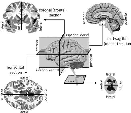
FIGURE 1.1 Planes of section and reference points of the human brain.
The brain surface is composed of numerous circumvolutions, which are the evolutionary result of the brain’s attempt to increase its cortical area, being confined to the skull. The protrusions are called gyri, and the grooves are called sulci; very deep sulci are called fissures. The exact pattern of gyri and sulci may vary considerably from individual to individual, but many features are common to all human brains.
By convention, the brain is divided into lobes, based on the overlying skull bones: the central sulcus separates the frontal lobe from the parietal3 lobe; and the lateral sulcus, or Sylvian fissure,4 separates the frontal lobe and the temporal5 lobe; and the occipital6 lobe is located on the caudal region of the brain, and is surrounded by the parietal and temporal lobes [1].
The temporal lobe receives and processes auditory information, which is related to object identification and naming. The frontal lobe (including the motor, the premotor, and prefrontal cortexes) is involved in planning actions and movements, as well as abstract thought. The parietal lobe is the primary somatosensory cortex and receives information about touch and pressure from thalamus, and the occipital lobe receives and processes visual information [2].
3 The term “parietal” is derived from Latin, “parietalis”, meaning wall.
4 Assigned in tribute to Franciscus Sylvius (1614–1672), who was a Dutch physician and scientist.
5 The term “temporal” arises from Latin “tempus” meaning time. The word “time” was used for this region because it is typically on the sides of the skull where hair first becomes gray, showing the ravages of time.
6 The term “occipital” means something situated near the “occiput”, which is derived from Latin prefix “ob” combined with “caput” meaning “at the back of head”.
The cerebral surface or cortex7 is organized like a patchwork quilt, which were first identified and numbered by Brodmann8 (Figure 1.2). The main areas related to the processing of the senses are the primary motor cortex, or M1 (area 4), the supplementary motor area (SMA), and premotor area (PMA) (area 6) in the frontal lobe; the primary somatosensory cortex, or S1 (areas 1, 2, and 3), and the primary gustatory cortex (area 43) in the parietal lobe; the primary auditory cortex, or A1 (areas 41 and 42), and the olfactory cortex in the temporal lobe; and the primary visual cortex, or V1 (area 17), in the occipital lobe.
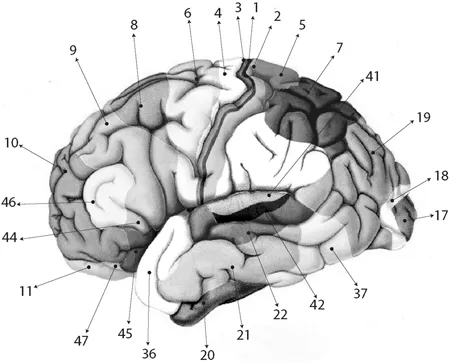
FIGURE 1.2 Brodmann’s cytoarchitectonic map. (Adapted from [1].)
In the context of the mental tasks addressed in this book, the areas 4 (M1) and 6 (SMA and PMA) are related to motor mental tasks, whereas areas 39 and 40 (Wernicke’s area), and 44 and 45 (Broca’s area) are related to the tasks of imagination of words, which are detailed in the next section. On the other hand, the areas 41 and 42 (A1) are related to music imagery tasks, and the area 17 (V1) is related to visual tasks (Figures 1.2 and 1.3).
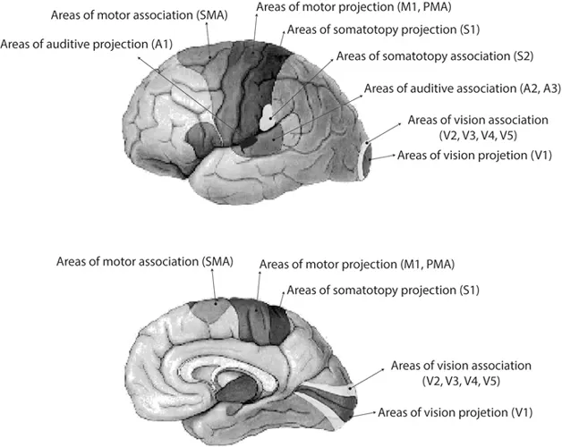
FIGURE 1.3 Main areas related to the processing of the senses. (Adapted from [1].)
1.2 Details of S1, M1, A1, V1, Wernicke’s, and Broca’s Areas
The primary motor cortex is directly responsible for the coordination of voluntary movements. The left side of Figure 1.4 shows the somatotopic9 map of M1, which correlates some M1 areas with the control of body parts. It is worth noting that more than a half of M1 comprises the control of muscles linked to hands and speech [2].
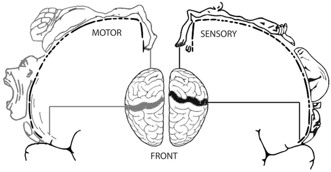
FIGURE 1.4 Left: Somatotopic map of human precentral gyrus (M1); right: somatotopic map of human postcentral gyrus (S1).
The right side of Figure 1.4 shows the somatotopic map of S1, correlating areas of the somatosensory cortex with the sensitivity of various areas of the body. Notice that the somatotopic organization of human precentral gyrus (M1) is very similar to that observed in somatosensory areas of postcentral gyrus (S1). Electrophysiological changes of areas corresponding to movements of hands and feet are located in the precentral gyrus, since the same area in the postcentral gyrus corresponds to the sensitivity of touch, pressure, and temperature of such limbs.
7 The term “cortex” is derived from Latin, meaning “bark” [1].
8 Korbinian Brodmann (1868–1918) was a German neurologist and psychiatrist responsible for the subdivision of the cerebral cortex in 47 functional areas, called Brodmann’s areas, which were numbered according to the sequence in which he studied them [1].
9 The mapping of body surface sensations or control of body movement in a CNS area is called somatotopy [1].
The PMA, or premotor cortex, has the function of supporting the movements generated by the primary motor cortex of both hemispheres, making possible the execution of a “motor imagery” task, which is a “simulation” of the muscular movement to be performed. The signals associated with this “motor imagery” task are directly conveyed from PMA to M1 to excite multiple muscle groups related to accomplishment of the task [1].
Human studies performed by the Danish neurologist Per Roland using positron emission tomography (PET) to track changes in cortical activation patterns that follow voluntary movements showed that performing finger movements increases blood flow in the following regions: somatosensory areas, posterior parietal cortex (PPC); portions of prefrontal cortex; and the areas M1, SMA, and PMA [3]. When participants were asked to just mentally imagine the movement without actually moving the fingers, the area of SMA and PMA remained active, while the area of M1 did not remain active [1].
The language processing, comprehension, and speech production occur in Broca’s area, whereas the association and interpretation of information occur in Wernicke’s area (Figure 1.5a), which plays a very important role during the chaining of the discourse. This area allows us to understand what others say and also provides the ability to organize the words in a way syntactically correct. Broca’s area is located in the left hemisphere in 95% of persons [2].
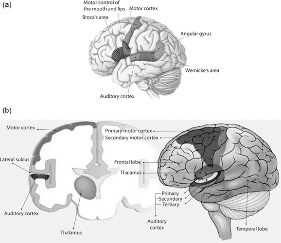
FIGURE 1.5 (a) Location of Broca’s area and Wernicke’s area. (Adapted from [1].) (b) Primary, secondary, and tertiary auditory cortexes.
The primary auditory cortex (A1) is the first ...
Table of contents
- Cover
- Half Title
- Title Page
- Copyright Page
- Table of Contents
- Preface
- Acknowledgments
- Editor
- Contributors
- Chapter 1 Review of the Human Brain and EEG Signals
- Chapter 2 Brain–Computer Interfaces (BCIs)
- Chapter 3 Applications of BCIs
- Chapter 4 Future of Non-Invasive BCIs
- Index
Frequently asked questions
Yes, you can cancel anytime from the Subscription tab in your account settings on the Perlego website. Your subscription will stay active until the end of your current billing period. Learn how to cancel your subscription
No, books cannot be downloaded as external files, such as PDFs, for use outside of Perlego. However, you can download books within the Perlego app for offline reading on mobile or tablet. Learn how to download books offline
We are an online textbook subscription service, where you can get access to an entire online library for less than the price of a single book per month. With over 1.5 million books across 990+ topics, we’ve got you covered! Learn about our mission
Look out for the read-aloud symbol on your next book to see if you can listen to it. The read-aloud tool reads text aloud for you, highlighting the text as it is being read. You can pause it, speed it up and slow it down. Learn more about Read Aloud
Yes! You can use the Perlego app on both iOS and Android devices to read anytime, anywhere — even offline. Perfect for commutes or when you’re on the go.
Please note we cannot support devices running on iOS 13 and Android 7 or earlier. Learn more about using the app
Please note we cannot support devices running on iOS 13 and Android 7 or earlier. Learn more about using the app
Yes, you can access Introduction to Non-Invasive EEG-Based Brain-Computer Interfaces for Assistive Technologies by Teodiano Bastos-Filho, Teodiano Freire Bastos-Filho,Teodiano Bastos-Filho, Teodiano Freire Bastos-Filho in PDF and/or ePUB format, as well as other popular books in Technology & Engineering & Human-Computer Interaction. We have over 1.5 million books available in our catalogue for you to explore.