- 173 pages
- English
- ePUB (mobile friendly)
- Available on iOS & Android
eBook - ePub
A Practical Guide to 3D Ultrasound
About this book
A Practical Guide to 3D Ultrasound was conceived with the beginner in mind. The guide summarizes the basics of 3D sonography in a concise manner and serves as a practical reference for daily practice. It is written in easy-to-read language and contains tables summarizing the step-by-step instructions for the techniques presented. Following introduc
Trusted by 375,005 students
Access to over 1.5 million titles for a fair monthly price.
Study more efficiently using our study tools.
Information
1 | Terminology and Basics |
INTRODUCTION
Welcome to the world of volume sonography, a world with added depth that enables you to obtain planes previously unattainable using conventional two-dimensional (2D) sonography. In volume sonography, the concept of the “voxel” replaces the “pixel,” where you now have three intersecting orthogonal or perpendicular planes with which you are working—the X, Y, and Z planes (Figure 1.1). Where these three planes intersect is the “reference dot,” an identifiable, locatable point of interest that can be defined through its relationship within the three planes. Within any acquired threedimensional (3D) volume is an infinite number of planes, stacked on top of each other, and containing within it all the information needed to analyze that specific area or organ of interest (Figure 1.2). For example, in the first trimester, a volume of the entire fetus may be obtained for analysis at any subsequent point in the future (Abu-Rustum et al. 2012). This volume, if obtained correctly, contains all the planes needed for a full evaluation of the first-trimester fetus (Figure 1.3). This also applies to a volume of the fetal heart that contains within it all the anatomic planes necessary for a complete assessment of the fetal heart and vessels (Abuhamad 2004). Once the volume of data is obtained and stored, it can subsequently be reformatted, post-processed, and displayed interchangeably in the multiplanar (Figure 1.4), surface-rendering mode (Figure 1.5), or in any other mode at any given point in the future.
BASIC CONCEPTS IN THE MULTIPLANAR MODE
1. Marker dot: reference dot
2. Address of the marker dot is determined by the intersection of the X, Y, Z axes (Figure 1.6)
The cornerstone of volume sonography is formed by three main concepts. These are addressed individually in the subsequent chapters.
BASIC CONCEPTS IN VOLUME SONOGRAPHY
1. Volume acquisition
2. Volume manipulation
3. Volume display: multiplanar or rendered
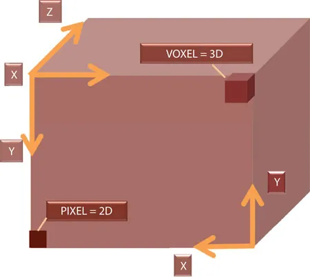
FIGURE 1.1 A cubic volume illustrating the three orthogonal planes along the X, Y, and Z axes, representative of an acquired 3D volume. At the bottom left-hand corner is a 2D rectangle along the X and Y axes, illustrating the 2D concept of a pixel. On the top right is a 3D rectangle along the X, Y, and Z axes, demonstrating the 3D concept of a voxel.
TERMINOLOGY
With volume sonography, there is a new vocabulary to learn (Table 1.1). Some of these terms are generic and others are specific to certain manufacturers. This is why one must become familiar with all the basic terms, their synonyms, and their meanings. It then becomes intuitive as to what is to be used where. The basic concept lies in obtaining what is called a static volume and then visualizing it in the three orthogonal planes in the multiplanar view. If it is subsequently decided to manipulate the volume in order to visualize the image using any of several display modes, this generates the rendered image. This can be surface mode (Figure 1.7), maximal mode (Figure 1.8), minimal mode (Figure 1.9), inversion mode (Figure 1.10), or any combination thereof, to name a few.
ADVANTAGES OF VOLUME SONOGRAPHY
With volume sonography, it is now possible to evaluate planes not previously accessible by 2D ultrasound. In addition, depth perception is now added. The stored volumes are available for educational purposes: they can be utilized for learning anatomy, and they facilitate off-line consultation with experts and over the web. With volume sonography, it is now possible to evaluate such areas as the top of the fetal head, the fetal sutures (Figure 1.11), and the mid-sagittal plane of the fetal head (Figure 1.12). In addition, beam steering allows the visualization of previously unattainable views such as the posterior aspect of structures. As such, the level of a neural tube defect may be localized, and skeletal malformations may be characterized.

FIGURE 1.2 A 3D volume of the chest of a 22w0d fetus displayed in the multiplanar mode. This volume contains within it all the 2D anatomical planes necessary for a complete assessment of the heart. These 2D planes exist in a defined spatial relationship with respect to each other, and they may be retrieved out of a standardized volume utilizing a specific navigational approach based on the established spatial relationships between them.
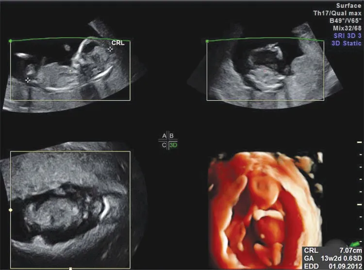
FIGURE 1.3 A 3D volume of a 13w2d fetus depicted in the multiplanar mode (three orthogonal planes A, B, and C) and surface rendered in the bottom right-hand corner, utilizing HDlive. This volume contains within it all the 2D planes necessary for a complete evaluation of this fetus. These 2D planes may be generated out of the volume by navigation along the three axes.
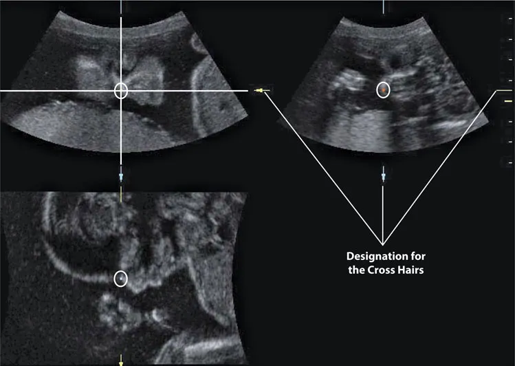
FIGURE 1.4 A 3D volume of a 20w6d fetal face displayed in the three orthogonal planes. Note the position of the reference dot (O). It is the intersection of the cross-hairs in each of the 3 planes.
Much in terms of fetal behavior can be studied as well by watching fetal movement, awake and sleep cycles, and eyelid movement, all of which further enhance fetal bonding. In gynecologic ultrasound, it is now possible to evaluate the coronal plane of the uterus (Figure 1.13), which enhances sensitivity in the detection of müllerian abnormalities (Bocca et al. 2012; Sakhel et al. 2013). Tumors may be ...
Table of contents
- Cover
- Half Title
- Title Page
- Copyright Page
- Dedication
- Table of Contents
- Foreword
- Preface
- Chapter 1 Terminology and Basics
- Chapter 2 Volume Acquisition
- Chapter 3 Volume Manipulation
- Chapter 4 Volume Display
- Chapter 5 Spatiotemporal Image Correlation
- Chapter 6 3D Tools
- Chapter 7 Clinical Applicability in the First Trimester
- Chapter 8 Clinical Applicability in the Fetal Face
- Chapter 9 Clinical Applicability in the Fetal Central Nervous System
- Chapter 10 Clinical Applicability in the Fetal Skeleton
- Chapter 11 Clinical Applicability in the Fetal Cardiovascular System
- Chapter 12 Clinical Applicability in the Fetal Chest
- Chapter 13 Clinical Applicability in the Fetal Gastrointestinal Tract
- Chapter 14 Clinical Applicability in the Fetal Genitourinary System
- Chapter 15 3D Applications in Obstetrics
- Chapter 16 3D Applications in Gynecology
- Chapter 17 Coding and Entertainment Ultrasound
- References
- Index
Frequently asked questions
Yes, you can cancel anytime from the Subscription tab in your account settings on the Perlego website. Your subscription will stay active until the end of your current billing period. Learn how to cancel your subscription
No, books cannot be downloaded as external files, such as PDFs, for use outside of Perlego. However, you can download books within the Perlego app for offline reading on mobile or tablet. Learn how to download books offline
Perlego offers two plans: Essential and Complete
- Essential is ideal for learners and professionals who enjoy exploring a wide range of subjects. Access the Essential Library with 800,000+ trusted titles and best-sellers across business, personal growth, and the humanities. Includes unlimited reading time and Standard Read Aloud voice.
- Complete: Perfect for advanced learners and researchers needing full, unrestricted access. Unlock 1.5M+ books across hundreds of subjects, including academic and specialized titles. The Complete Plan also includes advanced features like Premium Read Aloud and Research Assistant.
We are an online textbook subscription service, where you can get access to an entire online library for less than the price of a single book per month. With over 1.5 million books across 990+ topics, we’ve got you covered! Learn about our mission
Look out for the read-aloud symbol on your next book to see if you can listen to it. The read-aloud tool reads text aloud for you, highlighting the text as it is being read. You can pause it, speed it up and slow it down. Learn more about Read Aloud
Yes! You can use the Perlego app on both iOS and Android devices to read anytime, anywhere — even offline. Perfect for commutes or when you’re on the go.
Please note we cannot support devices running on iOS 13 and Android 7 or earlier. Learn more about using the app
Please note we cannot support devices running on iOS 13 and Android 7 or earlier. Learn more about using the app
Yes, you can access A Practical Guide to 3D Ultrasound by Reem S. Abu-Rustum in PDF and/or ePUB format, as well as other popular books in Medicine & Family Medicine & General Practice. We have over 1.5 million books available in our catalogue for you to explore.