- 192 pages
- English
- ePUB (mobile friendly)
- Available on iOS & Android
eBook - ePub
About this book
This book provides systematic coverage of small animal ophthalmology via randomized self-assessment case presentations: integrated questions, superb illustrations, color photos, imaging, diagrams, tables, and detailed explanatory answers. The authors have emphasize the more common ophthalmic conditions presented to veterinarians in practice with 25
Trusted by 375,005 students
Access to over 1.5 million titles for a fair monthly price.
Study more efficiently using our study tools.
Information
Topic
MedicineSubtopic
Veterinary MedicineSelf-Assessment Color Review Small Animal Ophthalmology
1, 2: Questions
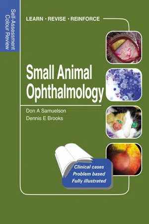
1 A nine-year-old domestic shorthaired cat was brought to the clinic for a complaint of the cat’s left nictitating membrane. The third eyelid conjunctiva of this cat exhibited protrusion, chemosis, and hyperemia (1). Rose bengal stain has been applied to the eye.
i. What does rose bengal stain evaluate?
ii. What are the differential diagnoses for the conjunctivitis in this cat?
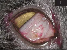
2 This 12-week-old Boston Terrier was presented for a puppy wellness examination. The owners explained that they were able to acquire the puppy for free because she had ‘funny eyes’ (2). They wanted to know what was wrong with the puppy’s eyes.
i. What do you tell the owners?
ii. Does this condition affect vision?
iii. What can be done to resolve the condition in this puppy?
1, 2: Answers
1 i. Tear film stability. The inner mucin layer of the tear film normally blocks staining of the surface epithelial cells and stroma. If the mucin layer is absent, rose bengal staining occurs. While it stains living cells, dead and degenerating cells, and mucus, this stain may have a dose-dependent ability to react with normal corneal and conjunctival epithelial cells. Rose bengal (dichloro-tetra-iodo-fluorescein) is available in solution form or impregnated paper strip. A lower concentration (0.5%) is often used, as higher concentrations (1.0% and greater) can be irritating.
ii. Herpesvirus, Chlamydophila, Mycoplasma, and bacterial infection. Conjunctivitis often accompanies viral respiratory diseases in cats. Herpesvirus is the major cause of respiratory disease with conjunctivitis in cats. Cats with chronic conjunctivitis may also be feline immunodeficiency virus positive. Often, more than one cat in a multi-cat household will be affected. Chemical and mechanical irritants may also cause conjunctivitis. Foreign bodies are frequently incriminated. Plant, upholstery, and carpet irritants may cause chemosis and conjunctivitis in cats. Household cleaners and soaps have been suspected as causes of conjunctivitis in cats. Lack of tear production is also a cause of conjunctivitis in cats. Other less common causes include hypersensitivity to topical ophthalmic preparations, parasites, and mycotic infections.
2 i. This puppy has congenital strabismus. Strabismus refers to a deviation in alignment of one globe in relation to the other globe. It may be constant or intermittent. The two eyes may be crossed (esotropia), out-turned (exotropia) as in this puppy, deviated up vertically (hyperopia), or deviated down vertically (hypotropia).
ii. Binocular vision is an acquired reflex that normally develops early in life. The development of binocular vision requires both eyes to have visual capability and to be properly aligned. Similar retinal images must project onto corresponding retinal areas of both eyes during the period of binocular vision development. Puppies with congenital or early-onset strabismus do not receive the essential visual retinal stimulation for development of binocular vision and thus lack true stereopsis. The two eyes fail to focus on the same image point, and the brain ignores the input from the deviated eye, resulting in a form of vision loss termed amblyopia.
iii. Surgical correction of the strabismus by rectus muscle transposition can be performed. Muscles can be weakened by moving the muscle insertion posteriorly or strengthened by shortening the muscle or advancing the insertion site anteriorly; alternatively, muscle insertions can be transposed to different locations in order to alter the functional pull of the muscles. Nothing but observation was done in this puppy and the strabismus self-corrected.
3, 4: Questions
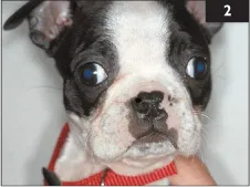
3 A seven-year-old female Husky is presented with a two-day history of blindness. Over the past month her iris color had changed from blue to brown (due to uveitis) (3a). She also has nasal depigmentation (3b) and retinal scarring (3c).
i. What is the most likely diagnosis?
ii. What breeds of dog are predisposed to this condition?
iii. What are the treatment options?
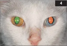
4 The owner of a seven-year-old Norwegian Forest cat had recently noticed a change in the color of her cat’s right eye (4). The irides of both eyes had been normally a light blue in this nearly albinotic individual. The iris of the right eye, however, had changed during the past week to a greenish-orange. The distinctly pigmented margin around the pupil had faded considerably. What are the two main differential diagnoses of iris color change?
3, 4: Answers
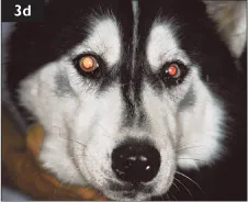
3 i. Uveodermatologic syndrome (UDS). This syndrome in the dog is similar to Vogt–Koyanagi–Harada (VKH) syndrome in humans. This immune-mediated disease against melanin is characterized by severe, bilateral panuveitis and hypotony, with secondary cataracts, glaucoma, retinal detachments, and blindness. Iris and retinal depigmentation, and poliosis/vitiligo of the face and muzzle are often noticed. Diagnosis is made from clinical lesions and breed of dog. A skin biopsy can help to confirm the condition.
ii. Originally described in the Akita, UDS has also been diagnosed in the Australian Shepherd Dog, Beagle, Brazilian Fila, Chow Chow, Dachshund, Golden Retriever, Irish Setter, Old English Sheepdog, Saint Bernard, Samoyed, Shetland Sheepdog, and Siberian Husky.
iii. The initial therapy for this condition is immunosuppressive doses of oral prednisone plus azathioprine or cyclophosphamide. After five weeks tapering, oral prednisone can begin. Most dogs require a low dose of both azathioprine and prednisone to control the disease. Topical anti-inflammatories and atropine are used to treat the uveitis (see case 12). The eye is carefully monitored for development of secondary glaucoma. In this case the nose repigmented and the uveitis quieted following therapy (3d).
4 Anterior uveitis and intraocular neoplasia. Eyes with anterior uveitis may also exhibit ocular hypotony, aqueous flare, miosis, chemosis, hypopyon, keratic precipitates, and/or synechiae formation. A complete physical and ocular examination is important in order to provide diagnostic clues to the etiology of the inflammation. Intraocular melanomas and lymphoma are common in the cat and may also cause iris color change.
5, 6: Questions
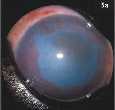
5 A nine-year-old spayed female dog was presented with this unilateral eye problem (5a).
i. Describe the clinical signs.
ii. What is your diagnosis?
iii. What are the possible causes?
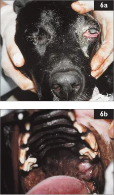
6 This nine-year-old Labrador Retriever was presented because of difficulty eating and an enlarged left eye (6a). There was epiphora and redness associated with the eye, and a corneal ulcer was present. The right eye appeared normal. A depigmented mass was present behind the last molar tooth of the exophthalmic side (6b).
i. What are the differential diagnoses?
ii. You discover that there is also a mass in the caudal aspect of the hard palate. What is the most likely diagnosis?
iii. How would you treat this condition?
iv. What is the likely prognosis?
5, 6: Answers
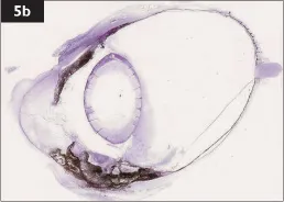
5 i. There is moderate conjunctival hyperemia with corneal vascularization at the whole corneal circumference (360°). Diffuse, severe corneal edema is noticed, but a dark mass can still be observed obliterating the anterior chamber. The Schirmer tear test is normal, there is no dazzle reflex or consensual light reflex, and the cornea is fluorescein negative.
ii. These clinical signs are consistent with glaucoma, which was confirmed by tonometry (intraocular pressure of 35 mmHg). Ocular ultrasonography revealed a mass in the anterior chamber attached to the iris. This mass was suspected to be a uveal melanoma, as this is the most common primary intraocular neoplasm in dogs. The eye was enucleated and histopathology confirmed the diagnosis (5b).
ii...
Table of contents
- Cover Page
- Self-Assessment Color Review Small Animal Ophthalmology
- Dedication
- Acknowledgements
- Copyright Page
- Preface
- Contributors
- Abbreviations
- Suggested further reading
- Classification of cases
- Self-Assessment Color Review Small Animal Ophthalmology
- Index
Frequently asked questions
Yes, you can cancel anytime from the Subscription tab in your account settings on the Perlego website. Your subscription will stay active until the end of your current billing period. Learn how to cancel your subscription
No, books cannot be downloaded as external files, such as PDFs, for use outside of Perlego. However, you can download books within the Perlego app for offline reading on mobile or tablet. Learn how to download books offline
Perlego offers two plans: Essential and Complete
- Essential is ideal for learners and professionals who enjoy exploring a wide range of subjects. Access the Essential Library with 800,000+ trusted titles and best-sellers across business, personal growth, and the humanities. Includes unlimited reading time and Standard Read Aloud voice.
- Complete: Perfect for advanced learners and researchers needing full, unrestricted access. Unlock 1.5M+ books across hundreds of subjects, including academic and specialized titles. The Complete Plan also includes advanced features like Premium Read Aloud and Research Assistant.
We are an online textbook subscription service, where you can get access to an entire online library for less than the price of a single book per month. With over 1.5 million books across 990+ topics, we’ve got you covered! Learn about our mission
Look out for the read-aloud symbol on your next book to see if you can listen to it. The read-aloud tool reads text aloud for you, highlighting the text as it is being read. You can pause it, speed it up and slow it down. Learn more about Read Aloud
Yes! You can use the Perlego app on both iOS and Android devices to read anytime, anywhere — even offline. Perfect for commutes or when you’re on the go.
Please note we cannot support devices running on iOS 13 and Android 7 or earlier. Learn more about using the app
Please note we cannot support devices running on iOS 13 and Android 7 or earlier. Learn more about using the app
Yes, you can access Small Animal Ophthalmology by Don Samuelson,Dennis Brooks in PDF and/or ePUB format, as well as other popular books in Medicine & Veterinary Medicine. We have over 1.5 million books available in our catalogue for you to explore.