Written by an experienced and well-respected physician and professor, this new volume combines the entire previous four books, Ultrasonic Topographical and Pathotopographical Anatomy, and its three sequels, also available from Wiley-Scrivener, presenings the ultrasonic topographical and pathotopographical anatomy of the entire body, offering further detail into these important areas for use by medical professionals.
This comprehensive and exhaustive medical atlas of topographic and pathotopographic human anatomy is a fundamental and practically important book designed for doctors of all specializations and students of medical schools. Here you can find almost everything that is connected with the topographic and pathotopographic human anatomy, including original graphs of logical structures of topographic anatomy and development of congenital abnormalities, topography of different areas in layers, pathotopography, computer and magnetic resonance imaging (MRI) of topographic and pathotopographic anatomy. You can also find here new theoretical and practical sections of topographic anatomy developed by the author himself which are published for the first time. They are practically important for mastering the technique of operative interventions and denying possibility of iatrogenic complications during operations.
This important new volume will be valuable to physicians, junior physicians, medical residents, lecturers in medicine, and medical students alike, either as a textbook or as a reference. It is a must-have for any physician's library.
Trusted by 375,005 students
Access to over 1.5 million titles for a fair monthly price.
Part 1 ULTRASONIC TOPOGRAPHICAL AND PATHOTOPOGRAPHICAL ANATOMY
1 Topography and Pathotopography of the Head
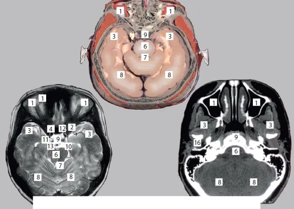
The chapter on the ultrasonic topography and pathotopographical anatomy of the head includes layer-by-layer topography of the visceral and cerebral craniums with the cross-sectional imaging of the head.
Ultrasonic images of external and internal bone lamellae, vessels of the subcutaneous layer, skin, and subcutaneous fat, depressed compression and linear fractures are demonstrated. Ultrasonic images of the medial cerebral artery, infundibulum, posterior communicating artery, pons cerebelli, medulla oblongata, anterior inferior cerebellar artery, basilar artery, anterior cerebral artery, posterior cerebral artery, and olfactory tract are verified based on the topographical anatomy of the basilar region of the cranium.
The deep facial area contains the internal wing muscle, mandibulum, and submandibular salivary gland; the oral cavity contains the tongue, peripharyngeal space, and posterior veil of the soft palate, as well as the superficial temporal artery, auriculotemporal nerve, maxillary artery, and middle meningeal artery. The ultrasonic images of the internal and external muscles are shown.
Images of the parotid gland, superficial cervical lymph nodes, and common carotid artery are presented.
Linear fracture is associated with the external bone lamella of the area of intact bone, with the intracranial space, and the hypoechogenic track. Under conditions of tamponade of the fourth ventricle of cerebrum with transition to the pons cerebelli, a blood clot is revealed in the vicinity of the clinoid plate at the pyramid apex of the temporal bone. The intraventricular blood clot can be pathotopographically associated with the left lateral ventricle, whereas liquid blood is observed at the lumen of the right lateral ventricle.
The atlas also contains images of the pathotopographical anatomy of the intraventricular hemorrhage, hematoma in the thalamus, frontobasal intracerebral hematoma, and acute epidural hematoma in the left parieto-occipital space accompanied by the phenomenon of the “boundary amplification”.
Thus, the ultrasonic topographic anatomy of the head provides the basis for the research into the pathotopographical anatomy of a given pathology and determines specific diagnostic features of injuries and/ or volume structures.
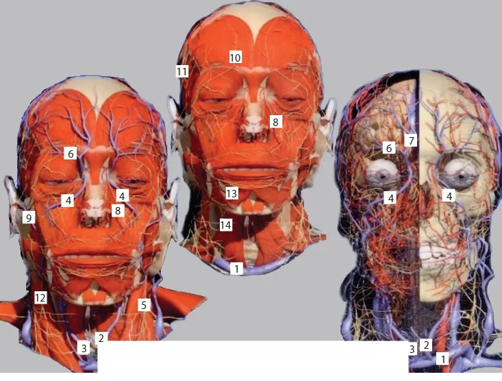
Figure 1 Layer-by-layer topography of the head.
1. v. brachiocephalica; 2. v. jugularis anterior sinistra; 3. v. jugularis anterior dextra; 4. v. angularis; 5. Plexus cervicalis; 6. v. supratrochlearis; 7. v. nasofrontalis; 8. a. angularis; 9. v. temporalis superlicialis; 10. n. supraorbitalis; 11. n. auriculotemporalis; 12. Plexus cervixalis; 13. m. depressor labii inferioris; 14. m. dieastricus (venter anterior).
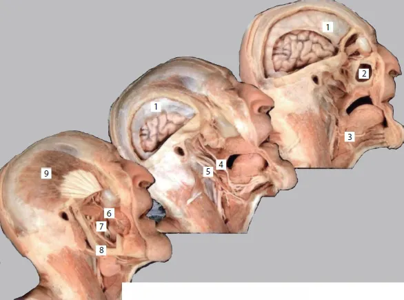
Figure 2 Layer-by-layer topography.
1. Falx cerebri; 2. Sinus maxillaries; 3. n. hypoglossus; 4. n. hypoglossus; 5. n. facialis; 6. n. lingualis; 7. n. alveolaris inferior; 8. Arcus maxillae inferioris; 9. m. temporalis
Part 1: ULTRASONIC TOPOGRAPHICAL AND PATHOTOPOGRAPHICAL ANATOMY
Part 2: TOPOGRAPHICAL AND PATHOTOPOGRAPHICAL MEDICAL ATLAS OF THE HEAD AND NECK
Part 3: TOPOGRAPHICAL AND PATHOTOPOGRAPHICAL MEDICAL ATLAS OF THE CHEST, ABDOMEN, LUMBAR REGION, AND RETROPERITONEAL SPACE
Part 4: TOPOGRAPHICAL AND PATHOTOPOGRAPHICAL MEDICAL ATLAS OF THE PELVIS, SPINE, AND LIMBS
Conclusion
Appendix A
Appendix B
End User License Agreement
Frequently asked questions
Yes, you can cancel anytime from the Subscription tab in your account settings on the Perlego website. Your subscription will stay active until the end of your current billing period. Learn how to cancel your subscription
No, books cannot be downloaded as external files, such as PDFs, for use outside of Perlego. However, you can download books within the Perlego app for offline reading on mobile or tablet. Learn how to download books offline
Perlego offers two plans: Essential and Complete
Essential is ideal for learners and professionals who enjoy exploring a wide range of subjects. Access the Essential Library with 800,000+ trusted titles and best-sellers across business, personal growth, and the humanities. Includes unlimited reading time and Standard Read Aloud voice.
Complete: Perfect for advanced learners and researchers needing full, unrestricted access. Unlock 1.5M+ books across hundreds of subjects, including academic and specialized titles. The Complete Plan also includes advanced features like Premium Read Aloud and Research Assistant.
Both plans are available with monthly, semester, or annual billing cycles.
We are an online textbook subscription service, where you can get access to an entire online library for less than the price of a single book per month. With over 1.5 million books across 990+ topics, we’ve got you covered! Learn about our mission
Look out for the read-aloud symbol on your next book to see if you can listen to it. The read-aloud tool reads text aloud for you, highlighting the text as it is being read. You can pause it, speed it up and slow it down. Learn more about Read Aloud
Yes! You can use the Perlego app on both iOS and Android devices to read anytime, anywhere — even offline. Perfect for commutes or when you’re on the go. Please note we cannot support devices running on iOS 13 and Android 7 or earlier. Learn more about using the app
Yes, you can access Topographical and Pathotopographical Medical Atlas of the Human Body by Z. M. Seagal in PDF and/or ePUB format, as well as other popular books in Medicine & Anatomy. We have over 1.5 million books available in our catalogue for you to explore.