- 288 pages
- English
- ePUB (mobile friendly)
- Available on iOS & Android
eBook - ePub
About this book
Finally - a guide to cytological techniques written specifically for the plant chromosome researcher and student. Plant Chromosomes: Laboratory Methods thoroughly covers all important approaches to the study of plant chromosomes. It reviews each specific approach and describes requisite experimental techniques. These practical descriptions cover basic, standard techniques as well as the most recent research advances and state-of-the-art technologies.
Plant Chromosomes: Laboratory Methods allows you to build on the knowledge of its expert authors, who have first-hand experience with the ins and outs of each approach. Through hundreds of trouble-shooting suggestions it also helps you avoid experimental pitfalls by providing invaluable tips at critical points in the experimental process. This book gives you the information you need to improve the power of your plant chromosome research - saving you time and effort in the process. No other single volume contains so much practical information on this topic.
Trusted by 375,005 students
Access to over 1.5 million titles for a fair monthly price.
Study more efficiently using our study tools.
Information
Chapter 1
Plant Chromosomes at Mitosis
Kiichi Fukui
Contents
- Outline of Mitotic Chromosomes
- A. Brief History of Mitotic Chromosome Research
- B. Mitotic Chromosomes and Their Terminology
- C. Two Categories of Plant Chromosomes
- Preparation of Small Chromosome Samples: Enzymatic Maceration/Air-Drying Method
- Preparation of Large Chromosome Samples: Squash Method
- Preparation of Chromosome Samples from Woody Plants
- Preparation of Chromosome Samples from Suspension Cells
- Preparation of Chromosome Samples from Flowers and Leaves
- Preparation on Permanent Chromosome Samples
Acknowledgments
References
I. Outline of Mitotic Chromosomes
A. Brief History of Mitotic Chromosome Research
Chromosomes were observed by Nägeli in 1842, soon after the discovery of the nucleus under the microscope, as objects that appeared at cell division. The behavior of chromosomes at cell division was described by Nägeli 2 years later, and his observation is now accepted as the first description of mitosis. A detailed sketch covering the whole process of mitosis was reported by Flemming in 1882; and the German term, Chromosomen (taken from the Greek, meaning colored body), was coined by von Waldeyer in 1888 because chromosomes could be stained with staining solutions.
The fact that chromosomes were transferred to the two new daughter cells convinced researchers that they constituted the materials carrying the information of inheritance. Studies in the early stages of chromosome research had already revealed a large variation in the number of chromosomes among plant species, and the determination of chromosome number thus became an important subject of the time. Early methods used for the precise determination of mitotic chromosome number were based on a smear or sectioning method to cut the tissues into thin slices by either hand or microtome. Following the appearance of all the chromosomal regions in the sliced sections, the number of chromosomes was determined. The chromosome number of rice was determined by Kuwada in 1910, and those of the majority of cultivated plants and popular wild species were determined during the same period. Chemical pretreatment methods, such as with colchicine, 8-hydroxyquinoline, and α-bromonaphthalene, were also developed to allow accumulated numbers of metaphase chromosomes.
A breakthrough in chromosome preparation techniques came with the development of the squash method, by which samples could be easily prepared while avoiding the time-consuming and laborious sectioning procedures and the difficulties inherent in the smear method for some hard materials. Total chromosomal morphology at mitosis could thus be observed easily under the microscope regardless of the plant material, although root tips became the most frequently used source for the collection of mitotic cells. Several associate techniques, such as pretreatment and softening methods, were subsequently integrated into the squash method. Although the squash method is still widely used among plant cytologists to prepare chromosome samples, it requires skill and experience to constantly obtain evenly spread chromosome samples on a glass slide.
In 1944, Emsweller and Stuart first suggested the possibility of using enzymatic maceration of plant tissues to prepare good chromosome samples. The most conspicuous difference between plant and animal cells lies in the fact that plant cells have thick cell walls, which interfere with the preparation of good chromosome samples. Several researchers in the mid-1940s and 1950s tested and demonstrated the effectiveness of the pectinase.1 Widespread use of the enzymatic maceration method, however, only came about when the less contaminated enzymes became available at more reasonable prices. The enzymatic maceration method, in combination with the air-drying method, which was once referred to as the evolutional technique in human cytology, is now widely employed in numerous laboratories. The Giemsa staining method, which is widely used for staining animal chromosomes, can also be applied to samples prepared by enzymatic maceration and air-drying; and together these constitute the current standard method for chromosome sample preparation.2-4 The main advantage of samples prepared by this method is that the chromosomes are free of cytoplasmic debris and are spread evenly on the glass slide, therefore allowing us to observe the fine structures of the chromosomes, with faintly stretched tails at the prometaphase stage, for the first time.5
B. Mitotic Chromosomes and Their Terminology
From a morphological point of view, chromosomes can be divided into the two phases of the cell cycle; i.e., the metabolic stage observed as a nucleus in the cell and the dividing stage at which time the characteristic morphology of the chromosomes of the species or individual appear. The dividing stage consists of mitosis and meiosis. Figure 1.1 shows the metabolic stage and mitosis of plant cells. Mitosis, in which the replicated chromosomes are evenly distributed to the two daughter cells, is morphologically classified into the five main stages of prophase, prometaphase, metaphase, anaphase, and telophase. Figure 1.2 shows a schematic representation of prometaphase and metaphase chromosomes.
The terminology used to describe chromosome morphology is often confused, as typically demonstrated by the term, telomere, which can mean either telomeric visible condensation appearing after banding treatment, or telomeric DNA sequences such as Tm(A)Gn visualized by in situ hybridization. Although it is essential to define all the chromosomal terms precisely, I leave that to future textbooks on chromosomes and define here only the terms necessary to conduct the experiments.
The primary constriction, or centromere, divides a chromosome into two arms, often a long and short arm. The types of chromosomes can be defined by the position of the centromere, the key parameter being the arm ratio which is the ratio of the short arm length to that of the long arm. Chromosomes with ratios ranging from 1.0 to 1.7 are classified as the median type; those with ratios over 7.0 are designated as the terminal type, while a ratio of 3.0 divides the submedian and subterminal chromosome types.6
The secondary constriction corresponds to the nucleolar-organizing region (nucleolus organizer region, NOR), where the 45S ribosomal RNA gene is tandemly clustered. NOR sometimes appears as a gap in a chromosome, and this end portion of the chromosome is called the satellite. While the relative position of the satellite is variable, it tends to be located close to the host chromosome as the cell cycle proceeds to metaphase. Other small constrictions in the chromosomes, called tertiary constrictions, have as yet unknown functions.
The telomere is a structure located specifically at the ends of both chromosomal arms. Originally, it referred to condensed chromosomal regions, or C-band positive sites, observed at the ends of the chromosomes as found in rye chromosomes. More often, however, it refers to a specific repeated nucleotide sequence, such as (TTTAGGG)n, located at both ends of chromosomal arms, which are thought to have protective functions.
Metaphase chromosomes are most usually studied after the inclusion of a pretreatment to accumulate the condensed chromosomes. Although chromosomes condense the most at the metaphase stage, when their morphology is thought to be stable, pretreatment makes them too condensed to detect the fine structures and may even modify their morphology.7
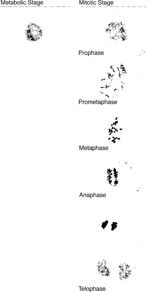
FIGURE 1.1
Rice chromosomes at the metabolic stage and mitosis.
Rice chromosomes at the metabolic stage and mitosis.
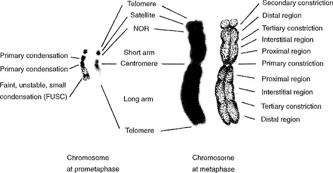
FIGURE 1.2
Schematic representation of a prometaphase chromosome in rice (left) and a metaphase chromosome in barley (right).
Schematic representation of a prometaphase chromosome in rice (left) and a metaphase chromosome in barley (right).
On the other hand, prometaphase chromosomes, especially the small plant chromosomes that condense to small dots or rods at the metaphase stage, have been known to contain critical information. Uneven staining patterns, characteristic of the prometaphase chromosomes, are caused by differential condensation of the chromatin fiber in small plant chromosomes and are thus called condensation pattern2,8,9 (Figure 1.3). Condensed regions at the proximal regions are referred to as primary condensations, whereas small condensations at the interstitial or terminal regions are termed as faint, unstable, small condensation (FUSC).
C. Two Categories of Plant Chromosomes
Plant chromosomes can be categorized into two types based on size; one is a large type (L-type) and the other is a small type (S-typ...
Table of contents
- Cover Page
- Title Page
- Copyright Page
- Preface
- The Editors
- Contributors
- Chapter 1. Plant Chromosomes at Mitosis
- Chapter 2. Plant Chromosomes at Meiosis
- Chapter 3. Plant Chromosomes at Metabolic Phase
- Chapter 4. Polytene Chromosomes
- Chapter 5. Flow Cytometry and Chromosome Sorting
- Chapter 6. Chromosome Dissection and Direct Cloning
- Chapter 7. Chromosome-Banding Methods
- Chapter 8. In Situ Hybridization
- Chapter 9. Sister Chromatid Exchange and Replication Banding
- Chapter 10. Replication of Chromosomes
- Chapter 11. Chromosome Manipulation in Wheat
- Chapter 12. Electron Microscopy and Plant Chromosomes
- Chapter 13. Analysis of Chromosome Information
- Appendix I. Buffer Solutions
- Appendix II. Tissue Culture Media
- Index
Frequently asked questions
Yes, you can cancel anytime from the Subscription tab in your account settings on the Perlego website. Your subscription will stay active until the end of your current billing period. Learn how to cancel your subscription
No, books cannot be downloaded as external files, such as PDFs, for use outside of Perlego. However, you can download books within the Perlego app for offline reading on mobile or tablet. Learn how to download books offline
Perlego offers two plans: Essential and Complete
- Essential is ideal for learners and professionals who enjoy exploring a wide range of subjects. Access the Essential Library with 800,000+ trusted titles and best-sellers across business, personal growth, and the humanities. Includes unlimited reading time and Standard Read Aloud voice.
- Complete: Perfect for advanced learners and researchers needing full, unrestricted access. Unlock 1.5M+ books across hundreds of subjects, including academic and specialized titles. The Complete Plan also includes advanced features like Premium Read Aloud and Research Assistant.
We are an online textbook subscription service, where you can get access to an entire online library for less than the price of a single book per month. With over 1.5 million books across 990+ topics, we’ve got you covered! Learn about our mission
Look out for the read-aloud symbol on your next book to see if you can listen to it. The read-aloud tool reads text aloud for you, highlighting the text as it is being read. You can pause it, speed it up and slow it down. Learn more about Read Aloud
Yes! You can use the Perlego app on both iOS and Android devices to read anytime, anywhere — even offline. Perfect for commutes or when you’re on the go.
Please note we cannot support devices running on iOS 13 and Android 7 or earlier. Learn more about using the app
Please note we cannot support devices running on iOS 13 and Android 7 or earlier. Learn more about using the app
Yes, you can access Plant Chromosomes by Kiichi Fukui,Shigeki Nakayama in PDF and/or ePUB format, as well as other popular books in Biological Sciences & Biology. We have over 1.5 million books available in our catalogue for you to explore.