- 440 pages
- English
- ePUB (mobile friendly)
- Available on iOS & Android
eBook - ePub
Human Embryology Made Easy
About this book
This book is a synopsis of the key facts and concepts of human development. It is intended for students who are taking a human embryology course. The book includes the underlying mechanisms involved in clinically important congenital anomalies that will prove useful to medical and nursing.
Trusted by 375,005 students
Access to over 1 million titles for a fair monthly price.
Study more efficiently using our study tools.
Information
Topic
MedicineCHAPTER
1
Cell Division
In a cell population that is constantly being renewed, individual cells divide periodically. A typical somatic cell division, mitosis, consists of an equal division of nuclear material, so that the two newly formed daughter cells receive exactly the number and kind of chromosomes that the parent cell had. This separation of nuclear material is then followed by division of the cytoplasm.
Before a cell can undergo division, it must increase its mass and contents, and double the mass of its DNA. All of this occurs during the growth period known as interphase. Following this is the Μ phase, during which nuclear division (mitosis) and cytoplasmic division (cytokinesis) take place. Duplicated DNA must be divided precisely between daughter cells.
Although the cell cycle is continuous, for simplicity and clarity, both interphase and Μ phase are subdivided into stages. The interphase is composed of the G1, S and G2 periods.
I. Interphase
A. G1 (Gap1) Period
After completion of cell division, the daughter cells enter the preduplication period, Gi (gapi). During this period there is synthesis of RNA and proteins, and total cell mass is increased.
After this, the cell is held at a restriction point. Any cell that passes this point will complete the rest of the stages of the cycle. A trigger or unstable protein (S phase activator) has been proposed. An accumulation of a threshold amount of this protein helps the cell to exceed the restriction point. The quiescent cells that do not accumulate this protein are arrested at the restriction point and are considered to be in the G0 period of interphase. This may be one of the mechanisms by which tissue growth is controlled. Crowding (contact inhibition) and starvation may also inhibit cell division. In many tissues, cells divide only when new cells are needed. Neoplastic cells appear to have lost these growth controls.
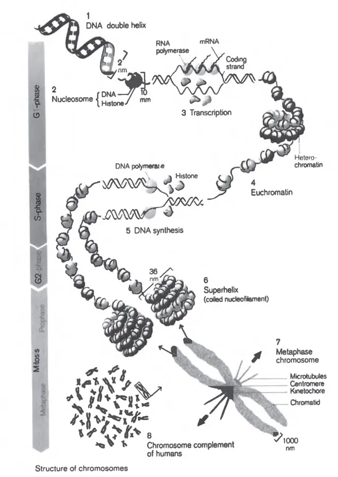
Figure 1.1
Chromosomes (see Color Plate 1.1). Reproduced with permission from Thieme Medical Publishers Inc., New York, 1995, Color Atlas of Embryology, Ulrich Drews, Chapter 1: Reproduction.
Chromosomes (see Color Plate 1.1). Reproduced with permission from Thieme Medical Publishers Inc., New York, 1995, Color Atlas of Embryology, Ulrich Drews, Chapter 1: Reproduction.
B. S (Synthesis) Period
DNA duplication, semiconservative in nature, occurs during period S, a period that is a constant characteristic of the cell type and growth conditions. The DNA is simultaneously replicated in discrete units called replicons. When all replicons have been duplicated, newly formed DNA segments join to complete the daughter molecule (Fig. 1.1).
The centriole pair separate from each other during late G1. Duplication of each centriole starts during the S period and is completed in G2. As DNA is replicated, new histones are synthesized during this period. Until DNA replication is complete, the M phase is delayed.
C. G2 Period
At the end of DNA duplication, the cell enters the preparatory period, G2. Some proteins essential for cell division are synthesized during this period. A kinase is detected that could be responsible for phosphorylation of proteins of nuclear membrane, which may in turn cause breakdown of nuclear lamins during the M phase. It may also cause phosphorylation of histone Hi molecules. In this period, components essential to form mitotic spindle are prepared. Most proteins and RNA molecules are synthesized continuously during interphase.
Three diffusible factors that may control events during interphase have been suggested: 1) an S phase activator begins DNA synthesis, 2) an M phase promoting factor (MPF) induces chromosomal condensation, and 3) an M phase delaying factor (MDF) inhibits production of MPF. There is a sequential relationship between the factors that control each successive step. For instance, DNA cannot start replicating unless the DNA re-replication block has been removed during G1. With appearance of an S phase activator and MDF, DNA synthesis continues until all the DNA has been replicated. MPF cannot be produced until the MDF has disappeared, and cells cannot enter mitosis until an MPF is produced. MPF concentration increases rapidly during early M phase. It is suggested that its surge may be triggered by an increase in the concentration of another protein, cyclin, whose concentration rises steadily. Its increase at threshold level during G2 activates MPF production. Both reach maximum concentration in the middle of M phase, when cyclin is abruptly destroyed, and MPF disappears. After this, the cyclin concentration again starts to increase steadily.
II. M Phase
M phase includes mitosis (division of nuclear material) and cytokinesis (division of cytoplasm).
A. Mitosis: Division of Nuclear Material
1. PROPHASE. The cell becomes spheroid and viscous because of breakdown of cytoskeleton. Dispersed chromatin becomes visible as delicate, longitudinally coiled filaments, known as chromosomes. DNA thread winds around a core that is formed by nucleosomes and appears as chromosomes. These delicate chromosomes undergo further condensation to form metaphase chromosomes. Each daughter centrosome acts as a microtubule organizer, and shows astral rays. As centrosomes begin to move apart, the microtubules in each aster elongate, keeping contact with both centrosomes, thus forming a mitotic spindle. Chromosomes move closer to the nuclear membrane and appear to be composed of two chromatids. They continue to become shorter and thicker. Meanwhile, the nucleolus elongates and disappears among chromosomes. Nuclear membrane disintegrates. Spindle apparatus assembly is initiated.
2. PROMETAPHASE. Prometaphase starts with disintegration of the nuclear membrane. The centrosomes reach opposite poles, and spindle microtubules enter the nuclear region. Each chromosome is seen as two sister chromatids held together at a centromere. A kinetochore, a protein complex, develops on each side of the centromere. Some of the spindle microtubules attach to the kinetochore. These kinetochore microtubules extend in opposite directions from the sister chromatids to one of the poles.
3. METAPHASE. Tension exerted by kinetochore tubules causes chromosomes to move toward the center of the cell and align at the middle of the spindle. The spindle shows polar microtubules extending from opposite poles. Kinetochore microtubules attach sister chromatids to opposite poles and to astral microtubules, which are incorporated in the spindle.
4. ANAPHASE. Anaphase starts abruptly. Shortening of the kinetochore microtubules causes separation of the kinetochore, and the centromere appears split. Cytosolic calcium is increased at this time. The sister chromatids start moving toward opposite poles. The polar microtubules elongate, moving the spindle asters farther apart. Elongated cells show a constriction in the middle.
5. TELOPHASE. Separated chromatids re...
Table of contents
- Cover
- Half Title Page
- Title Page
- Copyright Page
- Dedication
- Table of Contents
- Preface
- 1. Cell Division
- 2 Gametogenesis
- 3 Fertilization and Cleavage
- 4 Implantation
- 5 Placentation
- 6 Early Development of Embryo
- 7 Teratogenesis
- 8 Skeletomuscular System
- 9 Cardiovascular System
- 10 Respiratory System
- 11 Pharyngeal Apparatus
- 12 Craniofacial Development
- 13 Digestive System
- 14 Urinary System
- 15 Genital System
- 16 Endocrine System
- 17 Eye and Ear
- 18 Nervous System
Frequently asked questions
Yes, you can cancel anytime from the Subscription tab in your account settings on the Perlego website. Your subscription will stay active until the end of your current billing period. Learn how to cancel your subscription
No, books cannot be downloaded as external files, such as PDFs, for use outside of Perlego. However, you can download books within the Perlego app for offline reading on mobile or tablet. Learn how to download books offline
Perlego offers two plans: Essential and Complete
- Essential is ideal for learners and professionals who enjoy exploring a wide range of subjects. Access the Essential Library with 800,000+ trusted titles and best-sellers across business, personal growth, and the humanities. Includes unlimited reading time and Standard Read Aloud voice.
- Complete: Perfect for advanced learners and researchers needing full, unrestricted access. Unlock 1.4M+ books across hundreds of subjects, including academic and specialized titles. The Complete Plan also includes advanced features like Premium Read Aloud and Research Assistant.
We are an online textbook subscription service, where you can get access to an entire online library for less than the price of a single book per month. With over 1 million books across 990+ topics, we’ve got you covered! Learn about our mission
Look out for the read-aloud symbol on your next book to see if you can listen to it. The read-aloud tool reads text aloud for you, highlighting the text as it is being read. You can pause it, speed it up and slow it down. Learn more about Read Aloud
Yes! You can use the Perlego app on both iOS and Android devices to read anytime, anywhere — even offline. Perfect for commutes or when you’re on the go.
Please note we cannot support devices running on iOS 13 and Android 7 or earlier. Learn more about using the app
Please note we cannot support devices running on iOS 13 and Android 7 or earlier. Learn more about using the app
Yes, you can access Human Embryology Made Easy by Abdul Hamid Rana in PDF and/or ePUB format, as well as other popular books in Medicine & Gynecology, Obstetrics & Midwifery. We have over one million books available in our catalogue for you to explore.