Necessary for everything from reflexes to reading to running, it's no exaggeration to say that the brain and nervous system are responsible for nearly every endeavor of human activity. The sheer volume of information that the brain must process and respond to at every second of each day renders it one of the most remarkable systems of the human body. With illuminating diagrams and careful detail, this volume covers the amazing intricacies of this vital system as well as the effects of disease and damage.
- English
- ePUB (mobile friendly)
- Available on iOS & Android
eBook - ePub
The Brain and the Nervous System
About this book
Trusted by 375,005 students
Access to over 1.5 million titles for a fair monthly price.
Study more efficiently using our study tools.
Information
Subtopic
PhysiologyIndex
Biological SciencesCHAPTER 1
THE CENTRAL NERVOUS SYSTEM
The human nervous system functions as a high-speed anatomical and physiological unit. It controls the body’s movements, and through its ability to receive, process, and transmit information in the form of chemical and electrical signals, it can adjust and fine-tune its control. The integration of chemical and electrical signaling pathways in the brain provides us with cognitive abilities, such as perception, thought, memory, and emotion. The human nervous system can be divided into two main parts: the central nervous system and the peripheral nervous system. The central nervous system consists of the brain and spinal cord, while the peripheral system consists of all the neural (nerve) tracts that lie outside these central tissues and connect to the rest of the body.
The brain and spinal cord are both derived from the neural tube, a structure found in embryos. Both are surrounded by protective membranes called the meninges, and both float in a crystal-clear cerebrospinal fluid. The brain is encased in a bony vault, the neurocranium, while the cylindrical and elongated spinal cord lies in the vertebral canal, which is formed by successive vertebrae connected by dense ligaments.
THE BRAIN
The brain essentially serves as the body’s information processing centre. It receives signals from sensory neurons (nerve cell bodies and their axons and dendrites) in the central and peripheral nervous systems, and in response it generates and sends new signals that instruct the corresponding parts of the body to move or react in some way. It also integrates signals received from the body with signals from adjacent areas of the brain, giving rise to perception and consciousness.
The brain weighs about 1,500 grams (3 pounds) and constitutes about 2 percent of total body weight. It consists of three major divisions: (1) the massive paired hemispheres of the cerebrum, (2) the brainstem, consisting of the thalamus, hypothalamus, epithalamus, subthalamus, midbrain, pons, and medulla oblongata, and (3) the cerebellum.
CEREBRUM
The cerebrum is the largest, uppermost portion of the brain. It is involved with sensory integration, control of voluntary movement, and higher intellectual functions, such as speech and abstract thought. There are two cerebral hemispheres—one on the left and one on the right side of the brain. The outer layer of each of these duplicate cerebral hemispheres is composed of a convoluted (wrinkled) outer layer of gray matter, called the cerebral cortex.
Beneath the cerebral cortex is an inner core of white matter, which is composed of a special kind of nerve fibre called myelinated commissural nerve fibres. Nerve fibres, or axons, are long, thin strands of tissue that project from a nerve cell and carry electrical impulses to and from the brain. These fibres connect the two cerebral hemispheres via a thick band of white matter called the corpus callosum. Other fibres, called association fibres, connect different regions of a single hemisphere. Myelinated fibres (myelin is a fatty white material that forms a sheath around some nerve fibres) projecting to and from the cerebral cortex form a concentrated fan-shaped band, known as the internal capsule. The internal capsule consists of an anterior (forward) limb and a larger posterior limb and is abruptly curved, with the apex directed toward the centre of the brain; the junction is called the genu. The cerebrum also contains the basal ganglia, a mass of nerve fibre that helps to initiate and control matters of movement.
The cerebral hemispheres are partially separated from each other by a deep groove called the longitudinal fissure. At the base of the longitudinal fissure lies the corpus callosum, which provides a communication link between corresponding regions of the cerebral hemispheres.
Each cerebral hemisphere supplies motor function to the opposite, or contralateral, side of the body from which it receives sensory input. In other words, the left hemisphere controls the right half of the body, and vice versa. Each hemisphere also receives impulses conveying the senses of touch and vision, largely from the contralateral half of the body, while auditory input comes from both sides. Pathways conveying the senses of smell and taste to the cerebral cortex are ipsilateral (they do not cross to the opposite hemisphere).
In spite of this arrangement, the cerebral hemispheres are not functionally equal. In each individual, one hemisphere is dominant. The dominant hemisphere controls language, mathematical and analytical functions, and handedness. The nondominant hemisphere controls simple spatial concepts, recognition of faces, some auditory aspects, and emotion.
Lobes of the Cerebral Cortex
The cerebral cortex is highly convoluted. The crest of a single convolution is known as a gyrus, and the fissure between two gyri is known as a sulcus. Sulci and gyri form a more or less constant pattern, on the basis of which the surface of each cerebral hemisphere is commonly divided into four lobes: (1) frontal, (2) parietal, (3) temporal, and (4) occipital. Two major sulci located on the lateral, or side, surface of each hemisphere distinguish these lobes. The first one, the central sulcus, or fissure of Rolando, separates the frontal and parietal lobes. The second one, the deeper lateral sulcus, or fissure of Sylvius, forms the boundary between the temporal lobe and the frontal and parietal lobes.
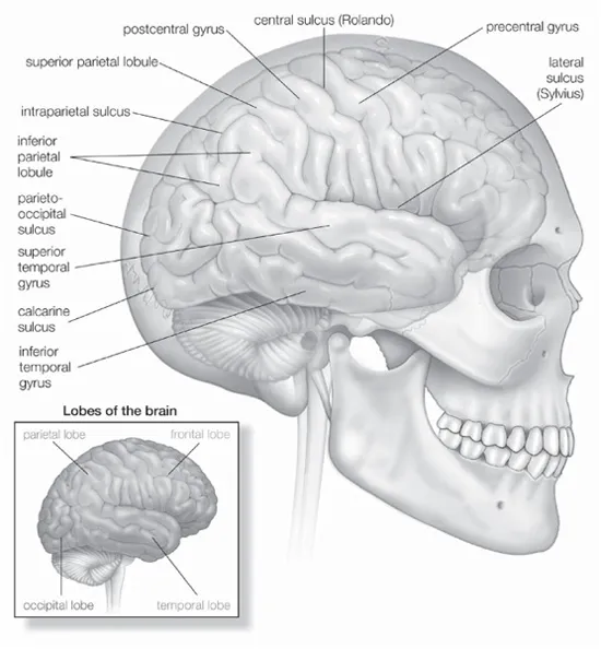
Lateral (side) view of the right cerebral hemisphere of the human brain, shown in situ within the skull. A number of convolutions (called gyri) and fissures (called sulci) in the surface define four lobes—the parietal, frontal, temporal, and occipital—that contain major functional areas of the brain. Encyclopædia Britannica, Inc.
The frontal lobe, the largest of the four cerebral lobes, lies rostral to the central sulcus (toward the nose from the sulcus). One important structure in the frontal lobe is the precentral gyrus, which constitutes the primary motor (motion) region of the brain. When parts of the gyrus are electrically stimulated in conscious patients who are under local anesthesia, they produce localized movements on the opposite side of the body that are interpreted by the patients as voluntary. Injury to parts of the precentral gyrus results in paralysis on the contralateral half of the body. Parts of the inferior frontal lobe constitute the Broca area, a region involved with speech.
The parietal lobe, which lies behind, or posterior, to the central sulcus, is divided into three parts: (1) the postcentral gyrus, (2) the superior parietal lobule, and (3) the inferior parietal lobule. The postcentral gyrus receives sensory input from the contralateral half of the body. The sequential representation is the same as in the primary motor area. Sensations from the head are represented in inferior (lower) parts of the gyrus and impulses from the lower extremities are represented in superior portions. The superior parietal lobule, located caudal to (below and behind) the postcentral gyrus, lies above the intraparietal sulcus. This lobule is regarded as an association cortex, an area that is not involved in either sensory or motor processing, although part of the superior parietal lobule may be concerned with motor function. The inferior parietal lobule (composed of the angular and supramarginal gyri) is a cortical—i.e; outer layer—region involved with the integration of multiple sensory signals.
In both the parietal and frontal lobes, each primary sensory or motor area is close to, or surrounded by, a smaller secondary area. The primary sensory area receives input only from the thalamus, while the secondary sensory area receives input from the thalamus, the primary sensory area, or both. The motor areas receive input from the thalamus as well as the sensory areas of the cerebral cortex.
The temporal lobe, which is below the lateral sulcus, fills the middle fossa, or hollow area, of the skull. The outer surface of the temporal lobe is an association area made up of the superior, middle, and inferior temporal gyri. An association area is a part of the cerebral cortex that is connected to both cerebral hemispheres and which helps link parts of the brain that are concerned with motor and sensory function. Near the margin of the lateral sulcus, two transverse (lying across) temporal gyri constitute the primary auditory area of the brain. The sensation of hearing is represented here in a tonotopic fashion—that is, with different frequencies of sound represented on different parts of the auditory area. The transverse gyri are surrounded by a less finely tuned secondary auditory area. A medial, or inner, protrusion near the underside, or ventral surface of the temporal lobe, known as the uncus, constitutes a large part of the primary olfactory area, concerning the sense of smell.
The occipital lobe, important to vision, lies toward the back of the brain, behind the parieto-occipital sulcus. The parieto-occipital sulcus joins another sulcus, the calcarine sulcus, in a Y-shaped formation. Cortex—a special kind of gray matter that makes up the outer layer of the cerebrum—on both banks of the calcarine sulcus constitutes the primary visual area, which receives input from the contralateral visual field via an information-carrying nerve fibre system called optic radiation. The visual field is represented near the calcarine sulcus with upper quadrants of the visual field laid out along the lower bank of the sulcus and lower quadrants of the visual field represented on the upper bank.
Aside from the four major cerebral lobes there are two other lobes worth noting. The insular lobe, or central lobe, is an invaginated (folded back in upon itself) triangular area on the medial surface of the lateral sulcus. Not visible from the surface of the cerebrum, it can be seen in the intact brain only by separating the frontal and parietal lobes from the temporal lobe. The insular lobe is thought to be involved in sensory and motor visceral functions as well as taste perception.
The limbic lobe is located on the medial margin (or limbus) of each hemisphere. Composed of adjacent portions of the frontal, parietal, and temporal lobes that surround the corpus callosum, the limbic lobe is involved with autonomic (involuntary) and related somatic (body) behavioral activities. The limbic lobe receives input from thalamic nuclei. A nucleus is a structure in the brain made up of a group of neurons. The thalamic nuclei, made up of neurons in the thalamus, are connected with and relay information from parts of the brain such as the hypothalamus. These neurons are also connected to the hippocampal formation.
Cerebral Ventricles
The hippocampal formation is located within one of the cerebral ventricles, cavities deep within the white matter of the cerebral hemispheres. These cavities, which are filled with cerebrospinal fluid, form the ventricular system. They include a pair of C-shaped lateral ventricles with anterior, inferior, and posterior “horns” protruding into the frontal, temporal, and occipital lobes, respectively. Most of the clear cerebrospinal fluid that flows both in the brain and the spinal column is produced in the ventricles, and about 70 percent of it is secreted by the choroid plexus, a collection of blood vessels in the walls of the lateral ventricles. The fluid drains via interventricular foramina, or openings, into a slitlike third ventricle, which, situated along the midline of the brain, separates the symmetrical halves of the thalamus and hypothalamus. From there the fluid passes through the cerebral aqueduct in the midbrain and into the fourth ventricle in the hindbrain. Openings in the fourth ventricle permit cerebrospinal fluid to enter certain regions called subarachnoid spaces surrounding both the brain and the spinal cord.
Basal Ganglia
Deep within the cerebral hemispheres, large gray masses of nerve cells, called nuclei, form components of the basal ganglia. Four basal ganglia can be distinguished: (1) the caudate nucleus, (2) the putamen, (3) the globus pallidus, and (4) the amygdala. Phylogenetically, the amygdala was the first to evolve and is the oldest of the basal ganglia.
The caudate nucleus and the putamen have similar cellular compositions, cytochemical features, and functions but slightly different connections. The putamen lies deep within the cortex of the insular lobe, while the caudate nucleus has a C-shaped configuration that parallels the lateral ventricle. The head of the caudate nucleus protrudes into the anterior horn of the lateral ventricle, the body lies above and lateral to the thalamus, and the tail is in the roof of the inferior horn of the lateral ventricle. The tail of the caudate nucleus ends in relationship to the amygdaloid nuclear complex, which lies in the temporal lobe beneath the cortex of the uncus.
There are an enormous number of neurons within the caudate nucleus and putamen; they are of two basic types: spiny and aspiny. Spiny striatal neurons are medium-size cells with radiating dendrites (small projections) that are studded with spines. The long, slender axons of these cells project beyond the caudate nucleus and putamen’s boundaries. All nerves providing input to these two kinds of basal ganglia terminate upon the dendrites of spiny striatal neurons. All output is via axons of the same neurons. Chemically, spiny striatal neurons are heterogeneous; that is, most contain more than one neurotransmitter (a chemical that moves nerve impulses). One kind of neurotransmitter, Gamma-aminobutyric acid (GABA) is the primary neurotransmitter contained in spiny striatal neurons. Other neurotransmitters found in spiny striatal neurons include substance P and enkephalin.
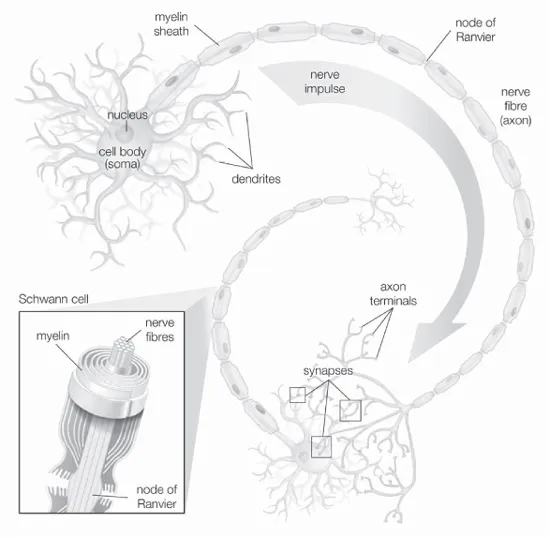
The structural features of a motor neuron include the cell body, the nerve fibre (or axon), and the dendrites. 2002 Encyclopædia Britannica, Inc
Aspiny striatal neurons have smooth dendrites and short axons confined to the caudate nucleus or putamen. Small aspiny striatal neurons secrete GABA, neuropeptide Y, somatostatin, or some combination of these. The largest aspiny neurons are evenly distributed neurons that also secrete neurotransmitters and are important in maintaining the balance of GABA and yet another kind of neurotransmitter called dopamine.
Because the caudate nucleus and putamen receive varied and diverse inputs from multiple sources that utilize different neurotransmitters, they are regarded as the receptive component of the corpus striatum (a unit of basal ganglia made up of the caudate nucleus, putamen, and globus pallidus). Most input originates from regions of the cerebral cortex, via connecting fibres called corticostriate fibres which contain the excitatory neurotransmitter glutamate. In addition, afferent fibres (which carry impulses to a nerve centre in the spinal cord or brain) project to the caudate nucleus or the putamen. These afferent fibres originate from a large nucleus located in the midbrain called the substantia nigra or from intralaminar thalamic nuclei. Neurons in the substantia nigra are known to synthesize dopamine, but the neurotransmitter secreted by thalamostriate neurons has not been identified. All striatal afferent systems terminate, or end, in patchy areas called strisomes; areas not receiving terminals are called the matrix. Spiny striatal neurons containing GABA, substance P, and enkephalin project in a specific pattern onto the globus pallidus and the substantia nigra.
The pattern is as follows: The globus pallidus, consisting of two cytologically (cellularly) similar wedge-shaped segments, the lateral and the medial, lies between the putamen and the internal capsule. Striatopallidal fibres from the caudate nucleus and putamen converge on the globus pallidus like spokes of a wheel. Both segments of the pallidum receive GABAergic terminals, but in addition the medial segment receives substance P fibres, and the lateral segment receives enkephalinergic projections. The output of the entire corpus striatum arises from GABAergic cells in the medial pallidal segment and in the substantia nigra, both of which receive fibres from the striatum. GABAergic cells in the medial pallidal segment and the substantia nigra project to different nuclei in the thalamus; these in turn influence distinct regions of the cerebral cortex involved with motor function. The lateral segment of the globus pallidus, on the other hand, projects almost exclusively to the subthalamic nucleus, from which it receives reciprocal input. No part of the corpus striatum projects fibres to spinal levels.
When basal ganglia don’t function properly, pathological processes result. These processes, involving the corpus striatum and related nuclei, are associated with a variety of specific diseases characterized by abnormal involuntary movements (collectively referred to as dyskinesia) and significant alterations of muscle tone. Parkinson disease and Huntington disease are among the more prevalent syndromes; each is associated with deficiencies in the synthesis of particular neurotransmitters.
The amygdala, located in the ...
Table of contents
- Cover Page
- Title Page
- Copyright Page
- Contents
- Introduction
- Chapter 1: The Central Nervous System
- Chapter 2: The Peripheral and Autonomic Nervous Systems
- Chapter 3: Functions of the Human Nervous System
- Chapter 4: The Nervous System in Motion
- Chapter 5: Perception, Sensation, and Neuroplasticity
- Chapter 6: Neurobiological Disorders of Development and Behaviour
- Chapter 7: Neurological and Neurodegenerative Disorders Affecting the Brain
- Chapter 8: Other Diseases of the Central and Peripheral Nervous Systems
- Conclusion
- Glossary
- Bibliography
- Index
Frequently asked questions
Yes, you can cancel anytime from the Subscription tab in your account settings on the Perlego website. Your subscription will stay active until the end of your current billing period. Learn how to cancel your subscription
No, books cannot be downloaded as external files, such as PDFs, for use outside of Perlego. However, you can download books within the Perlego app for offline reading on mobile or tablet. Learn how to download books offline
Perlego offers two plans: Essential and Complete
- Essential is ideal for learners and professionals who enjoy exploring a wide range of subjects. Access the Essential Library with 800,000+ trusted titles and best-sellers across business, personal growth, and the humanities. Includes unlimited reading time and Standard Read Aloud voice.
- Complete: Perfect for advanced learners and researchers needing full, unrestricted access. Unlock 1.5M+ books across hundreds of subjects, including academic and specialized titles. The Complete Plan also includes advanced features like Premium Read Aloud and Research Assistant.
We are an online textbook subscription service, where you can get access to an entire online library for less than the price of a single book per month. With over 1.5 million books across 990+ topics, we’ve got you covered! Learn about our mission
Look out for the read-aloud symbol on your next book to see if you can listen to it. The read-aloud tool reads text aloud for you, highlighting the text as it is being read. You can pause it, speed it up and slow it down. Learn more about Read Aloud
Yes! You can use the Perlego app on both iOS and Android devices to read anytime, anywhere — even offline. Perfect for commutes or when you’re on the go.
Please note we cannot support devices running on iOS 13 and Android 7 or earlier. Learn more about using the app
Please note we cannot support devices running on iOS 13 and Android 7 or earlier. Learn more about using the app
Yes, you can access The Brain and the Nervous System by Britannica Educational Publishing, Kara Rogers in PDF and/or ePUB format, as well as other popular books in Biological Sciences & Physiology. We have over 1.5 million books available in our catalogue for you to explore.