Though only about the size of a clenched fist, the human heart bears the immense burden of sustaining human life and activity. Functioning to circulate blood throughout the body, the heart is an organ on which all others intimately depend. This volume relates the anatomy of the heart and the effects of the diseases to which it is sometimes prone. Annotated diagrams and illustrations bolster the narrative and highlight significant aspects of cardiology and the incredible cardiovascular system.
Trusted by 375,005 students
Access to over 1.5 million titles for a fair monthly price.
The heart is one of the most vital organs in the human body. Its function is to circulate the blood by acting as a pump. With each heartbeat, blood is pushed into the arteries and through the veins. It then courses around the body in a one-way circuit so that it eventually returns to the heart to repeat the process. Driving this constant movement of the blood are the perpetual rhythmic contractions of the heart muscle. This defining characteristic of the heart underlies the body’s ability to routinely and reliably deliver oxygen and nutrients to organs, tissues, and cells.
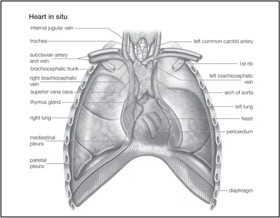
The human heart in situ. Encyclopædia Britannica, Inc.
With the exception of some invertebrates, the heart is an anatomical feature common to members of the animal kingdom. However, the shape and complexity of the heart varies greatly among the different groups of animals. It may be a straight tube, as in spiders and annelid worms, or a somewhat more elaborate structure with one or more receiving chambers (atria) and a main pumping chamber (ventricle), as in mollusks. In fishes the heart is a folded tube, with three or four enlarged areas that correspond to the chambers in the mammalian heart. In animals with lungs—amphibians, reptiles, birds, and mammals—the heart shows various stages of evolution from a single to a double pump that circulates blood (1) to the lungs and (2) to the body as a whole.
The human adult heart is normally slightly larger than a clenched fist with average dimensions of about 13 × 9 × 6 cm (5 × 3.5 × 2.5 inches) and weighing approximately 300 grams (10.5 ounces). It is cone-shaped, with the broad base directed upward and to the right and the apex pointing downward and to the left. It is located in the chest (thoracic) cavity behind the breastbone (sternum), in front of the windpipe (trachea), the esophagus, and the descending aorta, between the lungs, and above the diaphragm. About two-thirds of the heart lies to the left of the midline.
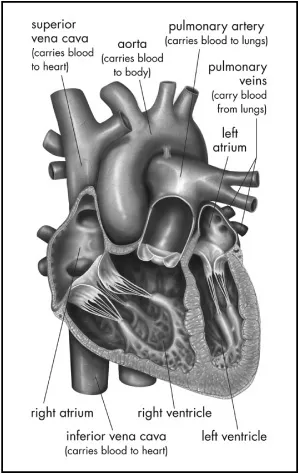
In humans and other mammals and in birds, the heart is a four-chambered system. The heart cavity is divided down the middle into a right and a left heart, which in turn are subdivided into two chambers. The upper chamber is called an atrium, and the lower chamber is called a ventricle. The two atria act as receiving chambers for blood entering the heart. The more muscular ventricles pump the blood out of the heart.
The right atrium receives venous blood from the head, chest, and arms via the large vein called the superior vena cava and receives blood from the abdomen, pelvic region, and legs via the inferior vena cava. Blood then passes through the tricuspid valve to the right ventricle, which propels it through the pulmonary artery to the lungs. In the lungs venous blood comes in contact with inhaled air, picks up oxygen, and loses carbon dioxide. Oxygenated blood is returned to the left atrium through the pulmonary veins. Valves in the heart allow blood to flow in one direction only and help maintain the pressure required to pump the blood.
Cross section of the human heart. Encyclopædia Britannica, Inc.
The low-pressure circuit from the heart (right atrium and right ventricle), through the lungs, and back to the heart (left atrium) constitutes the pulmonary circulation. Passage of blood through the left atrium, bicuspid valve, left ventricle, aorta, tissues of the body, and back to the right atrium constitutes the systemic circulation. Blood pressure is greatest in the left ventricle and in the aorta and its arterial branches. Pressure is reduced in the capillaries (vessels of minute diameter) and is reduced further in the veins returning blood to the right atrium.
The heart consists of a tough muscular wall, the myocardium. A thin layer of tissue, the pericardium, covers the outside of the myocardium, and another layer, the endocardium, lines the inside. The pumping of the heart, or the heartbeat, is caused by alternating contractions and relaxations of the myocardium.
The myocardial contractions are stimulated by electrical impulses from a natural pacemaker, the sinoatrial (or S-A) node located in the muscle of the right atrium. An impulse from the sinoatrial node causes the two atria to contract, forcing blood into the ventricles. Contraction of the ventricles is controlled by impulses from the atrioventricular (or A-V) node located at the junction of the two atria. Following contraction, the ventricles relax, and pressure within them falls. Blood again flows into the atria, and an impulse from the sinoatrial node starts the cycle over again. This process is called the cardiac cycle. The period of relaxation is called diastole. The period of contraction is called systole. Diastole is the longer of the two phases so that the heart can rest between contractions. In general, the rate of heartbeat varies inversely with the size of the animal. In elephants it averages 25 beats per minute, in canaries about 1,000. In humans the rate diminishes progressively from birth (when it averages 130) to adolescence but increases slightly in old age. The average adult rate is 70 beats at rest. The rate increases temporarily during exercise, emotional excitement, and fever and decreases during sleep. Rhythmic pulsation felt on the chest, coinciding with heartbeat, is called the apex beat. It is caused by pressure exerted on the chest wall at the outset of systole by the rounded and hardened ventricular wall.
ORIGIN AND DEVELOPMENT
In the embryo, formation of the heart begins in the pharyngeal, or throat, region. The first visible indication of the embryonic heart occurs in the undifferentiated mesoderm, the middle of the three primary layers in the embryo, as a thickening of invading cells. An endocardial (lining) tube of flattened cells subsequently forms and continues to differentiate until a young tube with forked anterior and posterior ends arises. As differentiation and growth progress, this primitive tube begins to fold upon itself, and constrictions along its length produce four primary chambers. These are called, from posterior to anterior, the sinus venosus, atrium, ventricle, and truncus arteriosus. The characteristic bending of the tube causes the ventricle to swing first to the right and then behind the atrium, the truncus coming to lie between the sideways dilations of the atrium. It is during this stage of development and growth that the first pulsations of heart activity begin.
Endocardial cushions (local thickenings of the endocardium, or heart lining) “pinch” the single opening between the atrium and the ventricle into two portions, thereby forming two openings. These cushions are also responsible for the formation of the two atrioventricular valves (the valves between atria and ventricles), which regulate the direction of blood flow through the heart.
The atrium becomes separated into right and left halves first by a primary partition with a perforation and later by a secondary partition, which, too, has a large opening, called the foramen ovale, in its lower part. Even though the two openings do not quite coincide in position, blood still passes through, from the right atrium to the left. At birth, increased blood pressure in the left atrium forces the primary partition against the secondary one, so that the two openings are blocked and the atria are completely separated. The two partitions eventually fuse.
The ventricle becomes partially divided into two chambers by an indentation of myocardium (heart muscle) at its tip. This developing partition is largely muscular and is supplemented by membranous connective tissue that develops in conjunction with the subdivision of the truncus arteriosus by a spiral partition into two channels, one for systemic and one for pulmonary circulation (the aorta and the pulmonary artery, respectively). At this time, the heart rotates clockwise and to the left so that it resides in the left thorax, with the left chambers posterior and the right chambers anterior. The greater portion of blood passing through the right side of the heart in the fetus is returned to the systemic circulation by the ductus arteriosus, a vessel connecting the pulmonary artery and the aorta. At birth this duct becomes closed by a violent contraction of its muscular wall. Thereafter, the blood in the right side of the heart is driven through the pulmonary arteries to the lungs for oxygenation and returned to the left side of the heart for ejection into the systemic circulation. A distinct median furrow at the apex of the ventricles marks the external subdivision of the ventricle into right and left chambers.
PERICARDIUM
The heart is suspended in its own membranous sac, the pericardium. The strong outer portion of the sac, or fibrous pericardium, is firmly attached to the diaphragm below, the mediastinal pleura on the side, and the sternum in front. It gradually blends with the coverings of the superior vena cava and the pulmonary (lung) arteries and veins leading to and from the heart. (The space between the lungs, the mediastinum, is bordered by the mediastinal pleura, a continuation of the membrane lining the chest. The superior vena cava is the principal channel for venous blood from the chest, arms, neck, and head.)
Smooth, serous (moisture-exuding) membrane lines the fibrous pericardium, then bends back and covers the heart. The portion of membrane lining the fibrous pericardium is known as the parietal serous layer (parietal pericardium), and the portion covering the heart is known as the visceral serous layer (visceral pericardium or epicardium).
The two layers of serous membrane are normally separated by only 10 to 15 ml (0.6 to 0.9 cubic inch) of pericardial fluid, which is secreted by the serous membranes. The slight space created by the separation is called the pericardial cavity. The pericardial fluid lubricates the two membranes with every beat of the heart as their surfaces glide over each other. Fluid is filtered into the pericardial space through both the visceral and parietal pericardia.
EXTERNAL SURFACE OF THE HEART
Shallow grooves called the interventricular sulci, containing blood vessels, mark the separation between ventricles on the front and back surfaces of the heart. There are two grooves on the external surface of the heart. One, the atrioventricular groove, is along the line where the right atrium and the right ventricle meet. It contains a branch of the right coronary artery (the coronary arteries deliver blood to the heart muscle). The other, the anterior interventricular sulcus, runs along the line between the right and left ventricles and contains a branch of the left coronary artery.
On the posterior side of the heart surface, a groove called the posterior longitudinal sulcus marks the division between the right and left ventricles. It contains another branch of a coronary artery. A fourth groove, between the left atrium and ventricle, holds the coronary sinus, a channel for venous blood.
CHAMBERS OF THE HEART
The right and left halves of the heart are divided by septa, or partitions, and each half is subdivided into two chambers, as noted previously. The upper chambers, the atria, are separated by a partition known as the interatrial septum. The lower chambers, the ventricles, are separated by the interventricular septum. The atria receive blood from various parts of the body and pass it into the ventricles. The ventricles, in turn, pump blood to the lungs and to the remainder of the body.
The right atrium, or right superior portion of the heart, is a thin-walled chamber receiving blood from all tissues except the lungs. Three veins empty into the right atrium, the superior and inferior venae cavae (previously noted), bringing blood from the upper and lower portions of the body, respectively, and the coronary sinus, draining blood from the heart itself. Blood flows from the right atrium to the right ventricle. The right ventricle, the right inferior portion of the heart, is the chamber from which the pulmonary artery carries blood to the lungs.
The left atrium, the left superior portion of the heart, is slightly smaller than the right atrium and has a thicker wall. The left atrium receives the four pulmonary veins, which bring oxygenated blood from the lungs. Blood flows from the left atrium into the left ventricle, as noted earlier. The left ventricle, the left inferior portion of the heart, has walls three times as thick as those of the right ventricle. Blood is forced from this chamber through the aorta to all parts of the body except the lungs.
ATRIA
The heart chambers that receive blood into the heart and drive it into the ventricles, the atria, have already been introduced. This section provides greater detail on the structure of these chambers. Fishes have one atrium; amphibians, reptiles, birds, and mammals have two.
In humans the atria are the two upper chambers of the heart. Each is roughly cube-shaped except for an ear-shaped projection called an auricle. (The term auricle is sometimes applied, incorrectly, to describe the entire atrium.) The major openings in the walls of the right atrium are (1) the points of entrance for the superior and inferior venae cavae (the great veins that return blood from the bodily tissues), and for the coronary sinus, the dilated terminal part of the cardiac vein, bearing venous blood from the heart muscle itself; and (2) the opening into the right ventricle. The principal openings into the left atrium are the points of entry of the pulmonary veins, bringing oxygenated blood from the lungs, and the opening into the left ventricle.
VENTRICLES
As discussed in earlier sections, the muscular chambers that pump blood out of the heart and into the circulatory system are known as the ventricles. Ventricles occur among some invertebrates. Among vertebrates, fishes and amphibians generally have a single ventricle, whereas reptiles, birds, and mammals have two. This section focuses on the structure of these chambers.
In humans, the ventricles are the two lower chambers of the heart. The walls of the chambers, and particularly the walls of the left ventricle, are far more heavily muscled than the walls of the atria because it is in the ventricles that the major force is exerted in the process of pumping the blood to the bodily tissues and to the lungs. Each opening leading into or away from the ventricles is guarded by a valve. These openings are the following: those from the two upper chambers; the opening from the right ventricle into the pulmonary artery, which transports blood to the lungs; and the opening from the left ventricle into the aorta, the main trunk by which oxygen-rich blood starts its course to the tissues. The interior surfaces of the ventricles are ridged with bundles and bands of muscle, called trabeculae carneae. The papillary muscles project like nipples into the cavities of the ventricles. They are attached by fine strands of tendon to the valves between the atria and ventricles and prevent the valves from opening when the ventricles contract.
VALVES OF THE HEART
To prevent backflow of blood, the heart is equipped with valves that permit the blood to flow in only one direction. There are two types of valves located in the heart: the atrioventricular valves (tricuspid and mitral) and the semilunar valves (pulmonary and aortic).
The atrioventricular valves are thin, leaflike structures located between the atria and the ventricles. The right atrioventricular opening is guarded by the tricuspid valve, so called because it consists of three irregularly shaped cusps, or flaps. The leaflets consist essentially of folds of endocardium (the membrane lining the heart) reinforced with a flat sheet of dense connective tissue. At the base of the leaflets, the middle supporting flat plate becomes continuous with that of the dense connective tissue of the ridge surrounding the openings.
Tendinous cords of dense tissue (chordae tendineae) covered by thin endocardium extend from the nipplelike papillary muscles to connect with the ventricular surface of the middle supporting layer of each leaflet. The chordae tendineae and the papillary muscles from which they arise limit the extent to which the portions of the valves near their free margin can billow toward the atria. The left atrioventricular opening is guarded by the mitral, or bicuspid, valve, so named because it consists of two flaps. The mitral valve is attached in the same manner as the tricuspid, but it is stronger and thicker because the left ventricle is by nature a more powerful pump working under high pressure.
Blood is propelled through the tricuspid and mitral valves as the atria contract. When the ventricles contract, blood is forced backward, passing between the flaps and walls of the ventricles. The flaps are thus pushed upward until they meet and unite, forming a complete partition between the atria and the ventricles. The expanded flaps of the valves are restrained by the chordae tendineae and papillary muscles from opening into the atria.
The semilunar valves are pocketlike structures attached at the point at which the pulmonary artery and the aorta leave the ventricles. The pulmonary valve guards the ori...
Table of contents
Cover Page
Title Page
Copyright Page
Contents
Introduction
Chapter 1: The Human Heart
Chapter 2: The Blood Vessels
Chapter 3: Congenital Heart Disease
Chapter 4: Acquired Heart Disease
Chapter 5: Diseases of Heart Tissues, Disturbances in Cardiac Rhythm, and Heart Failure
Chapter 6: Diseases of the Blood Vessels
Chapter 7: Hemodynamic Disorders and Shock
Chapter 8: Approaches to Cardiovascular Evaluation and Treatment
Conclusion
Glossary
Bibliography
Index
Frequently asked questions
Yes, you can cancel anytime from the Subscription tab in your account settings on the Perlego website. Your subscription will stay active until the end of your current billing period. Learn how to cancel your subscription
No, books cannot be downloaded as external files, such as PDFs, for use outside of Perlego. However, you can download books within the Perlego app for offline reading on mobile or tablet. Learn how to download books offline
Perlego offers two plans: Essential and Complete
Essential is ideal for learners and professionals who enjoy exploring a wide range of subjects. Access the Essential Library with 800,000+ trusted titles and best-sellers across business, personal growth, and the humanities. Includes unlimited reading time and Standard Read Aloud voice.
Complete: Perfect for advanced learners and researchers needing full, unrestricted access. Unlock 1.5M+ books across hundreds of subjects, including academic and specialized titles. The Complete Plan also includes advanced features like Premium Read Aloud and Research Assistant.
Both plans are available with monthly, semester, or annual billing cycles.
We are an online textbook subscription service, where you can get access to an entire online library for less than the price of a single book per month. With over 1.5 million books across 990+ topics, we’ve got you covered! Learn about our mission
Look out for the read-aloud symbol on your next book to see if you can listen to it. The read-aloud tool reads text aloud for you, highlighting the text as it is being read. You can pause it, speed it up and slow it down. Learn more about Read Aloud
Yes! You can use the Perlego app on both iOS and Android devices to read anytime, anywhere — even offline. Perfect for commutes or when you’re on the go. Please note we cannot support devices running on iOS 13 and Android 7 or earlier. Learn more about using the app
Yes, you can access The Cardiovascular System by Britannica Educational Publishing, Kara Rogers in PDF and/or ePUB format, as well as other popular books in Sciences biologiques & Physiologie. We have over 1.5 million books available in our catalogue for you to explore.