The human eye has been the subject of endless fascination. Indeed, the very nature of sensory perception is shaped by the visual cues the brain receives, thus rendering the eye one of the most crucial elements in our comprehension of the world. The structures and functions of the eye are examined in this exhaustive volume that describes how we are able to process what we observe and react accordingly. Detailed diagrams accompany the text and encourage the reader to consider all aspects of this beautiful and complex area of human anatomy.
eBook - ePub
The Eye: The Physiology of Human Perception
- English
- ePUB (mobile friendly)
- Available on iOS & Android
eBook - ePub
The Eye: The Physiology of Human Perception
About this book
Trusted by 375,005 students
Access to over 1.5 million titles for a fair monthly price.
Study more efficiently using our study tools.
Information
Topic
Sciences biologiquesSubtopic
Physiologie
CHAPTER 1
ANATOMY OF THE EYE

Close-up of a human eye. Shutterstock.com
The human eye is an amazingly complex structure that enables sight, one of the most important of the human senses. Sight underlies our ability to understand the world around us and to navigate within our environment. As we look at the world around us, our eyes are constantly taking in light, a component fundamental to the visual process. The front of the human eye contains a curved lens, through which light reflected from objects in the surrounding environment passes. The light travels deep into the eyeball, passing all the way to the back of the eye, where it converges to a point. A unique set of cells at the back of the eye receives the light, harnessing its energy by converting it into an electrical impulse, or signal, that then travels along neurons in the brain. The impulses are carried along a neuronal pathway that extends from the back of the eye all the way to the back of the brain, ultimately terminating in a region known as the visual cortex. There, the electrical signals from both eyes are processed and unified into a single image. The amount of time between the moment when light enters the eye and when a unified image is generated in the brain is near instantaneous, taking only fractions of a second.
Scientists’ knowledge of the intricacy and the complex associations of the vital structures within the human eye expanded tremendously in the 20th and early 21st centuries. Although some of this knowledge was obtained from studies of the eyes of animals, a significant amount of information was also gained from studies of diseases of the human eye. With this knowledge came an understanding of the function of each of the eye structures. Each structure contributes in a specific way to the visual process, and collectively they underlie a broad range of visual functions, from the perception of an object’s shape, size, and colour to the perception of distance.
THE EYE
The eyeball can be viewed as the fusing together of a small portion of a small, strongly curved sphere with a large portion of a large, not so strongly curved sphere. The small piece, occupying about one-sixth of the whole, has a radius of 8 mm (0.3 inch); it is transparent and is called the cornea; the remainder, called the scleral segment, is opaque and has a radius of 12 mm (0.5 inch). The ring where the two areas join is called the limbus. Thus, on looking directly into the eye from in front one sees the white sclera surrounding the cornea; because the latter is transparent one sees, instead of the cornea, a ring of tissue lying within the eye, the iris. The iris is the structure that determines the colour of the eye. The centre of this ring is called the pupil. It appears dark because the light passing into the eye is not reflected back to any great extent. By use of an ophthalmoscope, an instrument that permits the observer to illuminate the interior of the eyeball while observing through the pupil, the appearance of the interior lining of the globe can be made out. The lining, called the fundus oculi, is characterized by the large blood vessels that supply blood to the retina; these are especially distinct as they cross over the pale optic disk, or papilla, the region where the optic nerve fibres leave the globe.
The dimensions of the eye are reasonably constant, varying among healthy individuals by only a millimetre or two; the sagittal (vertical) diameter is about 24 mm (about 1 inch) and is usually less than the transverse (horizontal) diameter. At birth the sagittal diameter is about 16 to 17 mm (about 0.65 inch); it increases rapidly to about 22.5 to 23 mm (about 0.89 inch) by age 3; between ages 3 and 13 the globe attains its full size. The weight is about 7.5 grams (0.25 ounce), and its volume 6.5 mm (0.4 cubic inch).
The eye is made up of three coats, which enclose the optically clear aqueous humour, lens, and vitreous body. The outermost coat consists of the cornea and the sclera; the middle coat, or uvea, contains the main blood supply to the eye and consists, from the back forward, of the choroid, the ciliary body, and the iris. The innermost layer is the retina, lying on the choroid and receiving most of its nourishment from the vessels within the choroid, the remainder of its nourishment being derived from the retinal vessels that lie on its surface and are visible in the ophthalmoscope. The ciliary body and iris have a very thin covering, the ciliary epithelium and posterior epithelium of the iris, which is continuous with the retina.
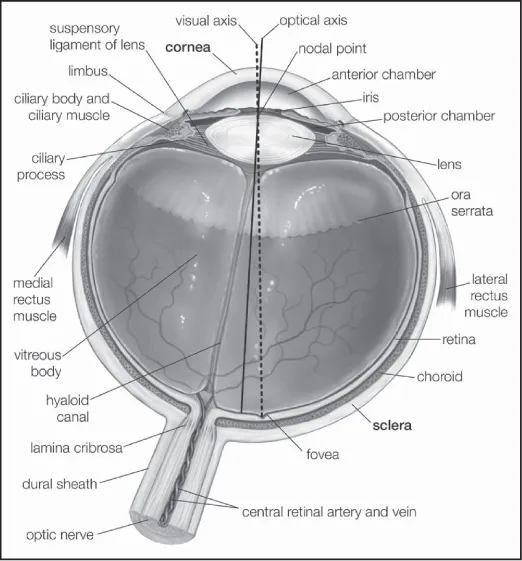
Horizontal cross section of the human eye, showing the structures of the eye, the visual axis (the central point of image focusing in the retina), and the optical axis (the axis about which the eye is rotated by the eye muscles). Encyclopædia Britannica, Inc.
Within the cavities formed by this triple-layered coat there are the crystalline lens, suspended by fine transparent fibres—the suspensory ligament or zonule of Zinn—from the ciliary body; the aqueous humour, a clear fluid filling the spaces between the cornea and the lens and iris; and the vitreous body, a clear jelly filling the much larger cavity enclosed by the sclera, the ciliary body, and the lens. The anterior chamber of the eye is defined as the space between the cornea and the forward surfaces of the iris and lens, while the posterior chamber is the much smaller space between the rear surface of the iris and the ciliary body, zonule, and lens; the two chambers both contain aqueous humour and are in connection through the pupil. This connection allows the aqueous humour to flow through the pupil from the posterior chamber to the anterior chamber. From there, the fluid flows out of the eye through the trabecular meshwork and Schlemm’s canal, which encircles the peripheral iris. Some aqueous humour also exits the eye directly through the ciliary body. The ciliary muscle attachments and the lens separate the aqueous humour in front from the vitreous humour behind.
The eye also contains special light receptors, called photoreceptors, and its construction is much like that of a simple camera. The retina, an extremely metabolically active layer of nerve tissue, is made up of millions of photoreceptors. Various structures of the eye function to focus light onto the retina, with the cornea providing the greatest focusing power of the eye. The cornea is curved to a much greater extent than the rest of the eyeball. However, the shape and focusing power of the cornea are not adjustable. This is in contrast to the lens, the shape of which is adjustable and is controlled by the action of the ciliary body, altering the focusing power of the lens as needed. The cornea and lens focus an image onto the retina at the back of the eye. If the image is projected too far in front of the retina, it causes the visual defect called myopia, or nearsightedness. If the image is theoretically focused “behind” the retina, the result is hyperopia, or farsightedness. If no deformation of the lens is present, the image is projected onto the fovea, a structure near the centre of the retina that contains a large number of cone photoreceptors and that provides the sharpest vision. When stimulated by light, retinal photoreceptor cells send signals to neighbouring cells in the retina that then relay the signals through the optic nerve to the visual centres of the brain.
OUTER TISSUES OF THE EYEBALL
The outermost coat of the eye is made up of the cornea and the sclera. Though these two structures are essentially extensions of the same layer of tissue in the eye, they have distinct functions, with the cornea playing a vital role in focusing light for photoreception and the sclera fulfilling an important role in the protection of the eyeball.
CORNEA
The cornea is the transparent window of the eye. It is about 12 mm (0.5 inch) in diameter and, except at its margins, contains no blood vessels. However, it does contain many nerves and is very sensitive to pain or touch. It is nourished and provided with oxygen anteriorly by tears and is bathed posteriorly by aqueous humour. It protects the pupil, the iris, and the inside of the eye from penetration by foreign bodies and is the first and most powerful element in the eye’s focusing system. As light passes through the cornea, it is partially refracted before reaching the lens. The curvature of the cornea, which is spherical in infancy but changes with age, gives it its focusing power; when the curve becomes irregular, it causes a focusing defect called astigmatism, in which images appear elongated or distorted.
The cornea contains five distinguishable layers: the epithelium, or outer covering; Bowman’s membrane; the stroma, or supporting structure; Descemet’s membrane; and the endothelium, or inner lining. Up to 90 percent of the thickness of the cornea is made up of the stroma. The epithelium, which is a continuation of the epithelium of the conjunctiva, is itself made up of about six layers of cells. The superficial layer is continuously being shed, and the layers are renewed by multiplication of the cells in the innermost, or basal, layer.
The stroma appears as a set of lamellae, or plates, running parallel with the surface and superimposed on each other like the leaves of a book; between the lamellae lie the corneal corpuscles, cells that synthesize new collagen (connective tissue protein) essential for the repair and maintenance of this layer. The lamellae are made up of microscopically visible fibres that run parallel to form sheets; in successive lamellae the fibres make a large angle with each other. The lamellae in humans are about 1.5 to 2.5 micrometres (1 micrometre = 0.001 millimetre) thick, so that there are about 200 lamellae in the human cornea. The fibrous basis of the stroma is collagen.
Immediately above the stroma, adjacent to the epithelium, is Bowman’s membrane, about 8 to 14 micrometres thick; with the electron microscope it is evident that it is really stroma, but with the collagen fibrils not arranged in the orderly fashion seen in the rest of the stroma.
Beneath the stroma are Descemet’s membrane and the endothelium. The former is about 5 to 10 micrometres thick and is made up of a different type of collagen from that in the stroma; it is secreted by the cells of the endothelium, which is a single layer of flattened cells. There is apparently no continuous renewal of these cells as with the epithelium, so that damage to this layer is a more serious matter.
The most obvious difference between the opaque sclera and the transparent cornea is the irregularity in the sizes and arrangement of the collagen fibrils in the sclera by contrast with the almost uniform thickness and strictly parallel array in the cornea; in addition, the cornea has a much higher percentage of mucopolysaccharide (a carbohydrate that has among its repeating units a nitrogenous sugar, hexosamine) as embedding material for the collagen fibrils. It has been shown that the regular arrangement of the fibrils is, in fact, the essential factor leading to the transparency of the cornea.
When the cornea is damaged—e.g., by a virus infection—the collagen laid down in the repair processes is not regularly arranged, with the result that an opaque patch called a leukoma, may occur. When an eye is removed or a person dies, the cornea soon loses its transparency, becoming hazy; this is due to the taking in of fluid from the aqueous humour, the cornea becoming thicker as it becomes hazier. The cornea can be made to reassume its transparency by maintaining it in a warm, well-aerated chamber, at about 31 °C (88 °F, its normal temperature); associated with this return of transparency is a loss of fluid.
The innermost layer of the cornea, the endothelium, plays a critical role in keeping the cornea from becoming swollen with excess fluid. Under normal conditions, the cornea tends to take in fluid and solutes, mainly from the aqueous humour and from the small blood vessels at the limbus. This is counteracted by the active transport of solutes from the cornea, which results in the movement of fluid from the cornea via osmotic gradients. The active transport of solutes depends on an adequate supply of energy, and any situation that prejudices this supply causes the cornea to swell—transport fails, or works so slowly that it cannot remove solutes and fluid as quickly as they enter.
When endothelial cells are lost, new cells are not produced; rather, existing cells expand to fill in the space left behind. Once loss of a critical number of endothelial cells has occurred, however, active transport of solutes decreases or becomes impaired. As a result, the cornea swells, causing decreased vision and, in severe cases, surface changes and pain. Endothelial cell loss can be accelerated via mechanical trauma or abnormal age-related endothelial cell death (called Fuchs endothelial dystrophy). Death of the eye causes complete transport failure, primarily because of the loss of temperature; if the dead eye is placed in a warm chamber, the reserves of metabolic energy it contains in the form of sugar and glycogen are adequate to keep the cornea transparent for 24 hours or more. When it is required to store corneas for grafting, as in an eye bank, the cornea is removed from the globe to prevent it from absorbing fluid from the aqueous humour.
The cornea is exquisitely sensitive to pain; for example, a corneal abrasion, or scratch, most often causes a sensation of something being on the eye and is accompanied by intense tearing, pain, and light sensitivity. The sensation of pain in the cornea is mediated by sensory nerve fibres, called ciliary nerves, that run just underneath the endothelium; they belong to the ophthalmic branch of the fifth cranial nerve, the large sensory nerve of the head. The ciliary nerves leave the globe through openings in the sclera, not in company with the optic nerve, which is concerned exclusively with responses of the retina to light.
The sclera is essentially the continuation backward of the cornea, the collagen fibres of the cornea being, in effect, continuous with those of the sclera. The sclera is pierced by numerous nerves and blood vessels; the largest of these holes is that formed by the optic nerve, the posterior scleral foramen. The outer two-thirds of the sclera in this region continue backward along the nerve to blend with its covering, or dural sheath—in fact, the sclera may be regarded as a continuation of the dura mater, the outer covering of the brain. The inner third of the sclera, combined with some choroidal tissue, stretches across the opening, and the sheet thus formed is perforated to permit the passage of fasciculi (bundles of fibres) of the optic nerve. This region is called the lamina cribrosa. The blood vessels of the sclera are largely confined to a superficial layer of tissue, and these, along with the conjunctival vessels, are responsible for the bright redness of the inflamed eye. As with the cornea, the innermost layer is a single layer of endothelial cells; above this is the lamina fusca, characterized by large numbers of pigment cells.
UVEAL TRACT
The middle coat of the eye is called the uvea (from the Latin for “grape”) because the eye looks like a reddish-blue grape when the outer coat has been dissected away. The posterior part of the uvea, the choroid, is essentially a layer of blood vessels and connective tissue sandwiched between the sclera and the retina. The forward portion of the uvea, the ciliary body and iris, is more complex, containing as it does the ciliary muscle and the sphincter and dilator of the pupil.
The blood supply responsible for nourishing the retina consists of the retinal and uveal circulations, both of which derive from branches of the ophthalmic artery. The two systems of blood vessels differ in that the retinal vessels, which supply nutrition to the innermost layers of the retina, derive from a branch of the ophthalmic artery, called the central artery of the retina, that enters the eye with the optic nerve, while the uveal circulation, which supplies the middle and outer layers of the retina as well as the uvea, is derived from branches of the ophthalmic artery that penetrate the globe independently of the optic nerve.
The ciliary body is the forward continuation of the choroid. It is a muscular ring, triangular in horizontal section, beginning at the region called the ora serrata and ending, in front, as the root of the iris. The surface is thrown into folds, called ciliary processes, the whole being covered by the ciliary epithelium, which is a double layer of cells; the layer next to the vitreous body, called the inner layer, is transparent, while the outer layer, which is continuous with the pigment epithelium of the retina, is heavily pigmented. These two layers are to be regarded embryologically as the forward continuation of the retina, which terminates at the ora serrata. Their function is to secrete the aqueous humour.
The ciliary muscle is an unstriped, involuntary, muscle concerned with alterations in the adjustments of focus—accommodation—of the optical system; the fibres run both across the muscle ring and circularly, and the effect of their contraction is to cause the whole body to move forward and to become fatter, so that the suspensory ligament that holds the lens in place is loosened.
Iris
The most anterior portion of the uvea is the iris. This is the only portion that is visible to superficial inspection, appearing as a perforated disc, the central perforation, or pupil, varying in size according to the surrounding illumination and other factors. A prominent feature is the collarette at the inner edge, representing the place of attachment of the embryonic pupillary membrane that, in embryonic life, covers the pupil. As with the ciliary body, with which it is anatomically continuous, the iris consists of several layers: namely, an anterior layer of endothelium, the stroma; and the posterior iris epithelium. The stroma contains the blood vessels and the two sheets of smooth muscle, the sphincter and dilator muscles, that control the contraction (constriction) and the expansion (dilation) of the iris, respectively. In addition, the stroma contains pigment cells that determine the colour of the eye.
Posteriorly, the stroma is covered by a double layer of epithelium, the continuation forward of the ciliary epithelium; here, however, both layers are heavily pigmented and serve to prevent light from passing through the iris tissue, confining...
Table of contents
- Cover Page
- Title Page
- Copyright Page
- Contents
- Introduction
- Chapter 1: Anatomy of the Eye
- Chapter 2: Protection and Movements of the Eye
- Chapter 3: Vision and the Retina
- Chapter 4: Electrophysiology of the Retina
- Chapter 5: Vision and the Brain
- Chapter 6: Diseases of the Outer Eye
- Chapter 7: Diseases of the Inner Eye
- Chapter 8: Visual Disorders and Eye Injuries
- Chapter 9: Diagnosis and Treatment of Eye Diseases
- Conclusion
- Glossary
- Bibliography
- Index
Frequently asked questions
Yes, you can cancel anytime from the Subscription tab in your account settings on the Perlego website. Your subscription will stay active until the end of your current billing period. Learn how to cancel your subscription
No, books cannot be downloaded as external files, such as PDFs, for use outside of Perlego. However, you can download books within the Perlego app for offline reading on mobile or tablet. Learn how to download books offline
Perlego offers two plans: Essential and Complete
- Essential is ideal for learners and professionals who enjoy exploring a wide range of subjects. Access the Essential Library with 800,000+ trusted titles and best-sellers across business, personal growth, and the humanities. Includes unlimited reading time and Standard Read Aloud voice.
- Complete: Perfect for advanced learners and researchers needing full, unrestricted access. Unlock 1.5M+ books across hundreds of subjects, including academic and specialized titles. The Complete Plan also includes advanced features like Premium Read Aloud and Research Assistant.
We are an online textbook subscription service, where you can get access to an entire online library for less than the price of a single book per month. With over 1.5 million books across 990+ topics, we’ve got you covered! Learn about our mission
Look out for the read-aloud symbol on your next book to see if you can listen to it. The read-aloud tool reads text aloud for you, highlighting the text as it is being read. You can pause it, speed it up and slow it down. Learn more about Read Aloud
Yes! You can use the Perlego app on both iOS and Android devices to read anytime, anywhere — even offline. Perfect for commutes or when you’re on the go.
Please note we cannot support devices running on iOS 13 and Android 7 or earlier. Learn more about using the app
Please note we cannot support devices running on iOS 13 and Android 7 or earlier. Learn more about using the app
Yes, you can access The Eye: The Physiology of Human Perception by Britannica Educational Publishing, Kara Rogers in PDF and/or ePUB format, as well as other popular books in Sciences biologiques & Physiologie. We have over 1.5 million books available in our catalogue for you to explore.