This textbook presents basic principles of local anesthesia and exodontia for undergraduate dental program students and dental surgeons in training. Readers will understand key concepts and points that prepare them for daily oral and maxillofacial surgery

eBook - ePub
Local Anesthesia and Extractions for Dental Students: Simple Notes and Guidelines
- English
- ePUB (mobile friendly)
- Available on iOS & Android
eBook - ePub
Local Anesthesia and Extractions for Dental Students: Simple Notes and Guidelines
About this book
Trusted by 375,005 students
Access to over 1.5 million titles for a fair monthly price.
Study more efficiently using our study tools.
Information
Topic
MedicineSubtopic
Oral Health & SurgeryPart I: The local Anesthesia in Dentistry
The Outline of the Trigeminal Nerve Anatomy
Esam Ahmad Z OmarFadi JarabWamiq Musheer Fareed
Abstract
The primary sensory nerve of the Oro-facial area is the trigeminal nerve. Understanding the anatomy of this nerve is essential for the dental practitioner to understand the pain mechanism and dental pain pathway. Most of the anatomy books discuss the details of the anatomical relation of Trigeminal nerve instead of an understanding of neural fibers carry by the nerve. This chapter discusses the anatomy, the neural fibers of each branch and the neural connection (nuclei) of the nerve.
Keywords: Anatomy, Central Bathway, Mandibular Nerve, Maxillary Nerve, Nuclie of trigeminal nerve, Ophthalamic Nerve, Sympathatic and Parasympathetic of trigeminal nerve, Trigeminal nerve.
The Trigeminal nerve is mixed Cranial nerve comprises principally of neurons for sensation. It enters the trigeminal ganglion after travelling parallel to the pons surface and exiting the brain. The trigeminal ganglion makes up the spinal nerve by acting as the dorsal root ganglion.
The trigeminal ganglion divides into three major branches, innervating different bone, teeth and facial dermatome. Every branch follows a different path and site to exit the cranium.
The Opthalmic nerve, the primary V1 branch, exits via the superior orbital fissure of the cranium, reaching the orbit to innervate the skin existing above the forehead and eye as well as the globe of the eye.
The Maxillary nerve makes the second V2 division, leaving via the foramen rotundum, into the pterygopalatine fossa, an area located posterior to the orbit. Thereafter, it again enters the inferior orbital fissure, making its way to the infraorbital foramen on the face, innervating the skin of the nose and cheek and below the eye.
The Mandibular nerve, the V3 third division, also has a motor component leave with the nerve and joining it at the foramen ovale (the motor root).
The different trigeminal nerve nuclei: Fig. (1.1)
- The sensory nucleus is primarily for touch and temperature, present in the pons.
- The spinal nucleus is responsible primarily for temperature and pain, and is a sensory nucleus.
- The sensory nucleus is ventromedial to motor nucleus.
The mesencephalic nucleus is the proprioceptive nucleus for all muscles of mastication.
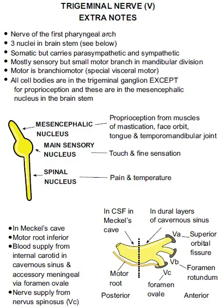
Trigeminal nerve nuclei (Acknowledgment: with appreciation and thanks to Instant Anatomy. 10 Summerfield Cambridge, CB3 9HE, UK. http://www.instantanatomy.net, for their permission for using this illustration).
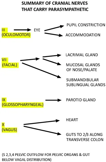
Nerves supplying parasympathetic fibers in head and neck area:
- Glossopharyngeal nerve (otic ganglion)
- Facial nerve (pterygopalatine ganglion, submandibular ganglion)
- Oculomotor nerve (ciliary ganglion)
The trigeminal nerve receives the parasympathetic and the special sensation (taste) from the following:
The nervus intermedius, or "nerve of Wrisberg" is the intermediate nerve of the facial nerve (cranial nerve VII) situated among the vestibulocochlear nerve (cranial nerve VIII) and the motor of the facial nerve.
The nucleus of nervus intermedius:
- Tractus solitaries (Taste nucleus): It contains the sensory taste (from tractus solitarius) which leaves the pons as nervus intermedius as a branch of facial. It gives branch to:
- Chorda tympani nerve travelling towards the submandibular ganglion.
- Greater petrosal nerve travelling towards the pterygopalatine ganglion.
- The superior salivary nucleus: parasympathetic secretory-motor fibers to:
- Submandibular, sublingual salivary gland to sublingual ganglion through the chorda tympani
- And to the minor salivary nucleus of nose and palate through the greater petrosal nerve which joins --- the ptrygo-palatine ganglion. It is made of parasympathetic fibers coming from the superior salivary nucleus and the tractus solitarius’ sensory taste and exiting alongside the facial nerve in the facial canal, joining it’s motor root at the geniculate ganglion. Parasympathetic axons originate from the superior salivatory nucleus. No synapse is present when these fibers pass the geniculate ganglion. Some of these preganglionic parasympathetic filaments, as they leave the geniculate ganglion, move within the greater petrosal nerve. This way they form a synapse with the pterygopalatine ganglion. These postganglionic neurons provide the lacrimal gland with parasympathetic innervation t through their axons.
The function of the tractus solitarius (solitary tract):
- Solitary tract are structures in the brainstem that receive and carry taste sensation from the visceral sensation from vagus, glossopharyngeal nerve (IX) and facial nerve through it,s fibres to trigeminal nerve (VII).
- Taste from the anterior part of the tongue, more specifically two third (2/3rd) area, through the facial nerve fibers given to the mandibular branch of the trigeminal nerve by chorda tympani The fibers of the facial nerve when leave the geniculate ganglion at the middle ear, they combine with the mandibular nerve at about one centimeter below the base of the skull by the chorda tympani. The posterior 1/3rd general and taste sensation through the glossopharyngeal nerve.
- Chemoreceptors in the carotid (by means of IX) and aortic body (through X).
- Stretch receptors from the aorta and carotid supply routes called blood vessel baroreceptors.
Petrosal nerve (Fig. 1.2) (a nerve going through the temporal bone, specifically the petrous part):
- Deep petrosal nerve
- Lesser petrosal nerve (also called the lesser superficial petrosal nerve)
- Greater petrosal nerve (also called as the greater superficial petrosal nerve)
- External superficial petrosal nerve
Petrosal nerve (Acknowledgments: with appreciation and thanks to Instant Anatomy. 10 Summerfield Cambridge, CB3 9HE, UK. http://www.instantanatomy.net, for their permission for using this illustration).
Deep Petrosal Nerve
Originating from the carotid plexus, the petrosus profundus or deep petrosal nerve travels within a canal positioned lateral to the internal carotid artery, known as the carotid canal.
It contains postganglionic sympathetic filaments supplied by the superior cervical ganglion.
As it passes through the cartilaginous substance of the foramen lacerum, it frames the nerve of the pterygoid canal (Vidian nerve) by combining with the greater petrosal nerve. At that point, without synapsing, it travels through the pterygopalatine ganglion and later joins the postganglionic parasympathetic filaments in supplying the lacrimal gland and nasal cavity.
The parasympathatic part of the large petrosal nerve, kn...
Table of contents
- Welcome
- Table of Contents
- Title
- BENTHAM SCIENCE PUBLISHERS LTD.
- FOREWORD
- PREFACE
- Part I: The local Anesthesia in Dentistry
- The Outline of the Trigeminal Nerve Anatomy
- Physiology of Pain
- The Outline of the Local Anesthesia - Pharmacology and Techniques
- Part II: Non-Surgical Dental Extractions
- Simple Extraction
- Complex Exodontia and Guidelines in Management of Medically Compromised Patients in Dental Chair
- Part III: Surgical Dental Extractions
- Wisdom Teeth & Maxillary Canine Impaction
Frequently asked questions
Yes, you can cancel anytime from the Subscription tab in your account settings on the Perlego website. Your subscription will stay active until the end of your current billing period. Learn how to cancel your subscription
No, books cannot be downloaded as external files, such as PDFs, for use outside of Perlego. However, you can download books within the Perlego app for offline reading on mobile or tablet. Learn how to download books offline
We are an online textbook subscription service, where you can get access to an entire online library for less than the price of a single book per month. With over 1.5 million books across 990+ topics, we’ve got you covered! Learn about our mission
Look out for the read-aloud symbol on your next book to see if you can listen to it. The read-aloud tool reads text aloud for you, highlighting the text as it is being read. You can pause it, speed it up and slow it down. Learn more about Read Aloud
Yes! You can use the Perlego app on both iOS and Android devices to read anytime, anywhere — even offline. Perfect for commutes or when you’re on the go.
Please note we cannot support devices running on iOS 13 and Android 7 or earlier. Learn more about using the app
Please note we cannot support devices running on iOS 13 and Android 7 or earlier. Learn more about using the app
Yes, you can access Local Anesthesia and Extractions for Dental Students: Simple Notes and Guidelines by Esam Omar,Fadi Jarab,Wamiq Fareed in PDF and/or ePUB format, as well as other popular books in Medicine & Oral Health & Surgery. We have over 1.5 million books available in our catalogue for you to explore.