This 3 volume set offers a comprehensive compilation which presents detailed information about ophthalmic (retinal, vitreous and macular) diseases. Key features of this set include:
Emphasis on practical features of clinical diagnosis
Concise and didactic presentation of key manifestations of diseases designed for rapid reference and target recall
A vast selection of illustrations to sharpen clinical problem-solving skills
Step by step treatment approaches to enhance the reader's ability to handle medical cases
Citations or relevant research articles in each chapter for further reading
The third volume of this set covers eye infections (bacterial and viral), inflammatory disorders and neoplasms. Written by a group of retina specialists, this book is an excellent resource for knowledge about retinal disorders. The streamlined format and evidence based medicine presented in the volume make this book the perfect reference for medical students, residents, general ophthalmologists and retina specialists.
eBook - ePub
Ophthalmology: Current and Future Developments: Volume 3: Diagnostic Atlas of Retinal Diseases
- English
- ePUB (mobile friendly)
- Available on iOS & Android
eBook - ePub
Ophthalmology: Current and Future Developments: Volume 3: Diagnostic Atlas of Retinal Diseases
About this book
Information
Topic
MedicinaSubtopic
Oftalmología y optometríaEndophthalmitis
ESSENTIALS OF DIAGNOSIS
Endophthalmitis (E): Severe intraocular inflammation associated with lid swelling, pain (absent in 26% of cases), anterior chamber with cells or hypopyon, vitritis and blurring or loss of vision, without involving the sclera and the extraocular orbital structure. “Panophthalmitis” involves outer layers (Figs. 1c and 6). Cases with an infectious etiology are the most devastating ones and carry guarded prognosis.
Classification (Table 1)
Table 1 Endophthalmitis Classification.
| Exogenous Endophthalmitis (EE) | • Postoperative Endophthalmitis * Acute. * Chronic. • Post-traumatic Endophthalmitis (Fig. 16) • Associated with infectious keratitis. |
| Endogenous Endophthalmitis (Ee) (Figs. 1, 21-23, 27, 28) | • Focal (anterior and posterior). • Diffuse (anterior and posterior). |
- Postoperative Endophthalmitis (PE):
E. is a potentially blinding complication with irreversible tissue damage after ocular surgery [1] (Figs. 2-5, 7-14, 26, 29). Most of the cases arise from cataract surgeries [2]. Early diagnosis and prompt treatment are therefore essential.
Commensal organisms found in the normal ocular flora are the most common cause. A mandatory step to reduce bacteria in the wound area is to apply povidone-iodine 5-10% to the cornea, conjunctival sac and periocular skin for a minimum of 3 minutes prior to surgery.
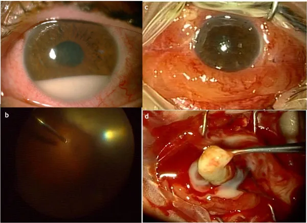
Fig. (1))
Klebsiella spp. Endogenous Endophthalmitis. 50-year-old woman. Diabetes. Klebsiella spp. hepatic abscess. a) Hypopyon and corneal haze. b) Identification of subretinal abscess during vitrectomy. c) Progression into panophthalmitis. d) Evisceration.
Klebsiella spp. Endogenous Endophthalmitis. 50-year-old woman. Diabetes. Klebsiella spp. hepatic abscess. a) Hypopyon and corneal haze. b) Identification of subretinal abscess during vitrectomy. c) Progression into panophthalmitis. d) Evisceration.
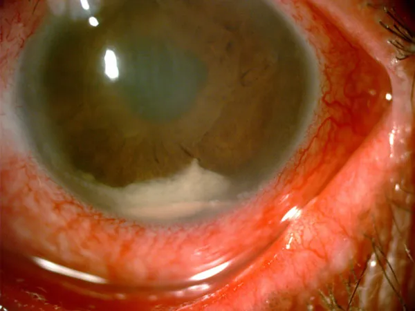
Fig. (2))
Postoperative Endophthalmitis: Hypopyon associated with fibrin clot. Hazy cornea, pupillary membrane. Gram-positive Coccus.
Postoperative Endophthalmitis: Hypopyon associated with fibrin clot. Hazy cornea, pupillary membrane. Gram-positive Coccus.
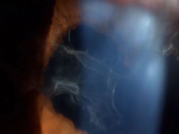
Fig. (3))
Exogenous Endophthalmitis. Anterior chamber fibrin.
Exogenous Endophthalmitis. Anterior chamber fibrin.
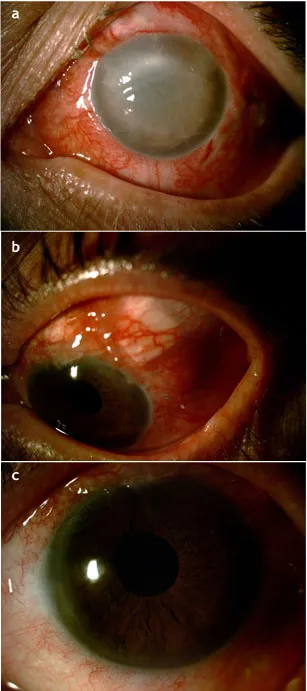
Fig. (4))
a) Late bleb-associated endophthalmitis. Not culture proven. Treated with intravitreal medication and vitrectomy. b) Scleral patch over trabeculectomy site. c) Good progress.
a) Late bleb-associated endophthalmitis. Not culture proven. Treated with intravitreal medication and vitrectomy. b) Scleral patch over trabeculectomy site. c) Good progress.
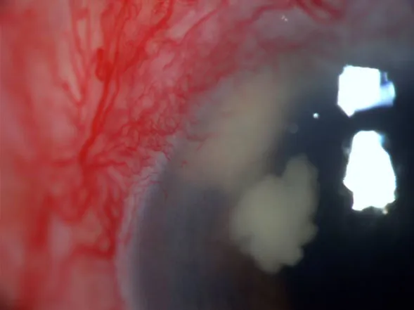
Fig. (5))
Corneal wound abscess after phacoemulsification.
Corneal wound abscess after phacoemulsification.

Fig. (6))
a) and b) Panophthalmitis.
a) and b) Panophthalmitis.

Fig. (7))
Acute-onset postoperative endophthalmitis. a) Hypopyon. b) After injection treatment and anterior chamber irrigation.
Acute-onset postoperative endophthalmitis. a) Hypopyon. b) After injection treatment and anterior chamber irrigation.
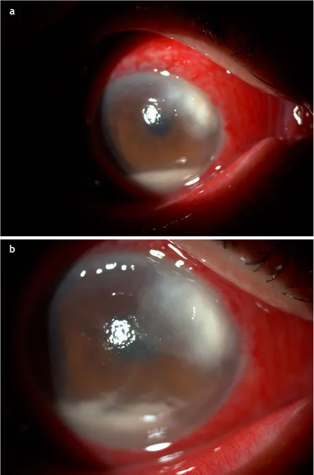
Fig. (8))
Acute-onset postoperative endophthalmitis. a) Hypopyon and corneal wound abscess. b) Negative progress. Bacterial source.
Acute-onset postoperative endophthalmitis. a) Hypopyon and corneal wound abscess. b) Negative progress. Bacterial source.
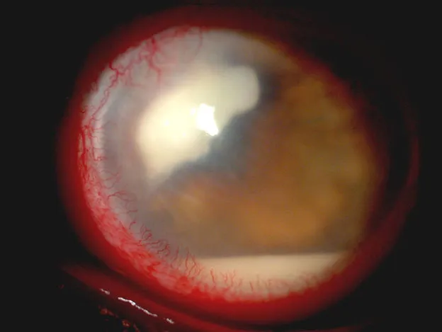
Fig. (9))
Exogenous fungal postoperative endophthalmitis. One month after phacoemulsification.
Exogenous fungal postoperative endophthalmitis. One month after phacoemulsification.
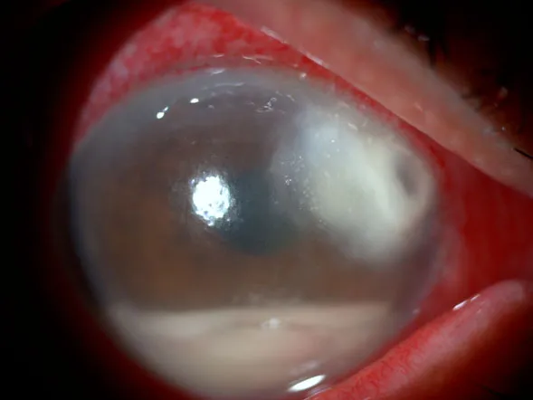
Fig. (10))
Postoperative bacterial endophthalmitis. Source of infection: phacoemulsification incision. One week after surgery.
Postoperative bacterial endophthalmitis. Source of infection: phacoemulsification incision. One week after surgery.

Fig. (11))
a) Acute endophthtalmitis after phacoemulsification. Germ: St. epidermidis. b) 48 hours post intravitreal treatment.
a) Acute endophthtalmitis after phacoemulsification. Germ: St. epidermidis. b) 48 hours post intravitreal treatment.

Fig. (12))
Acute-onset postoperative endophthalmitis after cataract extraction. St. epidermidis.
Acute-onset postoperative endophthalmitis after cataract extraction. St. epidermidis.
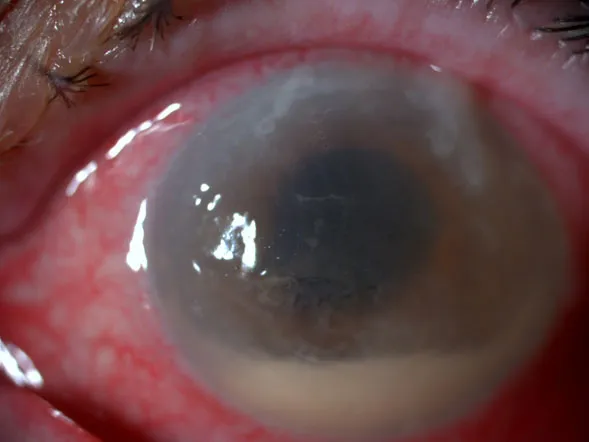
Fig. (13))
Acute postoperative endophthalmitis after phacoemulsification. Pseudomona aeruginosa.
Acute postoperative endophthalmitis after phacoemulsification. Pseudomona aeruginosa.

F...
Table of contents
- Welcome
- Table of Contents
- Title Page
- BENTHAM SCIENCE PUBLISHERS LTD.
- PREFACE
- List of Contributors
- Ocular Toxoplasmosis
- Ocular Tuberculosis
- Cytomegalovirus Retinitis
- Necrotizing Herpetic Retinopathies
- Ocular Syphilis
- HIV-Related Retinal Microangiopathy
- Neuroretinitis
- Endophthalmitis
- Acute Posterior Multifocal Placoid Pigment Epitheliopathy
- Multiple Evanescent White Dot Syndrome
- Multifocal Choroiditis
- Punctate Inner Choroidopathy
- Birdshot Retinochoroidopathy
- Serpiginous Choroiditis
- Diffuse Subretinal Fibrosis Syndrome
- Diffuse Unilateral Subacute Neuroretinitis
- Vogt Koyanagi Harada Disease
- Pars Planitis
- Sarcoidosis
- Retinoblastoma
- Cavernous Hemangioma of the Retina
- Von Hippel-Lindau Disease
- Astrocytoma Tuberous Sclerosis
- Retinal Vasoproliferative Tumor
- Melanocytoma
- Congenital Hypertrophy of the Retinal Pigment Epithelium
- Combined Hamartoma of Retina and Retinal Pigment Epithelium
- Choroidal Nevi
- Choroidal Melanoma
- Choroidal Metastasis
- Leukemic Retinopathy
- Primary Intraocular Lymphoma
- Idiopathic Uveal Effusion
- Hypotony Maculopathy
- Pregnancy-associated Retinal Diseases
Trusted by 375,005 students
Access to over 1.5 million titles for a fair monthly price.
Study more efficiently using our study tools.
Frequently asked questions
Yes, you can cancel anytime from the Subscription tab in your account settings on the Perlego website. Your subscription will stay active until the end of your current billing period. Learn how to cancel your subscription
No, books cannot be downloaded as external files, such as PDFs, for use outside of Perlego. However, you can download books within the Perlego app for offline reading on mobile or tablet. Learn how to download books offline
Perlego offers two plans: Essential and Complete
- Essential is ideal for learners and professionals who enjoy exploring a wide range of subjects. Access the Essential Library with 800,000+ trusted titles and best-sellers across business, personal growth, and the humanities. Includes unlimited reading time and Standard Read Aloud voice.
- Complete: Perfect for advanced learners and researchers needing full, unrestricted access. Unlock 1.5M+ books across hundreds of subjects, including academic and specialized titles. The Complete Plan also includes advanced features like Premium Read Aloud and Research Assistant.
We are an online textbook subscription service, where you can get access to an entire online library for less than the price of a single book per month. With over 1.5 million books across 990+ topics, we’ve got you covered! Learn about our mission
Look out for the read-aloud symbol on your next book to see if you can listen to it. The read-aloud tool reads text aloud for you, highlighting the text as it is being read. You can pause it, speed it up and slow it down. Learn more about Read Aloud
Yes! You can use the Perlego app on both iOS and Android devices to read anytime, anywhere — even offline. Perfect for commutes or when you’re on the go.
Please note we cannot support devices running on iOS 13 and Android 7 or earlier. Learn more about using the app
Please note we cannot support devices running on iOS 13 and Android 7 or earlier. Learn more about using the app
Yes, you can access Ophthalmology: Current and Future Developments: Volume 3: Diagnostic Atlas of Retinal Diseases by Mitzy E. Torres Soriano,Gerardo García Aguirre,Maximiliano Gordon,Veronica Kon Graversen, Mitzy E. Torres Soriano, Gerardo García Aguirre, Maximiliano Gordon, Veronica Kon Graversen in PDF and/or ePUB format, as well as other popular books in Medicina & Oftalmología y optometría. We have over 1.5 million books available in our catalogue for you to explore.