This innovative study charts the beginnings, history and fate of Interferon - one of modern medicine's most famous and infamous drugs. Interferon is part of the medical profession's armoury against viral infection, cancer and MS. The story of its development and use is one of survival in the face of remarkable cycles of promise and disappointment as a miracle drug. By telling this story, Toine Pieters' book provides insight into the research, manufacture, and marketing of new bio-molecules that mark modern medical science.
Pieters' closely argued book adopts a multi-disciplinary approach in seeking to trace the extraordinary voyage of interferon. Through the lens of interferon's voyage, the book explores the interaction of the broad range of actors driving medical science:
*biological and clinical researchers
*the pharmaceutical industry
*high-powered government agencies
*doctors and patients
*the media.
The book demonstrates how research on interferon led to new clinical definitions of cancer and a new rationale for therapeutic use of the drug. Interferon provides a marvellous insight into the development of one of the most controversial drugs of our time. It enhances our understanding of how medicine manufacture and marketing all played a part in pushing back the boundaries of research, from the post-penicillin era to the genetics revolution in medicine.
This study is of particular interest to undergraduates and postgraduates in the fields of History of Medicine, Pharmacology, Medical Genetics and History of Science.
- 284 pages
- English
- ePUB (mobile friendly)
- Available on iOS & Android
eBook - ePub
About this book
Trusted by 375,005 students
Access to over 1.5 million titles for a fair monthly price.
Study more efficiently using our study tools.
Information
Subtopic
Business GeneralIndex
Business1
Interferon’s birth 1
On an early Monday morning in June 1956, Jean Lindenmann arrived at the gates of the National Institute for Medical Research (NIMR) at Mill Hill, London, for the first time. The Institute’s enormous main entrance and seven-storeyed building, which overlooked both London’s northern suburbs and the adjacent countryside, made a profound impact on the young medical researcher from Switzerland (see Figure 1). On his way to the Division of Bacteriology and Virus Research on the second floor, Lindenmann became even more impressed when he glimpsed at the interior of some of the laboratories. All imaginable equipment seemed to be available. This was confirmed later on when Lindenmann was shown around the Institute by the head of the Division of Bacteriology and Virus Research, and Deputy Director of the NIMR, Christopher Andrewes.
Andrewes belonged to the group of ‘big names’ in biomedical research, which was accommodated at the NIMR, and formed part of a staff of approximately one hundred doctoral and post-doctoral workers, and an auxiliary workforce of about three hundred technicians, secretaries and others. The Institute ranked among the world’s best centres for biomedical research. It provided a state-of-the-art research environment aimed at stimulating internationally competitive research projects at the frontier of fundamental biomedical sciences. As a result there was great interest in doing research work at the NIMR.
The regular influx of visiting scientists was officially encouraged by the Director of the Institute, Sir Charles Harington. Harington believed that the resulting exchange of ideas would avoid scientific inbreeding, maintain a steady influx of new methods and techniques and keep researchers flexible in respect to incorporating new projects into their division’s research repertoire. Like most other Anglo-Saxon biomedical research centres, NIMR’s research agenda was primarily driven by motivations of scientific significance and impact, thereby favouring fundamental rather than applied research. At the same time the research agenda was often justified towards funding agencies for its potential medical therapeutic spin-off. 2
Lindenmann had never seen such a collection of sophisticated equipment like ultracentrifuges, freeze dryers, electric incubators and deep freezes, phase contrast microscopes and an electron microscope. The NIMR also had a special wing with large animal breeding facilities. The laboratory animals division assured a standardized supply of healthy laboratory animals such as dogs, ferrets, rabbits, rats, mice and poultry. Furthermore there was a well organized kitchen at the heart of the Institute on the lower ground floor, which was primarily responsible for cleaning and sterilizing glassware and preparing various standard solutions and culture media for the various research departments. The kitchen also produced special batches of media and solutions on request. This gave researchers a certain freedom to tinker with media and fluids during their experimental work. The kitchen was thus as much a central organ of the NIMR as the library on the fourth floor. This central storage for scientific paperwork was the last stop on their tour through the Institute.
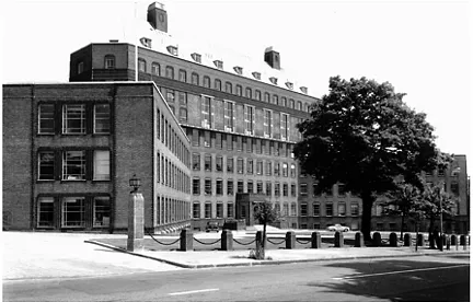
Figure 1 Front view of the NIMR. Courtesy of NIMR.
In comparison with the rest of the Institute, the Division of Bacteriology and Virus Research had relatively few highly sophisticated laboratory instruments and facilities at their immediate disposal. However, Andrewes—or ‘Cha’, as he was informally known in his department—was not at all bothered by this state of affairs. On the contrary, since his research group was mainly interested in the qualitative, phenomenological aspects of virus research, there simply was not that much need for high-tech equipment. With a regular supply of fertile eggs, laboratory animals, nutritive media and glassware, and facilities for cold and warm storage (hot and cold rooms, an electric deep freeze for biological specimens, incubators for eggs and tubes) and for sterile work (a hood for handling tissue cultures and viruses), the division was thought to provide the standard equipment and materials for work on animal viruses and bacteria. If on occasion one needed to do some work which required facilities and equipment for complicated procedures, such as ultracentrifuges, an electron microscope, or an arrangement for electrophoresis, one could always ask the divisions of Chemistry, and Biophysics and Optics, for help. Quite often researchers in Andrewes’s division would joke about their servants over there, who were so skillful in carrying out the quantitative jobs. Andrewes made no secret about the fact that he did not think a training in science was of any help for members of his division, who were predominantly graduates in medicine or veterinary medicine. 3
Lindenmann had been working under quite different conditions in Hermann Mooser’s laboratory that was part of the small institute of microbiology of the University of Zürich. The ‘Hygiene Institut’, as it was called, lagged years behind its counterparts in England with its rather old-fashioned laboratories and kitchen and no access to electron microscopes, ultracentrifuges and the like. In a way this is remarkable, since both Swiss and English science had come through the war relatively unharmed—without being cut off from a regular supply of American journals and without a dramatic drop in research activities. According to Lindenmann, Medical Microbiology was one of the sciences which remained ossified at a pre-war level until the 1950s, mainly due to the intellectual isolation of the leading scientists involved. 4
Unlike the Division of Bacteriology and Virus Research at the NIMR, the ‘Hygiene Institut’ did not have a virology section, as it was dominated by oldfashioned bacteriologists. They still regarded viruses as ultramicrobes: living, autonomous infectious entities, which, like bacteria, multiplied autonomically by a process of binary fission. 5 As such, viruses were thought to belong to the domain of bacteriology, thereby ignoring the international trend which recognized virology as an independent field of research. For the most part, the scientific staff at the ‘Hygiene Institut’ seemed to have missed the emerging consensus concerning the nature of viruses among American, British and French microbiologists. Viruses no longer were regarded as ultramicrobes but as infectious, potentially pathogenic, nucleic acid-containing entities of protein nature. Unlike bacteria, viruses were considered to be dependent upon the host cell for their reproduction. 6
These were anything but favourable circumstances for a young biomedical researcher with a vivid interest in the relatively new and dynamic field of animal virology—the study of viruses that prey on animals and human beings. So, after some unsatisfactory virus experiments in Zürich, Lindenmann asked permission to pursue his virus studies somewhere abroad. The head of the laboratory, the pre-eminent bacteriologist Mooser, was well aware of the limited possibilities for advanced virus study in the ‘Hygiene Institut’ and agreed to contact some British virologists whom he had met at an international microbiology meeting. One of the pioneers in the field of animal virology, Christopher Andrewes, who had acquired a worldwide reputation for innovative studies of animal viruses, agreed to let Lindenmann join his research group as a visiting worker for a period of one year. Subsequently, with Andrewes’s letter of intent and Mooser’s references, he was able to obtain a fellowship from the Swiss Academy of Medical Sciences and could start with preparations for his passage to England. 7
Starting a British/Swiss collaboration in a London laboratory
With the intent to learn as much about animal viruses as possible, Lindenmann started working in Andrewes’s division at the NIMR (see Figure 2). 8 As a relative novice to the field of virology he was assigned to a rather low-key research job which would give him the opportunity to learn a variety of viral techniques and at the same time make a contribution to one of the division’s research projects. Andrewes wanted him to work in his laboratory and do a series of experi- ments on the growth of polio virus in cultures of mouse and rabbit tissue. 9
In quite a number of laboratories around the world polio virus was produced on a daily basis in human and monkey tissue cultures for the large scale production of vaccines and for research purposes. 10 Since polio virus had been shown to be host-specific and not transmittable to mice or rabbits it had long been taken for granted that it would not grow in rabbit or mouse tissue culture either. However, with Donna Chaproniere, Andrewes had been able to demonstrate that myxomatosis virus did grow in guinea pig tissue cultures, in spite of the fact that the virus was known to be not transmittable to guinea pigs. 11 Andrewes had picked information up at a recent meeting of the Society for General Microbiology on successful experiments concerning the growth of polio virus in cultures of rabbit tissue, despite the fact that rabbits were not prone to the disease. This suggested the possibility that the specificity of viruses for particular hosts might be lost in tissue culture. Andrewes became interested in whether polio could indeed be grown in animals not prone to the disease. As a novice scientist at the frontier of virus research, Lindenmann was asked to show that polio virus would multiply in cultures of rabbit and mouse tissue. Lindenmann’s education as an M.D. and his training as a postgraduate student in diagnostic bacteriology was believed to provide him with enough resources and previous experience to tackle this new research problem. 12
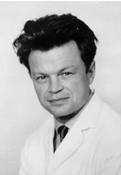
Figure 2 Jean Lindenmann (1957). Courtesy of NIMR.
Lindenmann’s first two months in Mill Hill were taken up by learning through handson apprenticeship the craft of working with viruses as it was being practised by workers in Andrewes’s division. At the same time, he initiated efforts to grow polio virus in nonspecific tissue cultures. Despite frequent changes of experimental conditions and procedures, Lindenmann was unable to find any evidence showing that polio virus multiplied in these tissues. In a first report to the Swiss Academy of Medical Sciences he stated rather optimistically: ‘Negative results in those experiments don’t mean a lot, as only small changes in the experimental conditions can make the difference between success and failure’. 13
However, to the best of Lindenmann’s memory the research project became increasingly frustrating. Despite numerous follow-up experiments, he still did not manage to produce any positive results: he could only show that polio virus did not multiply in his experimental arrangement. Andrewes strongly believed that Lindenmann’s unsuccessful attempts to replicate the claimed results regarding polio virus losing its host specificity in tissue cultures were due to an as-yet undetected artefact or some uncontrolled aspect of Lindenmann’s experimental set-up. 14 Andrewes therefore did not think much of the idea of abandoning the project in the face of the repeated negative results. As a visitor to the Institute and novice to the field of virology Lindenmann found it difficult to oppose Andrewes and to ask him to end the project.
Sometime during this period he was introduced to Alick Isaacs, a neighbouring researcher who had just returned from holidays. Only slightly older than Lindenmann, Isaacs at the age of 35 was already a distinguished virologist and an expert in influenza viruses who, like Lindenmann, had started his research career in a bacteriology department after being trained as a physician. Isaacs was in charge of the World Influenza Centre in room 215 which consisted of a large laboratory workspace with a rather small office corner for the necessary paperwork (see Figure 3). The World Influenza Centre had been set up in 1947 by Andrewes in collaboration with the United Nations’ World Health Organization (WHO) as the centre of a global ‘early warning’ network of collaborating laboratories to study and monitor outbreaks of influenza (‘flu’) epidemics throughout the world. The main task of the World Influenza Centre was to gather influenza virus specimens from all over the world and collect all sorts of data concerning the outbreak and spread of flu epidemics with the aim to study the nature and epidemiology of influenza. Ultimately, the research efforts of Isaacs and his collaborators were aimed at forecasting and controlling the outbreak of flu epidemics, thereby preventing a recurrence of the influenza pandemic of 1918 in which over twenty million people had died worldwide. 15
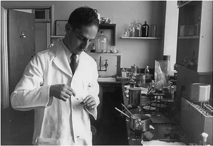
Figure 3 Alick Isaacs at work in his laboratory (1957). Courtesy of Dr S. IsaacsElmhirst.
During their first encounter, Isaacs asked Lindenmann about his research work in Zürich. Isaacs showed real interest the moment Lindenmann men- tioned his yet unpublished study of the virus interference phenomenon. 16 This biological phenomenon had first been named by the British scientists Gerald Findlay and Frank MacCallum in 1937. 17 Findlay and MacCallum had observed that infection with Rift Valley fever virus protected monkeys from infection with the unrelated yellow fever virus. Their phenomenon was defined as the interference of one virus with the pathogenic action of another, hence the name ‘interference phenomenon’. 18
Viral interference had been studied most extensively in the developing chicken egg since the 1940s when the German war refugees Werner and Gertrude Henle, who worked at the Children’s Hospital of Philadelphia, had shown that the interference phenomenon could be studied most conveniently in that test system. Laboratory animals often became cross-infected, carried undetected virus infections and were argued to have more complex immune reactions. Moreover, studying animal viruses in fertile hen’s eggs—or what were referred to as ‘embryonated eggs’ or simply as ‘eggs’—was more economical and allowed for more detailed study of viruses in their specific cellular environment. Christopher Andrewes called the fertile hen’s egg ‘the new experimental animal’ which yielded by far the best dividends’. 19 By the term ‘dividends’ Andrewes referred to the successful exploitation of chick embryo techniques both in viral research and in the large-scale production of viral vaccines—in particular, with reference to work on influenza virus and the development of an influenza vaccine (see Figure 4).
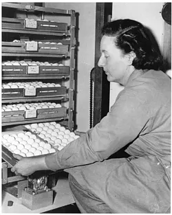
Figure 4 Hen’s egg incubator cabin (1950s). Courtesy of NIMR.
Isaacs had published a series of papers on the subject of viral interference. So he was eager to hear more about Lindenmann’s interference experiments. Isaacs probably then asked Lindenmann to join him for lunch. Since most researchers worked mainly within the confines of their laboratory and division, the lunch hall on the top floor was a prime marketplace for the exchange of information. Here researchers from all laboratories and research divisions mingled quite freely while communicating the latest shop talk and the Institute’s news. 20
Lindenmann told Isaacs that in May or June 1955 his boss in Zürich, Hermann Mooser, had seen a paper on the interference between strains of rickettsia, indicating that interference of one strain with the propagation or reproduction of the other was not brought about by competition for or blockade of a receptor. 21 Mooser thought it conceivable that the interference phenomenon between rickettsia might be rather similar to the interference between influenza viruses. However, to the best of his knowledge, the viral interference phenomenon was still explained in the literature as competition for or blockade of a cellular receptor. In the context of this view of the phenomenon of interference it seemed interesting to consider an interference experiment with influenza virus ...
Table of contents
- Cover Page
- Title Page
- Copyright Page
- Figures and tables
- Preface
- Acknowledgements
- Introduction
- 1: Interferon’s birth
- 2: Shaping a new field of research
- 3: Interferon on trial
- 4: Managing differences
- 5: About mice, malignancies and experimental therapies
- 6: Interferon, audiences and cancer
- 7: Yet another twist
- 8: Interferons in retrospect and prospect
- Appendix
- Notes
- Bibliography
Frequently asked questions
Yes, you can cancel anytime from the Subscription tab in your account settings on the Perlego website. Your subscription will stay active until the end of your current billing period. Learn how to cancel your subscription
No, books cannot be downloaded as external files, such as PDFs, for use outside of Perlego. However, you can download books within the Perlego app for offline reading on mobile or tablet. Learn how to download books offline
Perlego offers two plans: Essential and Complete
- Essential is ideal for learners and professionals who enjoy exploring a wide range of subjects. Access the Essential Library with 800,000+ trusted titles and best-sellers across business, personal growth, and the humanities. Includes unlimited reading time and Standard Read Aloud voice.
- Complete: Perfect for advanced learners and researchers needing full, unrestricted access. Unlock 1.5M+ books across hundreds of subjects, including academic and specialized titles. The Complete Plan also includes advanced features like Premium Read Aloud and Research Assistant.
We are an online textbook subscription service, where you can get access to an entire online library for less than the price of a single book per month. With over 1.5 million books across 990+ topics, we’ve got you covered! Learn about our mission
Look out for the read-aloud symbol on your next book to see if you can listen to it. The read-aloud tool reads text aloud for you, highlighting the text as it is being read. You can pause it, speed it up and slow it down. Learn more about Read Aloud
Yes! You can use the Perlego app on both iOS and Android devices to read anytime, anywhere — even offline. Perfect for commutes or when you’re on the go.
Please note we cannot support devices running on iOS 13 and Android 7 or earlier. Learn more about using the app
Please note we cannot support devices running on iOS 13 and Android 7 or earlier. Learn more about using the app
Yes, you can access Interferon by Toine Pieters in PDF and/or ePUB format, as well as other popular books in Business & Business General. We have over 1.5 million books available in our catalogue for you to explore.