- 288 pages
- English
- ePUB (mobile friendly)
- Available on iOS & Android
eBook - ePub
An Introduction to Cardiovascular Physiology
About this book
An Introduction to Cardiovascular Physiology is designed primarily for students of medicine and physiology. This introductory text is mostly didactic in teaching style and it attempts to show that knowledge of the circulatory system is derived from experimental observations. This book is organized into 15 chapters. The chapters provide a fuller account of microvascular physiology to reflect the explosion of microvascular research and include a discussion of the fundamental function of the cardiovascular system involving the transfer of nutrients from plasma to the tissue. They also cover major advances in cardiovascular physiology including biochemical events underlying Starling's law of the heart, nonadrenergic, non-cholinergic neurotransmission, the discovery of new vasoactive substances produced by endothelium and the novel concepts on the organization of the central nervous control of the circulation. This book is intended to medicine and physiology students.
Trusted by 375,005 students
Access to over 1.5 million titles for a fair monthly price.
Study more efficiently using our study tools.
Information
Chapter 1
Overview of the cardiovascular system
Publisher Summary
This chapter provides an overview of the cardiovascular system. The rate at which diffusional transport occurs is critically important because the supply of nutrients must keep up with cellular demand. The first and foremost function of cardiovascular system is the rapid convection of oxygen, glucose, amino acids, fatty acids, vitamins, drugs, and water to the tissues and the rapid washout of metabolic waste products like carbon dioxide, urea, and creatinine. The cardiac output is the volume of blood ejected by one ventricle during one minute, and this depends on both the volume ejected per contraction (the stroke volume) and the number of contractions per minute (heart rate). The behavior of the heart and the blood vessels has to be regulated to deal with varying environmental and internal stresses.
1.1 Diffusion: its virtues and limitations
1.2 Functions of the cardiovascular system
1.3 Circulation of blood
1.4 Cardiac output and its distribution
1.5 Introducing some hydraulic considerations: pressure and flow
1.6 Structure and functional classification of blood vessels
1.7 Plumbing of the vascular circuits
1.8 Central control of the cardiovascular system
The heart and blood vessels form a system for the rapid transport of oxygen, nutrients, waste products and heat around the body. Small primitive organisms lack such a system because their needs can be met by direct diffusion from the environment, and even in man diffusion remains the fundamental transport process between blood and cells. In order to appreciate fully the need for a cardiovascular system we must begin by considering some properties of the diffusion process.
1.1 Diffusion: its virtues and limitations
The ‘drunkard’s walk’ theory
Diffusion is a passive process in that it is not driven by metabolic energy but arises from the innate random thermal motion of molecules in a solution or gas. Although each individual movement of a solute molecule occurs in a random direction (the ‘drunkard’s walk’) this nevertheless produces a net movement of solute in the presence of a concentration gradient. Figure 1.1 illustrates how this happens. Notice that although the net transfer of solute is from compartment A into compartment B there is also a smaller backflux into compartment A. This can be proved by adding a trace of radiolabeled solute to compartment B; some labelled molecules appear in compartment A even though the net diffusion is from A to B.
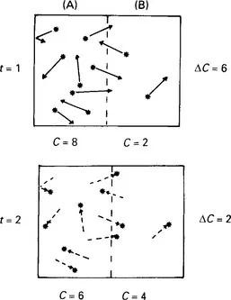
Figure 1.1 Sketch illustrating how random molecular steps result in a net movement of solute down a concentration gradient. At time 1 (upper sketch) there are 8 molecules per unit volume in (A) and 2 in (B). At time 2 (lower sketch) each molecule has moved a unit step in a random direction. Because there was a greater density of molecules in A there was a greater probability of random movement from A to B, resulting in a net ‘downhill’ flux
The importance of diffusion distance
The rate at which diffusional transport occurs is critically important because the supply of nutrients must keep up with cellular demand. However, as Albert Einstein showed the time (t) that it takes a randomly jumping particle to move a distance x in one specific direction increases with the square of distance:

(see footnote to Table 1.1); and as a result diffusional transport is extremely slow over large distances. While diffusion across a short distance, such as the neuromuscular gap (0.1 μm) takes only 5 millionths of a second, diffusion across the heart wall (approximately 1cm) is hopelessly slow, taking over half a day (Table 1.1). Sadly, Nature often proves the validity of Einstein’s equation and Figure 1.2 is an example of this: it shows the heart of a patient who suffered a coronary thrombosis (obstruction of the blood supply to the heart wall). The pale area in the wall is muscle which has died from lack of oxygen even though the adjacent cavity (the left ventricle) was fully of richly oxygenated blood; the patient died simply because a distance of a few millimetres reduced diffusional transport to an inadequate rate.
Table 1.1
Time taken for a glucose molecule to diffuse a specified distance in one direction
| Distance(x) | Time(t)* | Comparable distance in vivo |
| 0.1 μm | 0.000005 s | Neuromuscular gap |
| 1.0 μm | 0.0005 s | Capillary wall |
| 10.0 μm | 0.05 s | Cell to capillary |
| 1 mm | 9.26 min | Skin, artery wall |
| 1 cm | 15.4 h | Ventricle wall |
*Times are calculated by Einstein’s equation t = x2/2D. ‘D’ is the solute diffusion coefficient. For glucose in water at 37ºC, D is 0.9 × 10⁻⁵ cm2/s (Einstein, A. (1905) Theory of Brownian Movement (trans, and ed. by R. Furth and A. D. Cowper, 1956), Dover Publications, New York)
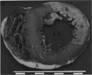
Figure 1.2 Section through the left ventricle of a human heart after a coronary thrombosis. The section is stained to show intracellular enzyme content. The pale area marked by asterisks is an infarct, an area of muscle severely damaged or killed by oxygen lack; the pallor is due to the intracellular enzyme having leaked out of the dying cells. The infarct was caused by a thrombus in a coronary artery, blocking the convectional delivery of oxygen. Diffusion of oxygen from the blood in the adjacent cavity of the left ventricle is unaffected yet only a thin rim of tissue (approximately 1 mm) can survive on this diffusional flux. (Courtesy of Professor M. Davies, St. George’s Hospital Medical School, London)
Convection for fast long-distance transport
Clearly then, for distances greater than approximately 0.1mm a faster transport system is needed and this is provided by the cardiovascular system (Figure 1.3). The cardiovascular system still relies on diffusion for the uptake of molecules at points of close proximity to the environment (e.g. oxygen uptake into lung capillaries) but it then transports them rapidly over large distances by sweeping them along in a stream of pumped fluid. This form of transport is called bulk flow or convective transport. Convective transport requires an energy input and this is provided by a pump, the heart. In man convection takes only 30 s to carry oxygen over a metre or more from the lungs to the smallest blood vessels of the limbs (capillaries). Over the final 10–20 microns separating the capillary from the cells, diffusion is again the main transport process.
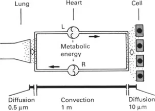
Figure 1.3 Schematic diagram of the mammalian cardiovascular system to illustrate the roles of diffusion and convection in oxygen transport. L, left side of h...
Table of contents
- Cover image
- Title page
- Table of Contents
- Copyright
- Preface
- Chapter 1: Overview of the cardiovascular system
- Chapter 2: Cardiac cycle
- Chapter 3: Cardiac excitation and contraction
- Chapter 4: Electrocardiography
- Chapter 5: Assessment of cardiac output
- Chapter 6: Control of stroke volume and cardiac output
- Chapter 7: Haemodynamics: pressure, flow and resistance
- Chapter 8: Solute transport between blood and tissue
- Chapter 9: Circulation of fluid between plasma, interstitium and lymph
- Chapter 10: Vascular smooth muscle
- Chapter 11: Control of blood vessels
- Chapter 12: Specialization in individual circulations
- Chapter 13: Cardiovascular receptors, reflexes and central control
- Chapter 14: Coordinated cardiovascular responses
- Chapter 15: Cardiovascular responses in pathological situations
- Technical appendix
- Index
Frequently asked questions
Yes, you can cancel anytime from the Subscription tab in your account settings on the Perlego website. Your subscription will stay active until the end of your current billing period. Learn how to cancel your subscription
No, books cannot be downloaded as external files, such as PDFs, for use outside of Perlego. However, you can download books within the Perlego app for offline reading on mobile or tablet. Learn how to download books offline
Perlego offers two plans: Essential and Complete
- Essential is ideal for learners and professionals who enjoy exploring a wide range of subjects. Access the Essential Library with 800,000+ trusted titles and best-sellers across business, personal growth, and the humanities. Includes unlimited reading time and Standard Read Aloud voice.
- Complete: Perfect for advanced learners and researchers needing full, unrestricted access. Unlock 1.5M+ books across hundreds of subjects, including academic and specialized titles. The Complete Plan also includes advanced features like Premium Read Aloud and Research Assistant.
We are an online textbook subscription service, where you can get access to an entire online library for less than the price of a single book per month. With over 1.5 million books across 990+ topics, we’ve got you covered! Learn about our mission
Look out for the read-aloud symbol on your next book to see if you can listen to it. The read-aloud tool reads text aloud for you, highlighting the text as it is being read. You can pause it, speed it up and slow it down. Learn more about Read Aloud
Yes! You can use the Perlego app on both iOS and Android devices to read anytime, anywhere — even offline. Perfect for commutes or when you’re on the go.
Please note we cannot support devices running on iOS 13 and Android 7 or earlier. Learn more about using the app
Please note we cannot support devices running on iOS 13 and Android 7 or earlier. Learn more about using the app
Yes, you can access An Introduction to Cardiovascular Physiology by J R Levick in PDF and/or ePUB format, as well as other popular books in Biological Sciences & Physiology. We have over 1.5 million books available in our catalogue for you to explore.