eBook - ePub
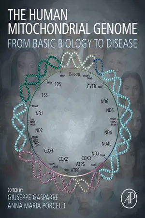
The Human Mitochondrial Genome
From Basic Biology to Disease
- 596 pages
- English
- ePUB (mobile friendly)
- Available on iOS & Android
eBook - ePub
About this book
The Human Mitochondrial Genome: From Basic Biology to Disease offers a comprehensive, up-to-date examination of human mitochondrial genomics, connecting basic research to translational medicine across a range of disease types. Here, international experts discuss the essential biology of human mitochondrial DNA (mtDNA), including its maintenance, repair, segregation, and heredity. Furthermore, mtDNA evolution and exploitation, mutations, methods, and models for functional studies of mtDNA are dealt with. Disease discussion is accompanied by approaches for treatment strategies, with disease areas discussed including cancer, neurodegenerative, age-related, mtDNA depletion, deletion, and point mutation diseases. Nucleosides supplementation, mitoTALENs, and mitoZNF nucleases are among the therapeutic approaches examined in-depth.
With increasing funding for mtDNA studies, many clinicians and clinician scientists are turning their attention to mtDNA disease association. This book provides the tools and background knowledge required to perform new, impactful research in this exciting space, from distinguishing a haplogroup-defining variant or disease-related mutation to exploring emerging therapeutic pathways.
- Fully examines recent advances and technological innovations in the field, enabling new mtDNA studies, variant and mutation identification, pathogenic assessment, and therapies
- Disease discussion accompanied by diagnostic and therapeutic strategies currently implemented clinically
- Outlines and discusses essential research protocols and perspectives for young scientists to pick up
- Features an international team of authoritative contributors from basic biologists to clinician-scientists
Trusted by 375,005 students
Access to over 1.5 million titles for a fair monthly price.
Study more efficiently using our study tools.
Information
Part 1
Biology of human mtDNA
Outline
Chapter 1
mtDNA replication, maintenance, and nucleoid organization
Mara Doimo, Annika Pfeiffer, Paulina H. Wanrooij and Sjoerd Wanrooij, Department of Medical Biochemistry and Biophysics, Umeå University, Umeå, Sweden
Abstract
Part of the genetic information in human cells resides in the mitochondria. Faithful maintenance of mitochondrial deoxyribonucleic acid (mtDNA) is crucial for the oxidative phosphorylation system that produces the majority of the cellular ATP, and therefore to life. This chapter provides an introduction into the characteristics of human mtDNA and summarizes the processes and factors required for the replication and maintenance of this small but essential genome. We also describe the organization of mtDNA in specialized nucleoprotein structures called nucleoids. Where applicable, we refer to human disease states that are caused by defects in the described factors or processes.
Keywords
Mitochondrial DNA; mtDNA; mtDNA maintenance; POL ɣ; Twinkle; mtSSB; POLRMT; TFAM; nucleoids; dNTPs
1.1 Human mitochondrial DNA
1.1.1 Characteristics of mitochondrial DNA
The evidence for mitochondrial deoxyribonucleic acid (mtDNA) dates back to the early 1960s when fibers with characteristics of DNA were first observed in the mitochondrial matrix of chick embryo mitochondria [1,2]. The human mitochondrial genome is a closed circular double-stranded DNA molecule with a length of about 5 μm [3,4]. MtDNA molecules mostly occur as monomers, but can also exist as catenated circles, which are most often dimers [4–6]. In accordance with its small size, mtDNA amounts to less than one percent of the total cellular DNA in animal cells [7]. Early estimations suggested that a single animal mitochondrion contains between two and more than ten mtDNA genomes [8], and—because there are multiple mitochondria per cell—human cells in general harbor from several hundred to over ten thousand mtDNA molecules per cell, depending on the cell type [9–12]. However, the mtDNA copy number of all cell types does not fit in this range, as, for example, human oocytes contain hundreds of thousands of mtDNA copies per cell [13–15].
Human mtDNA is maternally inherited [16]. A key characteristic of mtDNA that derives from its multicopy nature is that cells can contain more than one population of mtDNA genomes, a state referred to as heteroplasmy [17]. The level of heteroplasmy, that is, the frequency of a population of mutated mtDNA molecules, can determine the severity of symptoms in mitochondrial diseases that are caused by mutations in mtDNA [18]. Another peculiar feature that separates the mitochondrial genome from its nuclear counterpart is that mtDNA contains occasional embedded ribonucleotides, the building blocks of ribonucleic acid (RNA) [19,20].
1.1.2 Organization of the human mitochondrial genome
The human mitochondrial genome was the first fully sequenced mitochondrial genome [21]. It is 16.5 kilo base pairs (kbp) in length and encodes for 2 ribosomal RNAs (rRNAs), 22 transfer RNAs (tRNAs), and 13 protein-coding genes with very few or no noncoding bases between neighboring genes (Fig. 1.1A) [21]. All 13 protein-coding genes encode indispensable subunits of the ATP-producing oxidative phosphorylation (OXPHOS) system [21–26]. The two strands of mtDNA are designated as heavy (H) and light (L) due to differences in base composition (GT/CA ratio) that leads to their different buoyant densities on alkaline cesium chloride gradients [27]. Twelve of the protein-coding genes, the 12S and 16S rRNAs as well as fourteen tRNA genes are encoded on the H-strand, while the L-strand encodes one protein-coding gene and eight tRNA genes [21]. The H-strand is G-rich and thereby contains several sequence motifs with the potential to form specific secondary DNA structures called G-quadruplexes (G4s) [28,29]. These motifs often colocalize with mtDNA deletion break-points [29], suggesting that they may hamper the mtDNA replication process.

(A) The mitochondrial genome has a size of 16,568 base pairs in humans. The two strands are termed heavy (H-strand) and light strand (L-strand). The location of the displacement loop (D-loop) is indicated in the noncoding region between tRNAPro and tRNAPhe, and the position of the genes for complex I (NADH dehydrogenase; ND), complex III (cytochrome b; CYTB), complex IV (cytochrome c oxidase; COX), as well as complex V (ATP synthase; ATPase) subunits of the respiratory system are shown. Highlighted with gray boxes are the sites of replication priming in the OH region (B) and at OL (C). (B) Characteristic features of the noncoding region are the promoters HSP and LSP, the highly conserved sequence blocks (CSB 1–3), the single-termination sequence (TAS), and the DNA stretch (7S DNA) that forms the D-loop. Priming in the OH region is initiated with a RNA primer and RNA-to-DNA transition sites were mapped to the CSB region. (C) Priming at OL involves the formation of a DNA stem-loop structure and the synthesis of a short RNA primer by POLRMT. The RNA-to-DNA transition sites were mapped downstream of the OL region.
A circa 1100 bp long noncoding region (NCR), also referred to as the control region, is located between the tRNAPro and tRNAPhe genes [21]. The NCR contains the origin of replication for H-strand synthesis (OH) [21,30] as well as the H-strand promoter (HSP) and the L-strand promoter (LSP) for transcription of each mtDNA strand [31]. Although OH has traditionally been annotated at nucleotide position 191 [21], H-strand replication actually initiates at LSP, approximately 200 nucleotides (nt) upstream of OH [32]. We will therefore use the term “OH region” to refer to the region containing both LSP and OH [33]. The NCR also contains three highly conserved sequence blocks (CSB 1–3), which are considered to have a function in regulating replication [34], and a single termination–associated sequence (TAS) downstream of the three CSBs [35]. Early characterization of mtDNA in mouse [36–38] and later in human cells [39] revealed molecules with a short triple-stranded DNA region, the so-called displacement loop (D-loop), encompassing the OH region. The D-loop is the result of synthesis of a ~600 nt long DNA stretch, termed 7S DNA, that is complementary to the L-strand and is formed by replication initiated in the OH region followed by early termination at the TAS [35,40,41]. The second origin of replication on mtDNA, OL, directs L-strand synthesis. OL is located outside of the NCR, in a cluster of five tRNA genes two-thirds of the way around the genome from OH [42].
1.2 The process of mtDNA replication
1.2.1 Replication mechanisms
In comparison to the nuclear DNA duplication process, human mitochondrial replication is distinct in terms of both the mechanism(s) and the proteins involved. The proteins of the mitochondrial replication fork duplicate the organelle’s DNA by an asymmetric mechanism in which both strands are synthesized continuously [43]. This so-called strand-displacement model of mtDNA replication is based on early pulse and pulse-chase labeling experiments, electron microscopy and 5′-end characterization [7,44], as well as more recent biochemical reconstitution of mtDNA replication in vitro using recombinant proteins [45,46]. This mode of DNA replication is strikingly different from that of mammalian nuclear DNA, but it is not unique in biology since it is similar to replication of ColE1 plasmid DNA [47].
MtDNA replication starts with H-strand (leading strand) synthesis that initiates from the OH region. As the mtDNA replication machinery synthesizes a nascent H-strand, the parental H-strand is displaced, giving rise to the triple-stranded D-loop structure (Fig. 1.2). For yet unknown reasons, mtDNA replication often terminates around the TAS region and only a minority of the nascent H-strands are extended past this point. In the cases where H-strand replication continues beyond the TAS region, it continues unidirectionally, displacing the parental H-strand until it reaches OL. Unwinding of the DNA duplex at OL allows the formation of a characteristic hairpin structure that leads to activation of this origin [48]. L-strand (lagging strand) replication thus initiates and continues unidirectionally from OL in the opposite orientation to H-strand synthesis, and the synthesis of both strands proceeds until they have reached full-circle.

Mitochondrial DNA is replicated by a unique enzymatic machinery that includes the heterotrimeric DNA polymerase POL ɣ (pink), the mitochondrial single-stranded DNA-binding protein mtSSB (green), and the DNA helicase Twinkle (blue). (1) After initiation at the leading strand origin (OH),...
Table of contents
- Cover image
- Title page
- Table of Contents
- Copyright
- Dedication
- List of Contributors
- Editor’s biographies
- Preface
- Acknowledgments
- Part 1: Biology of human mtDNA
- Part 2: mtDNA evolution and exploitation
- Part 3: mtDNA mutations
- Part 4: mtDNA-determined diseases and therapies
- Index
Frequently asked questions
Yes, you can cancel anytime from the Subscription tab in your account settings on the Perlego website. Your subscription will stay active until the end of your current billing period. Learn how to cancel your subscription
No, books cannot be downloaded as external files, such as PDFs, for use outside of Perlego. However, you can download books within the Perlego app for offline reading on mobile or tablet. Learn how to download books offline
Perlego offers two plans: Essential and Complete
- Essential is ideal for learners and professionals who enjoy exploring a wide range of subjects. Access the Essential Library with 800,000+ trusted titles and best-sellers across business, personal growth, and the humanities. Includes unlimited reading time and Standard Read Aloud voice.
- Complete: Perfect for advanced learners and researchers needing full, unrestricted access. Unlock 1.5M+ books across hundreds of subjects, including academic and specialized titles. The Complete Plan also includes advanced features like Premium Read Aloud and Research Assistant.
We are an online textbook subscription service, where you can get access to an entire online library for less than the price of a single book per month. With over 1.5 million books across 990+ topics, we’ve got you covered! Learn about our mission
Look out for the read-aloud symbol on your next book to see if you can listen to it. The read-aloud tool reads text aloud for you, highlighting the text as it is being read. You can pause it, speed it up and slow it down. Learn more about Read Aloud
Yes! You can use the Perlego app on both iOS and Android devices to read anytime, anywhere — even offline. Perfect for commutes or when you’re on the go.
Please note we cannot support devices running on iOS 13 and Android 7 or earlier. Learn more about using the app
Please note we cannot support devices running on iOS 13 and Android 7 or earlier. Learn more about using the app
Yes, you can access The Human Mitochondrial Genome by Giuseppe Gasparre,Anna Maria Porcelli in PDF and/or ePUB format, as well as other popular books in Biological Sciences & Biochemistry. We have over 1.5 million books available in our catalogue for you to explore.