eBook - ePub
From Neuroscience to Neurology
Neuroscience, Molecular Medicine, and the Therapeutic Transformation of Neurology
- 552 pages
- English
- ePUB (mobile friendly)
- Available on iOS & Android
eBook - ePub
From Neuroscience to Neurology
Neuroscience, Molecular Medicine, and the Therapeutic Transformation of Neurology
About this book
The field of neurology is being transformed, from a therapeutically nihilistic discipline with few effective treatments, to a therapeutic specialty which offers new, effective treatments for disorders of the brain and spinal cord. This remarkable transformation has bridged neuroscience, molecular medicine, and clinical investigation, and represents a major triumph for biomedical research. This book, which contains chapters by more than 29 internationally recognized authorities who have made major contributions to neurotherapeutics, tells the stories of how new treatments for disabling disorders of the nervous system, such as stroke, multiple sclerosis, Parkinson's disease, and migraine, were developed, and explores evolving themes and technologies that offer hope for even more effective treatments and ultimately cures for currently untreatable disorders of the brain and spinal cord. The first part of this book reviews the development of new therapies in neurology, from their inception in terms of basic science to their introduction into the clinical world. It also explores evolving themes and new technologies. This book will be of interest to everyone – clinicians and basic scientists alike – interested in diseases of the brain and spinal cord, and in the quest for new treatments for these disorders.* Presents the evolution of the field of neurology into a therapeutic discipline * Discusses lessons learned from past successes and applications to ongoing work* Explores the future of this field
Trusted by 375,005 students
Access to over 1.5 million titles for a fair monthly price.
Study more efficiently using our study tools.
Information
Part I
Clinical Rewards From Neuroscience, Molecular Medicine, and Translational Research
Chapter 1
Seeing the Brain So We Can Save It: The Evolution of Magnetic Resonance Imaging as a Clinical Tool
Peter D. Schellinger, MD, PhD
Steven Warach, MD, PhD
Publisher Summary
Magnetic resonance imaging (MRI) has emerged as a clinical tool that has an impact on neurology. This chapter illustrates this transformation through three classical neurological diseases, namely, acute ischemic stroke, multiple sclerosis, and brain tumors. The advent of new MRI techniques such as diffusion- and perfusion-weighted imaging (DWI and PWI) has added another dimension to diagnostic imaging in stroke. Furthermore, MRI has changed not only clinical management because of a higher diagnostic yield but also daily neurological and neurosurgical practice. Not all tumors are candidates for surgical resection; however, confirmation of the pathology is critical for treatment decisions or enrollment in a clinical trial. Imaging and the development of intraoperative MRI suites have played an increasing role in assisting with biopsy guidance as part of frameless navigation systems. It is exciting to observe that, with an increasing number of clinical therapeutic trials being designed; MRI may not only function as a diagnostic tool but may also have prognostic strength and thus serve as a surrogate endpoint for the development of new therapies.
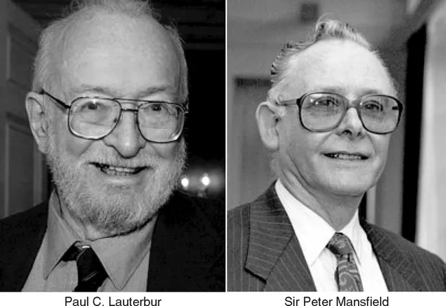
“MRI researchers win Nobel Prize for medicine” – CBC News 10/2003
STOCKHOLM, SWEDEN -Two scientists whose discoveries led to the development of medical imaging of the body’s inner organs have won the 2003 Nobel Prize for medicine. The Swedish Academy said the discoveries of American Paul C. Lauterbur and Briton Sir Peter Mansfield represent “a breakthrough in medical diagnostics and research.” The last time the medicine prize was awarded in the field of diagnostics was for the development of computer assisted tomography in 1979. (See Figure 1.1.)
The last, but most important, step for a new technology is its establishment in clinical practice. The evolution from tentative reports and early research activity to the definition of possible clinical benefits and acceptance into practice (Jackson, 2001) is often protracted in complex technologies such as magnetic resonance imaging (MRI). Lauterbur (1973) published his landmark article about the derivation of position-dependent information by nuclear magnetic resonance and magnetic gradients in the early 1970s. Despite the work that followed by Sir Peter Mansfield (Mansfield and Maudsley, 1976, 1977) and other groups, the first clinical MRI system was not available until 1984 followed in 1987 by the first paramagnetic MRI contrast agent. The last 20 years have realized several advances in computer technology and software development, and these changes have continuously improved the accessibility and quality of MRI. The earliest scans—T2-weighted, T1-weighted, and (proton density) PD-weighted sequence without contrast—took hours to complete. Today, multiparametric protocols can assess within 15 minutes the most complex pathophysiological processes, which has allowed a dramatic shift in the evaluation and treatment of neurological illness. As the clinical discipline of neurology has evolved from a diagnostic to a therapeutic specialty, MRI is being transformed into a clinical tool that has an impact on neurology at the bedside. This chapter illustrates this transformation through three classical neurological diseases that have gained growing interest with regard to therapeutic options only within the last 30 years: acute ischemic stroke, multiple sclerosis, and brain tumors. It is exciting to observe that, with an increasing number of clinical therapeutic trials being designed, MRI may not only function as a diagnostic tool but may also have prognostic strength and thus serve as a surrogate endpoint for the development of new therapies (Warach, 2001b).
ADVANCES IN MRI
Once a technology is accepted into clinical practice, it is inevitable that the demands of clinical specialties stimulate the evolution of the technology and the extension of its functionality to new applications (Jackson, 2001). For instance, interest in functional imaging led to developments such as higher gradients, allowing rapid and repetitive image acquisition with high spatial and temporal resolution. Echo planar imaging (EPI) was found useful for expanding the capabilities of MRI in the routine acquisition of T2-weighted images (Heiland et al., 1998; Pierpaoli et al., 1996; Warach et al., 1995) and allowing formerly time-consuming sequences such as fat- (STIR, short tau inversion recovery) and fluid-suppressed sequences (FLAIR, fluid attenuated inversion recovery) to become practical in the clinical setting (Bydder and Young, 1985; De Coene et al., 1992). FLAIR images have substantially improved the diagnostic yield in diseases such as stroke and multiple sclerosis, where chronic vascular lesions and foci of demyelination are in close proximity to cerebrospinal fluid (CSF) spaces (Hajnal et al., 1992). Other examples of technical progress stimulated by clinical needs include magnetic resonance angiography (Brant-Zawadzki and Heiserman, 1997; Fujita et al., 1994; Johnson et al., 1994) and magnetization transfer imaging (Haehnel et al., 2000; Knauth et al., 1996; Kurki et al., 1994).
Two techniques that have received much attention in the last 15 years are diffusion- and perfusion-weighted imaging (DWI and PWI) (Heiland et al., 1997; Moseley et al., 1991; Ostergaard et al., 1996; Warach et al., 1992). The phenomenon of indirectly measuring brownian molecular motion of water with DWI was first described in 1965 (Stejskal and Tanner, 1965). The measurement of water diffusion renders pathophysiological tissue information, especially in ischemic, inflammatory, and neoplastic disease, that cannot be obtained with standard MRI sequences (Le Bihan et al., 1986; Le Bihan et al., 1992). In addition to the qualitative measurements of diffusion, the apparent diffusion coefficient (ADC) can be quantified from measurements at different b-values (Tanner and Stejskal, 1968) on a per pixel basis, within a region of interest, or to generate ADC parameter maps. For structures, such as nerve fibers or tracts, that have a predominant direction in space, the ADC depends on the direction of the diffusion gradient. Whereas water diffusion is only mildly impaired in the longitudinal direction, water diffusion in a perpendicular direction to the main axis is decreased (Basser and Pierpaoli, 1996); this is referred to as anisotropy. In the brain, anisotropy is most pronounced in the white matter, especially in densely packed fiber structures such as the corpus callosum and the corona radiata. The effects of anisotropy can be used to obtain information of the regional fascicular or fiber tract anatomy, which may be helpful for neurosurgical procedures. The expansion of these principles has led to the development of diffusion tensor imaging, which has been used to characterize myelination and demyelination (Le Bihan et al., 2001; Neil et al., 1998; Ono et al., 1995; Pierpaoli et al., 1996).
PWI uses a dynamic bolus (paramagnetic contrast) tracking technique to derive concentration time curves, allowing the qualitative assessment of various hemodynamic parameters relative to areas of normal parenchyma (Rosen et al., 1989, 1990; Villringer et al., 1988). Typical parameters are mean transit time (MTT), cerebral blood volume (CBV), cerebral blood flow, and time to peak (TTP). The actual quantification of hemodynamic information has been limited by postprocessing capabilities and methods of calculating the arterial input function. Nevertheless, areas of relative hypoperfusion can be reliably demonstrated by current methods, which is of special relevance in ischemic stroke (Schlaug et al., 1999). Other applications, such as functional imaging with the BOLD (blood oxygen level dependence) technique and spectroscopic imaging, exceed the scope of this chapter. It suffices to say that substantial technical and programming refinements have supplied neuroscience with a comprehensive diagnostic armament to guide our daily clinical practice. The growing interest in neurology for physiological data, the development of new treatment paradigms, and the definition of parameters that predict disease course will cont...
Table of contents
- Cover image
- Title page
- Table of Contents
- Preface
- Contributors
- Part I: Clinical Rewards From Neuroscience, Molecular Medicine, and Translational Research
- Part II: Moving Toward the Clinic: Evolving Themes and Technologies
- Index
Frequently asked questions
Yes, you can cancel anytime from the Subscription tab in your account settings on the Perlego website. Your subscription will stay active until the end of your current billing period. Learn how to cancel your subscription
No, books cannot be downloaded as external files, such as PDFs, for use outside of Perlego. However, you can download books within the Perlego app for offline reading on mobile or tablet. Learn how to download books offline
Perlego offers two plans: Essential and Complete
- Essential is ideal for learners and professionals who enjoy exploring a wide range of subjects. Access the Essential Library with 800,000+ trusted titles and best-sellers across business, personal growth, and the humanities. Includes unlimited reading time and Standard Read Aloud voice.
- Complete: Perfect for advanced learners and researchers needing full, unrestricted access. Unlock 1.5M+ books across hundreds of subjects, including academic and specialized titles. The Complete Plan also includes advanced features like Premium Read Aloud and Research Assistant.
We are an online textbook subscription service, where you can get access to an entire online library for less than the price of a single book per month. With over 1.5 million books across 990+ topics, we’ve got you covered! Learn about our mission
Look out for the read-aloud symbol on your next book to see if you can listen to it. The read-aloud tool reads text aloud for you, highlighting the text as it is being read. You can pause it, speed it up and slow it down. Learn more about Read Aloud
Yes! You can use the Perlego app on both iOS and Android devices to read anytime, anywhere — even offline. Perfect for commutes or when you’re on the go.
Please note we cannot support devices running on iOS 13 and Android 7 or earlier. Learn more about using the app
Please note we cannot support devices running on iOS 13 and Android 7 or earlier. Learn more about using the app
Yes, you can access From Neuroscience to Neurology by Stephen Waxman in PDF and/or ePUB format, as well as other popular books in Medicine & Neurology. We have over 1.5 million books available in our catalogue for you to explore.