eBook - ePub
MRI Atlas of Pituitary Pathology
- 60 pages
- English
- ePUB (mobile friendly)
- Available on iOS & Android
eBook - ePub
MRI Atlas of Pituitary Pathology
About this book
MRI Atlas of Pituitary Imaging focuses on magnetic resonance imaging (MRI), the imaging modality of choice for the evaluation of pituitary disorders, since it provides a detailed anatomy of the pituitary gland and surrounding structures, particularly the soft tissues. A basic understanding and interpretation of MRI is important for many clinicians outside of the field of radiology, especially endocrinologists who may receive limited formal training in such areas.
This concise Atlas includes a brief review of the principles of magnetic resonance imaging and then reinforces these principles by utilizing a case-based approach to review various pituitary pathologies. The Atlas serves as a strong clinical teaching aid for endocrinologists, radiologists, and neurosurgeons in training. It also serves as a great reference for physicians who are currently in practice.
- Provides readers with a simple, visual approach for the evaluation of pituitary images
- Features 160 high-resolution images of the most common to the rarest of disorders affecting the pituitary
- Serves an audience of fellows, residents, and clinicians in endocrinology, radiology, neurosurgery, and anyone involved in the multidisciplinary diagnosis of pituitary disease
Trusted by 375,005 students
Access to over 1.5 million titles for a fair monthly price.
Study more efficiently using our study tools.
Information
Topic
MedicinaSubtopic
Endocrinología y metabolismoAtlas of Pituitary Imaging
The basic understanding and interpretation of magnetic resonance imaging (MRI) is important for many clinicians outside of the field of radiology, especially for endocrinologists who may have received limited formal training in such areas. Combining a detailed clinical history, lab data, and images may provide the endocrinologist an upper hand in evaluating patients with pituitary disorders. Accordingly, it is important that endocrinologists be familiar with reviewing pituitary images. The purpose of this atlas is to provide readers with a simple approach for the evaluation of pituitary images. This review does not preclude timely review of pituitary cases with a neuroradiologist. It needs to be emphasized that the best patient outcome usually requires a multidisciplinary team of endocrinologists, radiation oncologists, and neurosurgeons.
Keywords
Adenoma; computed tomography (CT); CSF; magnetic resonance imaging (MRI); pituitary; pre- and postcontrast; sellar mass; T1- and T2-weighted
Introduction
The basic understanding and interpretation of magnetic resonance imaging (MRI) is important for many clinicians outside of the field of radiology, especially for endocrinologists who may have received limited formal training in such areas. Combining a detailed clinical history, lab data, and images may provide the endocrinologist with an upper hand in evaluating patients with pituitary disorders. Accordingly, it is important that endocrinologists be familiar with reviewing pituitary images. The purpose of this atlas is to provide readers with a simple approach for the evaluation of pituitary images. This review does not preclude timely review of pituitary cases with a neuroradiologist. It needs to be emphasized that the best patient outcome usually requires a multidisciplinary team of endocrinologists, radiation oncologists, and neurosurgeons.
MRI is the imaging modality of choice to evaluate pituitary disorders since it provides a detailed anatomy of the pituitary gland and surrounding structures, particularly the soft tissues. MRI utilizes the application of a strong static magnetic field to align some of the protons within water molecules, and radiofrequency fields to alter their alignments and produce a signal that is detectable outside the body. Various characteristics of the subsequent relaxation process of these protons are used to construct detailed images of the anatomy. Since different tissues have different relaxation properties, images can be generated with exquisite tissue contrast. Furthermore, different MRI sequences can produce different styles of tissue contrast – for example, T1-weighted or T2-weighted images.
Computed tomography (CT) of the sella will be discussed briefly since an MRI may not be appropriate in all patients (e.g., those with a cardiac pacemaker). At the same time, CT may provide the necessary imaging for some patients at lower cost, provide high-resolution and sensitive visualization of any calcifications associated with lesions, and provide superior distinction of osseous anatomy to help guide medical decision-making or surgical procedures.
Normal Pituitary Gland Anatomy
The pituitary gland projects from the inferior aspect of the hypothalamus, maintaining a functional connection via the infundibulum (pituitary stalk) (Figure 1.1). It resides in a saddle-like bone cavity referred to as the sella turcica, and is covered by a dural fold (diaphragm sella). Superior to the pituitary gland is the suprasellar cistern containing the optic chiasm. Lateral to the sella turcica are the cavernous sinuses (a large, thin-walled venous plexus), which contain the internal carotid artery and cranial nerves III (oculomotor), IV (trochlear), V [trigeminal, branches V1 (ophthalmic) and V2 (maxillary)], and VI (abducens). See Figure 1.1 for a schematic representation.

The pituitary gland is comprised of anterior and posterior lobes. The intermediate lobe, located between the anterior and posterior pituitary, is rudimentary in humans, and usually absent after birth. The anterior pituitary secretes hormones under the influence of the hypothalamus, the main hormones being growth hormone (GH), thyroid-stimulating hormone (TSH), adrenocorticotropic hormone (ACTH), prolactin (PRL), and the gonadotropins [luteinizing hormone (LH) and follicle-stimulating hormone (FSH)]. The posterior pituitary secretes oxytocin and antidiuretic hormone.
MRI Interpretation
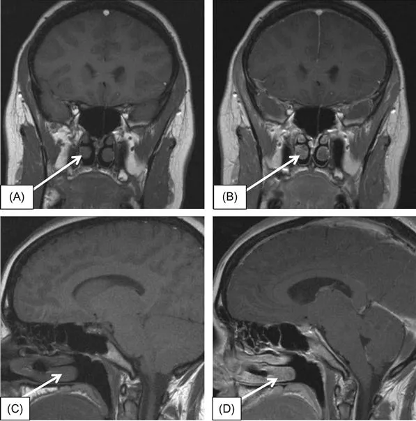
I. The first step in interpreting MR imaging is to correctly identify T1- and T2-weighted (Figure 1.2A and B) and pre- and postcontrast images (Figure 1.3A–D). These different images complement one another and are both useful in characterizing sellar anatomy and pathology.
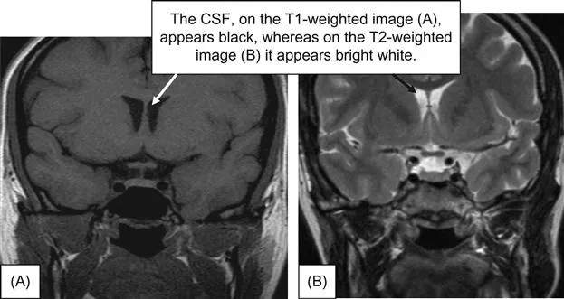
T1-weighted images can easily differentiate fat and water from other common intracranial tissues: fat appears brighter, water appears darker. T2-weighted images have a similar capacity but with opposite features: fat appears darker, and water appears brighter. Thus, within the head, the most common T2-weighted bright object is the cerebrospinal fluid (CSF), and this is particularly evident in the cisterns surrounding the sella. In practice, directing one’s attention immediately to the lateral cerebral ventricles will easily assist the viewer in differentiating T1- and T2-weighted images; the CSF will appear black on T1-weighted images (Figure 1.2A) and white on T2-weighted (Figure 1.2B) images. Both T1- and T2-weighted images contrast the gray and white matter. Figure 1.2A was obtained from a previous publication by our group [1].
II. After distinguishing T1- and T2-weighted images, the next step is to identify the pre- and postcontrast T1-weighted images. For physicians with less familiarity with pituitary imaging, looking at the nasal conchae is an easy way to differentiate between pre- and postcontrast images. In precontrast images (Figure 1.3A and C), the nasal conchae have intensity similar to the brain gray matter (isointense); however, on postcontrast images (Figure 1.3B and D), the nasal conchae appear brighter than the gray matter (hyperintense). Note that the pachymeninges (dura mater) and falx also show conspicuous brightening after gadolinium administration. MRI contrast agents (which use a water-soluble gadolinium chelate...
Table of contents
- Cover image
- Title page
- Table of Contents
- Copyright
- Atlas of Pituitary Imaging
- Index
Frequently asked questions
Yes, you can cancel anytime from the Subscription tab in your account settings on the Perlego website. Your subscription will stay active until the end of your current billing period. Learn how to cancel your subscription
No, books cannot be downloaded as external files, such as PDFs, for use outside of Perlego. However, you can download books within the Perlego app for offline reading on mobile or tablet. Learn how to download books offline
Perlego offers two plans: Essential and Complete
- Essential is ideal for learners and professionals who enjoy exploring a wide range of subjects. Access the Essential Library with 800,000+ trusted titles and best-sellers across business, personal growth, and the humanities. Includes unlimited reading time and Standard Read Aloud voice.
- Complete: Perfect for advanced learners and researchers needing full, unrestricted access. Unlock 1.5M+ books across hundreds of subjects, including academic and specialized titles. The Complete Plan also includes advanced features like Premium Read Aloud and Research Assistant.
We are an online textbook subscription service, where you can get access to an entire online library for less than the price of a single book per month. With over 1.5 million books across 990+ topics, we’ve got you covered! Learn about our mission
Look out for the read-aloud symbol on your next book to see if you can listen to it. The read-aloud tool reads text aloud for you, highlighting the text as it is being read. You can pause it, speed it up and slow it down. Learn more about Read Aloud
Yes! You can use the Perlego app on both iOS and Android devices to read anytime, anywhere — even offline. Perfect for commutes or when you’re on the go.
Please note we cannot support devices running on iOS 13 and Android 7 or earlier. Learn more about using the app
Please note we cannot support devices running on iOS 13 and Android 7 or earlier. Learn more about using the app
Yes, you can access MRI Atlas of Pituitary Pathology by Kevin M. Pantalone,Stephen E. Jones,Robert J. Weil,Amir H. Hamrahian in PDF and/or ePUB format, as well as other popular books in Medicina & Endocrinología y metabolismo. We have over 1.5 million books available in our catalogue for you to explore.