- 298 pages
- English
- ePUB (mobile friendly)
- Available on iOS & Android
eBook - ePub
Handbook of Amygdala Structure and Function
About this book
Handbook of Amygdala Structure and Function, Volume 26, provides an updated overview on the functional neuroanatomy of amygdala nuclei, with an emphasis on interconnections (basolateral, central amygdala, medial amygdala) and their integration into related networks/circuits (prefrontal cortex, bed nucleus, nucleus accumbens). The design of this volume builds upon the foundations of functional neural circuits and the corresponding (cellular) electrophysiology important for the homeostatic control of amygdala function. This volume contains a dedicated section on the anatomical organization of the amygdala nuclei, emphasizing the role of neurotransmitters and neuropeptides that integrate signals and regulate behavior.
Additional chapters discuss cellular physiology, plasticity and the integration of electrical signals that contribute to neural activity. The final section of the book connects the role of amygdala dysfunction and the development of disorders in human health and disease.
- Emphasizes a comparative and multidisciplinary approach on the topic of the amygdala
- Discusses, in detail, the role of amygdala dysfunction and the development of disorders in human health and disease
- Examines the current state of research in cellular physiology, plasticity and the integration of electrical signals
- Includes a dedicated section on neuropeptides, neurotransmitters and cannabinoids that links to behavior control
Trusted by 375,005 students
Access to over 1.5 million titles for a fair monthly price.
Study more efficiently using our study tools.
Information
Chapter 1
Functional neuroanatomy of the basolateral amygdala: Neurons, neurotransmitters, and circuits
Alexander J. McDonald Department of Pharmacology, Physiology and Neuroscience, University of South Carolina School of Medicine, Columbia, SC, United States
Abstract
The basolateral nuclear complex (BNC) of the amygdala consists of lateral, basolateral, and basomedial nuclei in the rat, and homologous nuclei in the monkey (lateral, basal, and accessory basal). There are two main types of neurons in the BNC: pyramidal neurons and nonpyramidal neurons. Although these cells do not exhibit a laminar organization, their morphology, synaptology, electrophysiology, and pharmacology are remarkably similar to their counterparts in the cerebral cortex. Like the cortex, there are several distinct interneuronal subpopulations in the BNC. Collectively, the nuclei of the BNC receive information from all sensory modalities, and via projections to the central amygdalar nucleus, bed nucleus of the stria terminalis, striatum, hypothalamus, and prefrontal cortex elicit emotional behavior. The BNC also plays a critical role in the formation and extinction of emotional memories. This review summarizes the organization of the local and extrinsic connections of BNC neurons that underly these functions.
Keywords
Neuroanatomy; Pyramidal cells; Interneurons; Afferents; Efferents; Monoamines; Basal forebrain; Glutamate; GABA; Peptides
Introduction
The amygdala is a nuclear complex found in the cerebral hemispheres of all vertebrates. It is located in the temporal lobe of primates, and in the caudoventral forebrain of rodents and other mammals that do not have a well-developed temporal lobe. It consists of over a dozen nuclei, each of which exhibit several subdivisions. Each nuclear subdivision is characterized by distinct connections (Pitkänen, 2000). The cortical and medial amygdalar nuclei are distinguished by interconnections with the olfactory system and the hypothalamus. The central nucleus is the only nucleus with extensive projections to the brainstem. The basolateral nuclear complex (BNC), the subject of this chapter, is characterized by extensive interconnections with higher order cortical areas in the prefrontal, temporal, insular, and hippocampal cortices. The size of the BNC increases throughout phylogeny, in concert with the increase in size and differentiation of the neocortices with which it has connections (Janak & Tye, 2015).
Early studies demonstrated that bilateral lesions of the anterior temporal lobes that included the amygdala make monkeys remarkably tame and hypoemotional (Brown & Schafer, 1888; Klüver & Bucy, 1939). These animals exhibited “psychic blindness,” a specific type of visual agnosia characterized by the inability to recognize the emotional or behavioral significance of sensory stimuli. Subsequent studies indicated that lesions limited to the amygdala could produce similar deficits (Aggleton & Passingham, 1981; Weiszkrantz, 1956). Weiskrantz famously stated that the effect of amygdalectomy “is to make it difficult for reinforcing stimuli, whether positive or negative, to become established or recognized as such.” Thus, the amygdala constitutes an essential link between brain regions that process sensory stimuli, such as the cerebral cortex and thalamus, and brain regions responsible for producing emotional and motivational responses, such as the hypothalamus and brainstem. It has thus been called the “sensory gateway to the emotions” (Aggleton & Mishkin, 1986). The BNC is the main target of sensory inputs. It then projects to the central nucleus, bed nucleus of the stria terminalis (BNST) and ventral striatum, which in turn activate the hypothalamus, brainstem, and other regions to generate somatomotor, autonomic, and endocrine components of emotional and motivational behavior.
Circuits involving the BNC are critical for aversive behavior, including fear conditioning, fear extinction, and anxiety (Herry et al., 2010; LeDoux, 2000; Pape & Paré, 2010; Tovote, Fadok, & Lüthi, 2015). In addition, although largely overlooked until recently, the BNC is also important for appetitive conditioning (Janak & Tye, 2015). Understanding the amygdalar circuitry underlying these functions, and how the activity of these circuits may be disrupted in disease, will be critical for developing therapies for neurological and neuropsychiatric disorders involving the amygdala, including anxiety disorders such as posttraumatic stress disorder (PTSD), depression, Alzheimer disease, temporal lobe epilepsy, and drug addiction. Most of the studies on basic systems neuroanatomy of the amygdala were completed by the turn of the century (see reviews by McDonald, 1998; Pitkänen, 2000). Over the last two decades, further details of amygdalar circuitry were clarified at the neuronal and synaptic levels. This review discusses the functional neuroanatomy of the BNC, and emphasizes the roles of individual cell types such as interneuronal subpopulations and projection-specific principal neurons. While the focus is on investigations in rodents, since these species have been heavily studied, the review also provides information on nonhuman primates, since it is anticipated that finer details of these connections in the latter species may more closely resemble those of humans. This information should be useful for designing electrophysiological and behavioral investigations using recently developed optogenetic techniques to selectively activate/inactivate different components of amygdalar circuits in brain slices and conscious behaving animals.
Nuclei of the basolateral amygdala
The cortical (Co) and medial nuclei (M) are located along the ventral and medial surfaces of the amygdala in most mammals including rodents (Fig. 1A). The central amygdalar nucleus (CM/CL; Fig. 1A) is located dorsolateral to the medial nucleus. The BNC is located deep into the cortical and central nuclei. It consists of three main nuclei, the lateral (LA), basolateral (BL), and basomedial nuclei (BM), arranged from dorsal to ventral, respectively. Each nucleus has several subdivisions [LA: dorsolateral (Ldl), ventrolateral (Lvl), and ventromedial (Lvm) subdivisions; BL: anterior (BLa), posterior (BLp), and ventral (BLv) subdivisions; BM: anterior (Bma), and posterior (BMp) subdivisions of Paxinos & Watson, 1997] (Fig. 1A). Price and coworkers recognized these same nuclei in the rat but used an alternative nomenclature (Price et al., 1987) (Table 1).
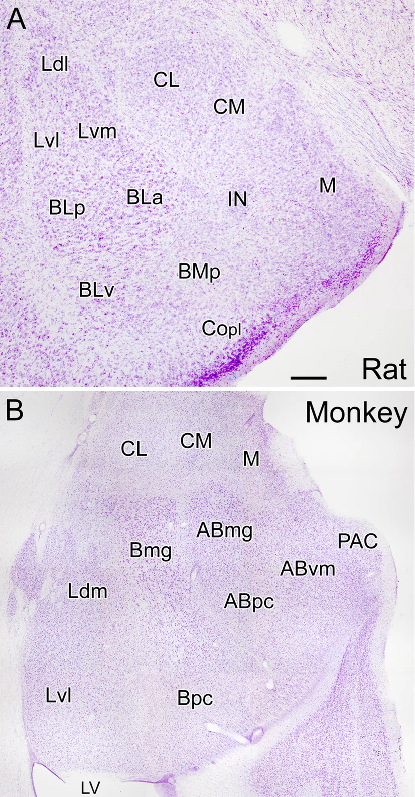
Table 1
Table of contents
- Cover image
- Title page
- Table of Contents
- Copyright
- Contributors
- Preface
- Chapter 1: Functional neuroanatomy of the basolateral amygdala: Neurons, neurotransmitters, and circuits
- Chapter 2: Structure and function of the medial amygdala
- Chapter 3: Neuronal diversity of the amygdala and the bed nucleus of the stria terminalis
- Chapter 4: Amygdala physiology in pain
- Chapter 5: Neural plasticity of the amygdala
- Chapter 6: Plasticity of amygdala neurons underlying fear learning and extinction
- Chapter 7: Neuropeptide Y and amygdala circuitry: Modulation of stress-related behavior
- Chapter 8: The amygdalar opioid system
- Chapter 9: Noradrenergic regulation of the basolateral amygdala
- Chapter 10: Pituitary adenylate cyclase-activating polypeptide (PACAP) in stress, pain, and learning
- Chapter 11: Protect and harm: Effects of stress on the amygdala
- Chapter 12: Sex differences in amygdala structure and function: From rodents to humans
- Index
Frequently asked questions
Yes, you can cancel anytime from the Subscription tab in your account settings on the Perlego website. Your subscription will stay active until the end of your current billing period. Learn how to cancel your subscription
No, books cannot be downloaded as external files, such as PDFs, for use outside of Perlego. However, you can download books within the Perlego app for offline reading on mobile or tablet. Learn how to download books offline
Perlego offers two plans: Essential and Complete
- Essential is ideal for learners and professionals who enjoy exploring a wide range of subjects. Access the Essential Library with 800,000+ trusted titles and best-sellers across business, personal growth, and the humanities. Includes unlimited reading time and Standard Read Aloud voice.
- Complete: Perfect for advanced learners and researchers needing full, unrestricted access. Unlock 1.5M+ books across hundreds of subjects, including academic and specialized titles. The Complete Plan also includes advanced features like Premium Read Aloud and Research Assistant.
We are an online textbook subscription service, where you can get access to an entire online library for less than the price of a single book per month. With over 1.5 million books across 990+ topics, we’ve got you covered! Learn about our mission
Look out for the read-aloud symbol on your next book to see if you can listen to it. The read-aloud tool reads text aloud for you, highlighting the text as it is being read. You can pause it, speed it up and slow it down. Learn more about Read Aloud
Yes! You can use the Perlego app on both iOS and Android devices to read anytime, anywhere — even offline. Perfect for commutes or when you’re on the go.
Please note we cannot support devices running on iOS 13 and Android 7 or earlier. Learn more about using the app
Please note we cannot support devices running on iOS 13 and Android 7 or earlier. Learn more about using the app
Yes, you can access Handbook of Amygdala Structure and Function by Janice H. Urban,J. Amiel Rosenkranz in PDF and/or ePUB format, as well as other popular books in Psychology & Neuroscience. We have over 1.5 million books available in our catalogue for you to explore.