
- 416 pages
- English
- ePUB (mobile friendly)
- Available on iOS & Android
eBook - ePub
Handbook of Footwear Design and Manufacture
About this book
Understanding footwear design and manufacture is vital for improving the functionality, aesthetics and marketability of a product. The Handbook of footwear design and manufacture provides a comprehensive review of footwear production and design and explores how these processes are used across a variety of application areas.Part one, an introductory section, reviews the fundamentals of footwear anatomy; chapters discuss the anatomy of the human foot, biomechanics and gait, foot models and measurements, the development of the foot in childhood and adolescence, and foot problems and their implications for footwear design. Part two examines footwear design including the development of shoe design, foot sketch templates, and footwear drawing templates. Aspects of footwear manufacture are highlighted in part three including the design, manufacture, and sizing and grading of shoe lasts. Further chapters focus on the footwear business, advertising, and the environmental impact of footwear manufacture. Part four explores the design and manufacture of footwear for specific applications and includes chapters on footwear for cold weather, textiles and other materials used in the production of protective military and orthopaedic footwear, and design issues in geriatric footwear.The Handbook of footwear design and manufacture is a wide-ranging and technical resource for footwear designers, materials scientists and researchers involved in the production of footwear, and professionals in the footwear industry looking to expand their knowledge of design and manufacture processes.
- Discusses foot anatomy in detail and considers its implications for footwear design
- Looks at design issues from foot and footwear drawing templates to shoe last design and footwear manufacture
- Specific chapters focus on the footwear business, advertising and the environmental impact of footwear manufacture
Trusted by 375,005 students
Access to over 1.5 million titles for a fair monthly price.
Study more efficiently using our study tools.
Information
Subtopic
Fashion & Textile IndustryIndex
BusinessPart I
Fundamentals of footwear anatomy
1
The anatomy of the human foot
L.K. Chan, The University of Hong Kong, P. R. China
Abstract:
This chapter describes the anatomy of the human foot; first the bones and joints, followed by muscles and tendons. The arches of the human foot are then dealt with. Finally, the chapter describes the neurovasculature and surface anatomy of the foot.
Key words
bones
joints
muscles
tendons
neurovasculature
surface anatomy
1.1 Introduction
The primary purpose of the human foot is supporting weight. The human foot is never used for grasping, unlike the feet of many other primates. This specialization is reflected in its anatomy. The following sections describe parts and aspects of the foot anatomy:
– Bones
– Joints
– Muscles and tendons
– Arches
– Neurovasculature
– Surface anatomy.
1.2 The bones of the human foot
The bones of the foot can be divided into three groups: the tarsal bones, the metatarsals, and the phalanges (see Fig. 1.1).
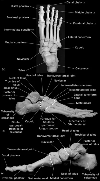
1.1 Articulated foot bones (right foot). (a) Superior view. (b) Lateral view. (c) Medial view.
1.2.1 The tarsal bones
The tibia and fibula in the leg articulate with only the talus of the foot, at the ankle joint. The talus thus receives the weight of the whole body. The talus sits on the calcaneus, which touches the ground at its posterior tuberosity, and therefore part of the body weight is transmitted through the calcaneus to the ground. The body weight is further distributed from the talus and calcaneus, through other tarsal bones, and then to the metatarsals, the heads of which also touch the ground and support the body weight.
The calcaneus
Of all the seven tarsal bones, the calcaneous is the largest and is the only one that touches the ground (see Fig. 1.1b and 1.1c). It articulates with the talus above it and the cuboid anterior to it. It is shaped somewhat like a rectangular box, with a shelf-like protrusion from the medial upper surface called the sustentaculum tali, which is partly responsible for supporting the talus (see Fig. 1.2a and 1.2b). The inferior surface of the sustentaculum tali is grooved by the tendon of the flexor hallucis longus, the long flexor of the big toe (see Fig. 1.2b).
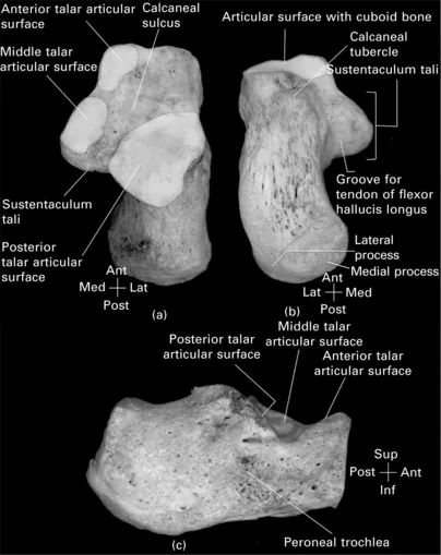
1.2 The right calcaneus. (a) Superior view. (b) Inferior view. (c) Lateral view.
The superior surface of the calcaneus has three articular surfaces, all of which are located on its anterior half (see Fig. 1.2a):
– The anterior talar articular surface. A small concave articular surface on the superior surface of the anteromedial corner of the calcaneus.
– The middle talar articular surface. A concave articular surface on the superior surface of sustentaculum tali, close to or even fused with the anterior talar articular surface. The anterior and middle talar articular surfaces articulate with the head of the talus, in the talocalcaneonavicular joint, a ball-and-socket synovial joint formed by the head of the talus (the ball) and the calcaneus and navicular (the socket).
– The posterior talar articular surface. A convex articulating surface, for articulating with a concave articular surface on the inferior aspect of the talus. The joint thus formed is the subtalar joint.
The calcaneal sulcus is the deep groove separating the anterior and middle articular surfaces from the posterior articular surface. The calcaneal sulcus, together with the talar sulcus on the inferior surface of the talus, forms the tarsal canal, which widens laterally into the tarsal sinus. The two bones are firmly attached to each other by the talocalcaneal interosseous ligament in the talar canal and the ligamentum cervicis tali in the tarsal sinus (see Fig. 1.3).
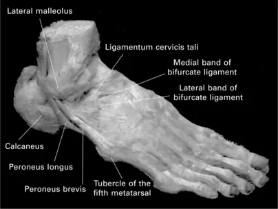
1.3 Dorsolateral view of an osteoligamentous specimen of the human right foot, with all tendons and muscles on the foot dorsum removed.
The part of the calcaneus posterior to the articulations with the talus is the calcaneal tuberosity (see Fig. 1.1b). The posterior surface of the tuberosity receives the calcaneal tendon (Achilles tendon). Superior to the attachment of the calcaneal tendon, there may be a smooth area for a bursa between the deep surface of the tendon and the calcaneus. On the inferior surface of the calcaneal tuberosity are medial and lateral tubercles or processes, separated by a V-shaped notch (see Fig. 1.2b). These are the weight-bearing areas of the calcaneus. Further anteriorly, on the inferior surface of the most anterior part of the calcaneus, is the calcaneal tubercle, to which the longer plantar ligament attaches, on its way from the medial and lateral calcaneal tubercles to the inferior surfaces of the middle three metatarsals.
On the lateral surface of the calcaneus (see Fig. 1.2c), there is a small bony prominence, the peroneal trochlea. The peroneus brevis tendon passes above the trochlea with the peroneus longus tendon below it, and is bound down to the calcaneus by the inferior peroneal retinaculum.
The talus
The talus receives the weight of the whole body from the tibia and fibula, and transmits the weight to the calcaneus and navicular. It has a body, which is projected anteriorly into the talar head. Between the body and the head is a short neck (see Fig. 1.4a).
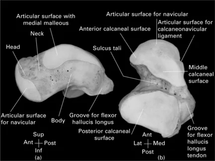
1.4 The right talus. (a) Medial view. (b) Inferior view.
The body has large articular surfaces on its superior and inferior surfaces. The inferior articular surface, called the posterior calcaneal articular surface, articulates with the convex, posterior talar articular surface on the superior surface of the calcaneus (see Fig. 1.4b). The superior articular surface, called the trochlea, is a dome-shaped surface for articulating with the tibia (see Fig. 1.1). The trochlea is broader anteriorly than posteriorly. It extends onto the medial and lateral side of the body for articulating with the medial (from the tibia) and the lateral malleoli (from the fibula). The surface on the medial side of the body is comma-shaped, with the tail directed posteriorly (see Fig. 1.4a). The surface on the lateral side is triangular in shape, much larger, and covers almost the entire lateral surface of the body.
The body is projected posteriorly as the posterior process, which is grooved by the tendon of the flexor hallucis longus (see Fig. 1.4a,b). The bony prominences of the medial and lateral side of the groove are called the medial and lateral tubercles, respectively.
The head of the talus is covered by a large articular surface which is subdivided into different parts for articulating with different structures (see Fig. 1.4b). The anterior part of the head is convex and articulates with the concave, posterior surface of the navicular. The very lateral part of this convex surface also articulates with the medial band of the bifurcate ligament (both the medial and lateral bands arise from the superior surface of the calcaneus just behind the calcaneocuboid joint, with the medial band going to the navicular and the lateral band to the cuboid) (see Fig. 1.3).
The inferior part of the head bears two small, flat articular areas for the anterior and middle talar articular surfaces on the body of the calcaneus and the sustentaculum tali (see Fig. 1.4b). They are respectively called the anterior and middle calcaneal articular surfaces. Between the navicular surface and the calcaneal surfaces on the talar head is another triangular articular surface for the spring (or calcaneonavicular) ligament (see Fig. 1.4b), which closes the gap between the calcaneus and navicular (see Fig. 1.1c), so that the weight-bearing talus will not separate the two bones, and touches the group.
The navicular
Navicular means 'boat-shaped', and this bone is so named because of its anterior convexity and posterior concavity. Th...
Table of contents
- Cover image
- Title page
- Table of Contents
- Copyright
- Contributor contact details
- Woodhead Publishing Series in Textiles
- Part I: Fundamentals of footwear anatomy
- Part II: Footwear design
- Part III: Shoe lasts and other aspects of footwear manufacture
- Part IV: Applications
- Index
Frequently asked questions
Yes, you can cancel anytime from the Subscription tab in your account settings on the Perlego website. Your subscription will stay active until the end of your current billing period. Learn how to cancel your subscription
No, books cannot be downloaded as external files, such as PDFs, for use outside of Perlego. However, you can download books within the Perlego app for offline reading on mobile or tablet. Learn how to download books offline
We are an online textbook subscription service, where you can get access to an entire online library for less than the price of a single book per month. With over 1.5 million books across 990+ topics, we’ve got you covered! Learn about our mission
Look out for the read-aloud symbol on your next book to see if you can listen to it. The read-aloud tool reads text aloud for you, highlighting the text as it is being read. You can pause it, speed it up and slow it down. Learn more about Read Aloud
Yes! You can use the Perlego app on both iOS and Android devices to read anytime, anywhere — even offline. Perfect for commutes or when you’re on the go.
Please note we cannot support devices running on iOS 13 and Android 7 or earlier. Learn more about using the app
Please note we cannot support devices running on iOS 13 and Android 7 or earlier. Learn more about using the app
Yes, you can access Handbook of Footwear Design and Manufacture by A. Luximon in PDF and/or ePUB format, as well as other popular books in Business & Fashion & Textile Industry. We have over 1.5 million books available in our catalogue for you to explore.