
- 622 pages
- English
- ePUB (mobile friendly)
- Available on iOS & Android
eBook - ePub
About this book
Clinical Neurophysiology: Basis and Technical Aspects, the latest release in the Handbook of Clinical Neurology series, is organized into sections on basic physiological concepts, on the function and limitations of modern instrumentation, and on other fundamental or methodologic aspects related to the recording of various bioelectric signals from the nervous system for clinical or investigative purposes. There is discussion of the EEG, nerve conduction studies, needle electromyography, intra-operative clinical neurophysiology, sleep physiology and studies, the autonomic nervous system, various sensory evoked potentials, and cognitive neurophysiology.
- Provides an up-to-date review on the practice of neurophysiological techniques in the assessment of neurological disease
- Explores the electrophysiological techniques used to better understand neurological function and dysfunction, first in the area of consciousness and epilepsy, then in the areas of the peripheral nervous system and sleep
- Focuses on new techniques, including electrocorticography, functional mapping, stereo EEG, motor evoked potentials, magnetoencephalography, laser evoked potentials, and transcranial magnetic stimulation
Trusted by 375,005 students
Access to over 1.5 million titles for a fair monthly price.
Study more efficiently using our study tools.
Section II
EEG: Technologic aspects and basic rhythms
Chapter 6
EEG source localization
Christoph M. Michel1,2,*; Bin He3 1 Department of Basic Neurosciences, University of Geneva, Geneva, Switzerland
2 Center for Biomedical Imaging (CIBM) Lausanne-Geneva, Geneva, Switzerland
3 Department of Biomedical Engineering, Carnegie Mellon University, Pittsburgh, PA, United States
* Correspondence to: Professor Christoph M. Michel, Ph.D., Functional Brain Mapping Laboratory, Department of Basic Neurosciences, Campus Biotech 9, chemin des Mines, Geneva 1202, Switzerland. Tel: + 41-22-379-54-57 email address: [email protected]
Abstract
Since the discovery of electroencephalography (EEG), when it was hoped that EEG would offer “a window into the brain,” researchers and clinicians have attempted to localize the neuronal activity in the brain that generates the scalp potentials measured noninvasively with EEG. Early explorations in the 1950s using electric field theory to infer the location and orientation of the current dipole in the brain from the scalp potential distribution triggered considerable efforts to quantitatively deduce these sources. Initially, dipole fitting, or dipole localization, was the method of choice and many studies used this approach in experimental and clinical studies with remarkable success. Later on, new methods were proposed that attempted to overcome the problem of having to fix the number of sources a priori; these methods are known as distributed source imaging techniques. The introduction and increasing availability of magnetic resonance imaging, allowing detailed realistic anatomy of the brain and head to be incorporated in source localization methods, has drastically increased the precision of such approaches. Today, source localization of EEG (and magnetoencephalography, or MEG) has reached a level of consistency and precision that allows these methods to be placed in the family of brain imaging techniques. The particular advantage that they have over other imaging methods is their high temporal resolution, which allows the origin of activity to be distinguished from its propagation and information flow in large-scale brain networks to be examined. This chapter gives an overview of these methods and illustrates them with several examples, thereby focusing on EEG source imaging in epilepsy and presurgical planning, as clinical applications with remarkable maturation.
Keywords
EEG source imaging; Forward problem; Inverse problem; Connectivity; Epilepsy; Surgical planning
Introduction
It is generally understood that the main generators of the electroencephalograph (EEG) are the postsynaptic potentials that take place on the pyramidal cortical neurons (Mitzdorf, 1985; Lopes da Silva, 1991). Synchronized activity of these synaptic currents leads to current flows in the head volume. Because the head is a conducting medium, volume conduction allows the propagation of these current flows to the scalp surface, where they give rise to electric potential differences between electrodes placed on different positions on the scalp (Brazier, 1949). By recording these potentials using an array of electrodes, topographical maps can be constructed that display the distribution of the scalp potential produced by the active neuronal population at any given moment in time. If only one brain area is active, the potential distribution on the scalp is rather simple and dipolar. However, if several brain areas are simultaneously active, complex patterns of scalp potentials arise, and the deduction of the underlying sources becomes a nontrivial task. In general, a priori assumptions are required, preferentially incorporating anatomical, physiological, and biophysical knowledge. An important development was to introduce anatomical constraints of the head to facilitate solving the EEG source localization problem (He et al., 1987; Hämäläinen and Sarvas, 1989). Further development has introduced physiological constraints of cortical sources to facilitate solving the distributed source imaging (Dale and Sereno, 1993; Pascual-Marqui et al., 1994). Such a priori constraints greatly improve the solvability and precision of the EEG source localization, albeit only an estimation of the underlying sources.
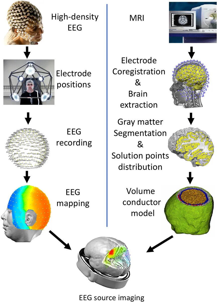
Historically, the first attempts of EEG source localization were based on the strong a priori assumption that only one source is active at a certain time point, that the head can be approximated as a sphere, and that the conductivity is homogeneous throughout the brain. In this case, nonlinear multidimensional optimization procedures allowed the position, orientation, and strength of an equivalent dipole in the brain to be found that best explained the observed scalp potential measurements. Soon, the conductivity difference between different tissues were incorporated in multilayer spherical head models, and finally realistic geometry head models based on magnetic resonance (MR) images were developed using boundary or finite element reconstruction of the scalp and the different tissues. Also, the inverse model developed from the strong constraint of one or a few dipoles with time-varying amplitudes to distributed source reconstruction methods that estimate the three-dimensional (3D) current density distribution in the whole brain volume. In the following text, the concepts and principles of EEG source localization are reviewed. More detailed methodological reviews can be found in He and Ding (2013), Pascual-Marqui et al. (2009), Michel et al. (2004b), Michel and He (2011). An illustration of the full pipeline for modern EEG source imaging is given in Fig. 6.1.
The EEG Forward Problem
The electric potential generated by synchronized postsynaptic potentials does not propagate homogeneously through the brain. Different tissues such as the scalp, skull, cerebrospinal fluid, and brain have different conductivity characteristics and therefore attenuate the current to a different extent. If the conductivity parameters are known and are correctly taken into account, Poisson's equation allows the potential to be determined ...
Table of contents
- Cover image
- Title page
- Table of Contents
- Copyright
- Handbook of Clinical Neurology 3rd Series
- Foreword
- Preface
- Contributors
- Section I: Basic physiological and recording concepts
- Section II: EEG: Technologic aspects and basic rhythms
- Section III: Nerve conduction studies, methods and techniques
- Section IV: Needle electromyography, methods and techniques
- Section V: Intraoperative clinical neurophysiology, MEP, SSEP
- Section VI: Sleep physiology and studies
- Section VII: Autonomic nervous system: Basic and technical aspects
- Section VIII: Auditory, visual and somatosensory evoked potentials
- Section IX: Cognitive neurophysiology
- Index
Frequently asked questions
Yes, you can cancel anytime from the Subscription tab in your account settings on the Perlego website. Your subscription will stay active until the end of your current billing period. Learn how to cancel your subscription
No, books cannot be downloaded as external files, such as PDFs, for use outside of Perlego. However, you can download books within the Perlego app for offline reading on mobile or tablet. Learn how to download books offline
We are an online textbook subscription service, where you can get access to an entire online library for less than the price of a single book per month. With over 1.5 million books across 990+ topics, we’ve got you covered! Learn about our mission
Look out for the read-aloud symbol on your next book to see if you can listen to it. The read-aloud tool reads text aloud for you, highlighting the text as it is being read. You can pause it, speed it up and slow it down. Learn more about Read Aloud
Yes! You can use the Perlego app on both iOS and Android devices to read anytime, anywhere — even offline. Perfect for commutes or when you’re on the go.
Please note we cannot support devices running on iOS 13 and Android 7 or earlier. Learn more about using the app
Please note we cannot support devices running on iOS 13 and Android 7 or earlier. Learn more about using the app
Yes, you can access Clinical Neurophysiology: Basis and Technical Aspects by in PDF and/or ePUB format, as well as other popular books in Medicine & Neurology. We have over 1.5 million books available in our catalogue for you to explore.