eBook - ePub
About this book
Human Prion Diseases, Volume 153 is designed to update the reader on the latest advances and clinical aspects of prion diseases. The book is organized into five sections, including the pathophysiology of prions and a description of animal and human diseases. This is followed by detailed reports on recent advances in diagnosis strategies for the development of novel anti-prion molecules and possible designs of clinical trials in such a rare disease. An introductory chapter gives an extensive historical background of prion research, with a final chapter highlighting recent progress, and more importantly, unsolved problems.
- Offers an authoritative overview of prion diseases in humans, detailing the pathogenesis of the disease, clinical investigations, and the diagnosis of both the genetic and acquired forms
- Provides clarity and context by presenting prion diseases in relation to other neurodegenerative diseases in humans
- Emphasizes the unique properties of prion diseases and consequent problems they can cause, both clinically and in public health terms
Trusted by 375,005 students
Access to over 1.5 million titles for a fair monthly price.
Study more efficiently using our study tools.
Chapter 1
Human transmissible spongiform encephalopathies: historic view
David M. Asher*; Luisa Gregori Laboratory of Bacterial and Transmissible Spongiform Encephalopathy Agents, Division of Emerging and Transfusion-Transmitted Diseases, Center for Biologics Evaluation and Research, Food and Drug Administration, Silver Spring, MD, United States
* Correspondence to: David Michael Asher, MD, Building 72, Room 4332, 10903 New Hampshire Avenue, Silver Spring MD 20993, United States. Tel: + 1-240-402-9367 email address: [email protected]
* Correspondence to: David Michael Asher, MD, Building 72, Room 4332, 10903 New Hampshire Avenue, Silver Spring MD 20993, United States. Tel: + 1-240-402-9367 email address: [email protected]
Abstract
The first of several pivotal moments leading to current understanding of human transmissible spongiform encephalopathies (TSEs) occurred in 1959 when veterinary pathologist W.J. Hadlow first recognized several similarities between scrapie—a slow infection of sheep caused by an unusual infectious agent—and kuru, a fatal exotic neurodegenerative disease affecting only people of a single language group in the remote mountainous interior of New Guinea, described two years earlier by D.C. Gajdusek and V. Zigas. Based on the knowledge of scrapie, Gajdusek, C.J. Gibbs, Jr., and M.P. Alpers soon initiated efforts to transmit kuru by inoculating kuru brain tissue into non-human primates, that—although requiring several years—ultimately proved successful. In the same year that Hadlow first proposed that kuru and scrapie might have similar etiology, I. Klatzo noted that kuru's histopathology resembled that of Creutzfeldt-Jakob disease (CJD), another progressive fatal neurodegenerative disease of unknown etiology that A.M. Jakob had first described in 1921. Gajdusek and colleagues went on to demonstrate that not only the more common sporadic form of CJD but also familial CJD and a generally similar familial brain disease (Gerstmann-Sträussler-Scheinker syndrome) were also transmissible, first to non-human primates and later to other animals. (Other investigators later transmitted an even rarer brain disease, fatal familial insomnia, to animals.) Iatrogenic CJD (spread by human pituitary-derived hormones and tissue grafts) was also transmitted to animals. Much later, in 1996, a new variant of CJD was attributed to human infection with the agent of bovine spongiform encephalopathy; vCJD itself caused an iatrogenic TSE spread by blood transfusion (and probably by a human-plasma-derived clotting factor). Starting in the 1930s, the scrapie agent was found to have a unique constellation of physical properties (marked resistance to inactivation by chemicals, heat and radiation), eventually interpreted as suggesting that it might be an unconventional self-replicating pathogen based on protein and containing no nucleic acid. The work of S.B. Prusiner led to the recognition in the early 1980s that a misfolded form of a ubiquitous normal host protein was usually if not always detectable in tissues containing TSE agents, greatly facilitating the diagnosis and TSEs and understanding their pathogenesis. Prusiner proposed that the TSE agent was likely to be composed partly if not entirely of the abnormal protein, for which he coined the term “prion” protein and “prion” for the agent. Expression of the prion protein by animals—while not essential for life—was later found to be obligatory to infect them with TSEs, and a variety of mutations in the protein clearly tracked with TSEs in families, explaining the autosomal dominant pattern of disease and confirming a central role for the protein in pathogenesis. Prusiner's terminology and the prion hypothesis came to be widely though not universally accepted. A popular corollary proposal, that prions arise by spontaneous misfolding of normal prion protein leading to sporadic cases of CJD, BSE, and scrapie, is more problematic and may serve to discourage continued search for environmental sources of exposure to TSE agents.
Keywords
historic view; transmissible spongiform encephalopathy; kuru; Creutzfeldt–Jakob disease; Gerstmann–Sträussler–Scheinker syndrome; fatal familial insomnia; scrapie; prion
Introduction, including the concept of slow infections
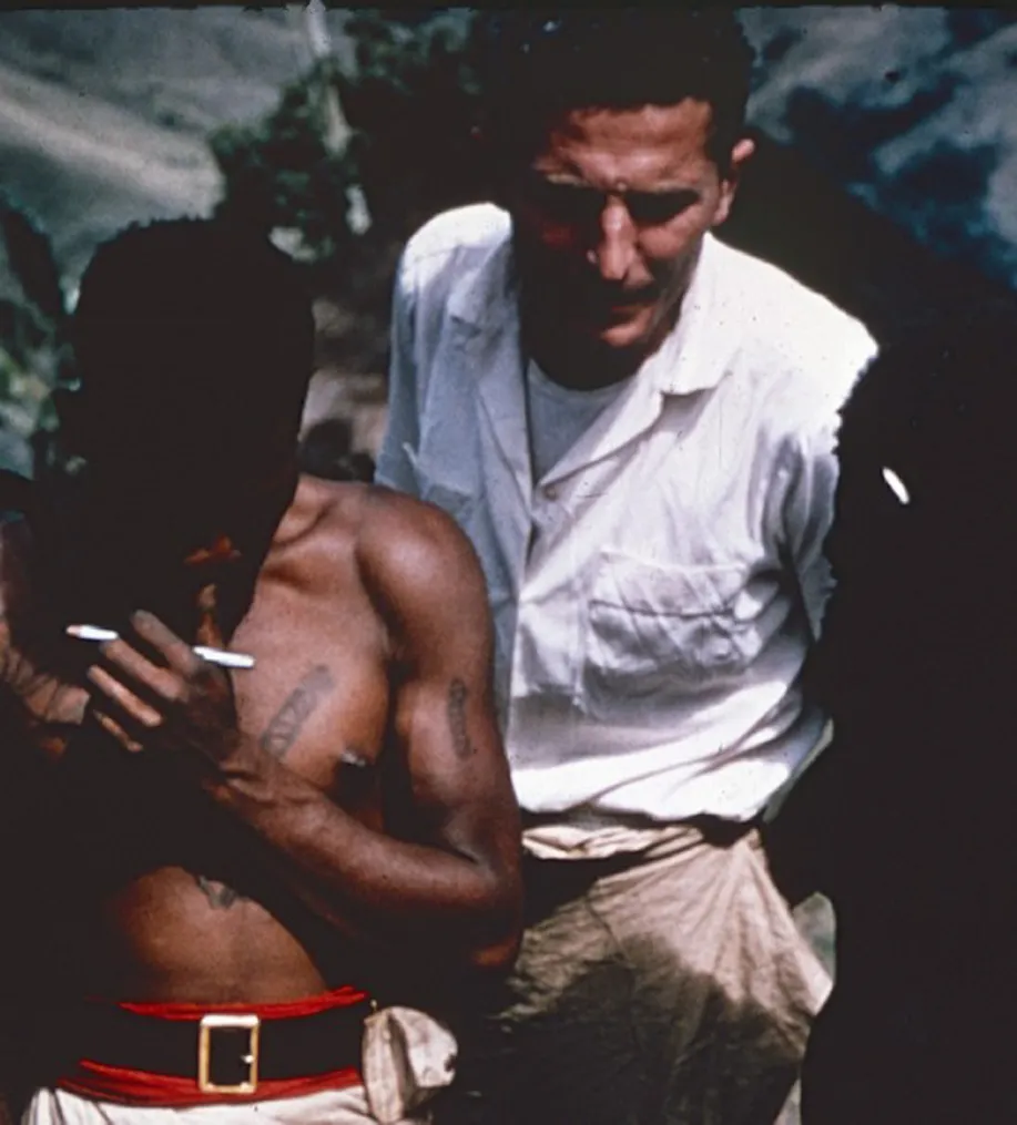
Our narrative begins with the story of kuru, the first human disease recognized to be a transmissible spongiform encephalopathy (TSE). Kuru caused shivering tremors (called kuru in the language of the Foré peoples of the Eastern Highland Region of the Australian Trust Territory of New Guinea, where the disease was epidemic in the mid 20th century), ataxia, and progressive motor dysfunction, leading to death, usually within a year of onset. Kuru patients had dramatic emotional lability – most often inappropriate hilarity – but not frank dementia. Credit should be assigned to Vincent Zigas (Zigas, 1979, 1990; Zigas and Gajdusek, 1957) – ethnic Lithuanian born in 1920 to a Russian-speaking family in Tallinn (then Reval), Estonia, physician educated mainly in Germany during World War II, and expatriate “bush” doctor at the Okapa Patrol Post (Fig. 1.1), Australian Trust Territory of New Guinea (now Papua New Guinea). Zigas first recognized that kuru was a previously unreported degenerative disease of the brain. He was also first to suspect that kuru might be an infection. In October 1956, Zigas sent a brain and 26 serum samples from kuru patients to S.G. Anderson in the laboratories of Sir F. Macfarlane Burnet, world-renowned virologist (Nobel laureate 1960) at the Walter and Eliza Hall Institute, Melbourne, Australia (Zigas, 1990); Anderson's conventional attempts to detect an infectious agent by inoculating mice and embryonated eggs failed. But Zigas's description of kuru captured the interest of a restless American visiting fellow, Daniel Carleton Gajdusek (Nobel laureate 1976) who was studying antibodies in sera of patients with hepatitis. Gajdusek (Fig. 1.2) joined Zigas in March of 1957, and together they conducted a thorough study of kuru. In September 1957, they published clinical descriptions of 114 kuru cases in two joint reports (Gajdusek and Zigas, 1957; Zigas and Gajdusek, 1957), including clinical descriptions and laboratory data, but referred only briefly to postmortem findings from six autopsies, noting widespread neuronal degeneration, most severe in the cerebellum and extrapyramidal system. Although uncertain of kuru's etiology, Gajdusek and Zigas dismissed the hypothesis that kuru was likely to be an acquired infection, favoring instead a possible genetic or toxic etiology.

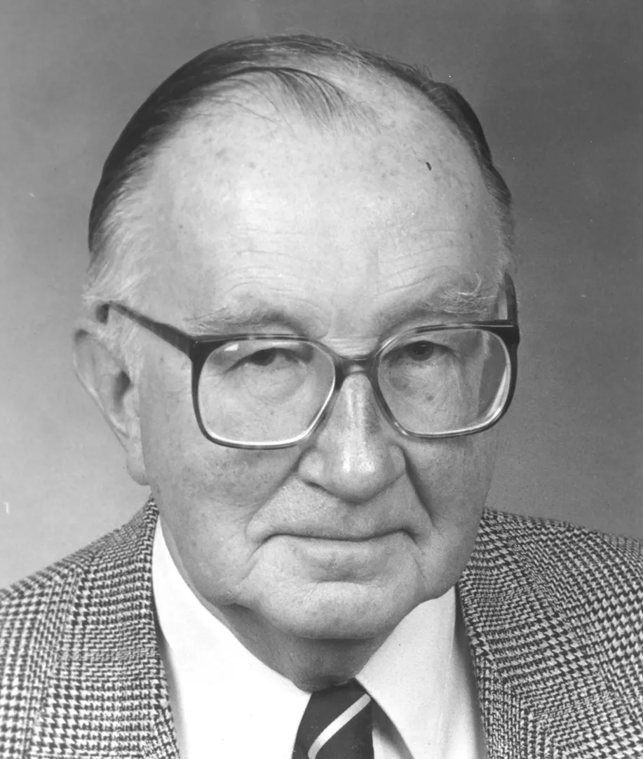
In February 1959, Malcolm Fowler and E. Graeme Robertson, in Australia, first described histopathologic findings in brains of five cases of kuru, noting diffuse prominent neuronal degeneration with peculiar vacuolation that was unlikely to be an artifact and not typical of any other human brain disease they knew. Later that year, Igor Klatzo (Fig. 1.3), at the National Institutes of Health (NIH) with Gajdusek and Zigas (Klatzo et al., 1959), described similar neuronal degeneration and vacuolation in brains of 12 kuru cases; Klatzo also noted microglial and astroglial proliferation and hypertrophy plus “remarkable plaque-like structures…between 20 and 60 microns in diameter…most frequently in the cerebellum [but] also in the basal ganglia and cerebral cortex.” Klatzo further remarked that “Creutzfeldt–Jacob [sic] disease appears to be closest in resemblance [to kuru]” (Klatzo et al., 1959). (More about Creutzfeldt–Jakob disease and the protein in plaques later.)
The next important event in the history of human TSEs was serendipitous, resulting from an almost chance encounter in 1959 between a veterinary pathologist and a public display of kuru histopathology. American William J. Hadlow (Fig. 1.4) had been sent by the US Department of Agriculture in 1958 to study the animal disease scrapie – a progressive degenerative neurologic disease of sheep and goats – at the UK Agricultural Research Council Field Station in Compton, England. Scrapie was thought by most investigators and veterinarians to be an infectious disease, and 20 years earlier two French investigators – J. Cuillé and P-L. Chelle (1936, 1939) – had transmitted scrapie from sick sheep to healthy sheep and goats by an agent that passed through a Chamberland L3 filter, fulfilling what were then criteria for an infection caused by a “filterable virus.”
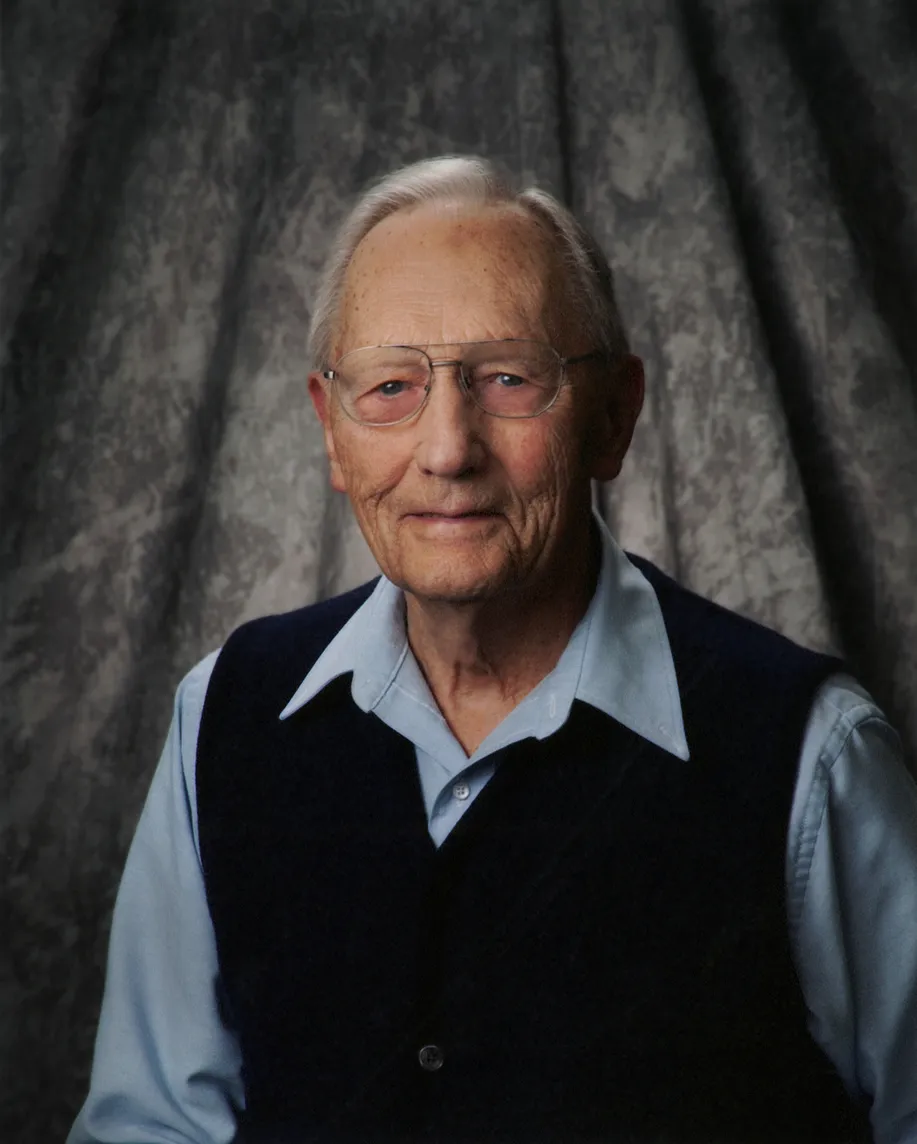
Scrapie had long been endemic in the United Kingdom (Brown and Bradley, 1998) but was relatively new in Canada and the United States, where it was slowly spreading. Hadlow (1992) recounted later that William Jellison, American parasitologist visiting him at Compton, had seen an exhibit on kuru mounted by Gajdusek at the Wellcome Medical Museum in London. Jellison suggested that Hadlow, because he was studying a degenerative neurologic disease of animals, might find the exhibit interesting. Hadlow traveled to London and viewed the exhibit, which displayed some of Klatzo's micrographs. Hadlow was immediately struck by similarities between the histopathology of kuru and scrapie; kuru brains displayed a typical scrapie “triad” of spongiform changes in gray matter, neuronal degeneration, and astrocytosis, but the unusual vacuolated neurons were especially striking. Later that day he read several articles about kuru in the library of the Royal Society of Medicine and concluded that the resemblance of “epidemiologic features, general clinical pattern and neurohistologic changes” between kuru and scrapie was “uncanny.” He summarized his observations in a brief seminal letter sent, on July 18, 1959, to the editor of The Lancet (and shared with Gajdusek a few days later). The letter was published almost unaltered on September 5 (Hadlow, 1959).
In his 1959 letter, Hadlow, not content s...
Table of contents
- Cover image
- Title page
- Table of Contents
- Copyright
- Handbook of Clinical Neurology 3rd Series
- Foreword
- Preface
- Contributors
- Chapter 1: Human transmissible spongiform encephalopathies: historic view
- Section I: Pathophysiology of prions
- Section II: Animal prion diseases (clinical, epidemiology, neuropathological, biochemical, biomarker, and genotypes)
- Section III: Human prion diseases (clinical, epidemiology, neuropathological, biochemical, biomarker, and genotypes)
- Section IV: Prion-like mechanisms in other neurodegenerative diseases
- Section V: Diagnosis and treatment
- Section VI: Public health issues
- Index
Frequently asked questions
Yes, you can cancel anytime from the Subscription tab in your account settings on the Perlego website. Your subscription will stay active until the end of your current billing period. Learn how to cancel your subscription
No, books cannot be downloaded as external files, such as PDFs, for use outside of Perlego. However, you can download books within the Perlego app for offline reading on mobile or tablet. Learn how to download books offline
Perlego offers two plans: Essential and Complete
- Essential is ideal for learners and professionals who enjoy exploring a wide range of subjects. Access the Essential Library with 800,000+ trusted titles and best-sellers across business, personal growth, and the humanities. Includes unlimited reading time and Standard Read Aloud voice.
- Complete: Perfect for advanced learners and researchers needing full, unrestricted access. Unlock 1.5M+ books across hundreds of subjects, including academic and specialized titles. The Complete Plan also includes advanced features like Premium Read Aloud and Research Assistant.
We are an online textbook subscription service, where you can get access to an entire online library for less than the price of a single book per month. With over 1.5 million books across 990+ topics, we’ve got you covered! Learn about our mission
Look out for the read-aloud symbol on your next book to see if you can listen to it. The read-aloud tool reads text aloud for you, highlighting the text as it is being read. You can pause it, speed it up and slow it down. Learn more about Read Aloud
Yes! You can use the Perlego app on both iOS and Android devices to read anytime, anywhere — even offline. Perfect for commutes or when you’re on the go.
Please note we cannot support devices running on iOS 13 and Android 7 or earlier. Learn more about using the app
Please note we cannot support devices running on iOS 13 and Android 7 or earlier. Learn more about using the app
Yes, you can access Human Prion Diseases by in PDF and/or ePUB format, as well as other popular books in Medicine & Neurology. We have over 1.5 million books available in our catalogue for you to explore.