eBook - ePub
Atlas of the Human Body
Central Nervous System and Vascularization
- 278 pages
- English
- ePUB (mobile friendly)
- Available on iOS & Android
eBook - ePub
Atlas of the Human Body
Central Nervous System and Vascularization
About this book
Atlas of Human Body: Central Nervous System and Vascularization is a multidisciplinary approach to the technical coverage of anatomical structures and relationships. It contains surface and 3D dissection images, native and colored cross sectional views made in different planes, MRI comparisons, demonstrations of cranial nerve origins, distribution of blood vessels by dissection, and systematic presentation of arterial distribution from the precapillary level, using the methyl metacrylate injection and subsequent tissue digestion method.
Included throughout are late prenatal (fetal) and early postnatal images to contribute to a better understanding of structure/relationship specificity of differentiation at various developmental intervals (conduits, organs, somatic, or branchial derivatives). Each chapter features clinical correlations providing a unique perspective of side-by side comparisons of dissection images, magnetic resonance imaging and computed tomography. Created after many years of professional and scientific cooperation between the authors and their parent institutions, this important resource will serve researchers, students, and doctors in their professional work.
- Contains over 700 color photos of ideal anatomical preparations and sections of each part of the body that have been prepared, recorded, and processed by the authors
- Covers existing gaps including developmental and prenatal periods, detailed vascular anatomy, and neuro anatomy
- Features a comprehensive alphabetical index of structures for ease of use
- Features a companion website which contains access to all images within the book
Trusted by 375,005 students
Access to over 1.5 million titles for a fair monthly price.
Study more efficiently using our study tools.
Information
Chapter 1
Upper Limb and Vascularization
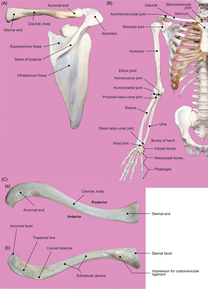
Figure 1.1 Skeleton of the upper limb.
(A) Posterior view of the right clavicle and scapula. (B) Bones of the shoulder region. (C) Right clavicle: (a) superior and (b) inferior views.
(A) Posterior view of the right clavicle and scapula. (B) Bones of the shoulder region. (C) Right clavicle: (a) superior and (b) inferior views.
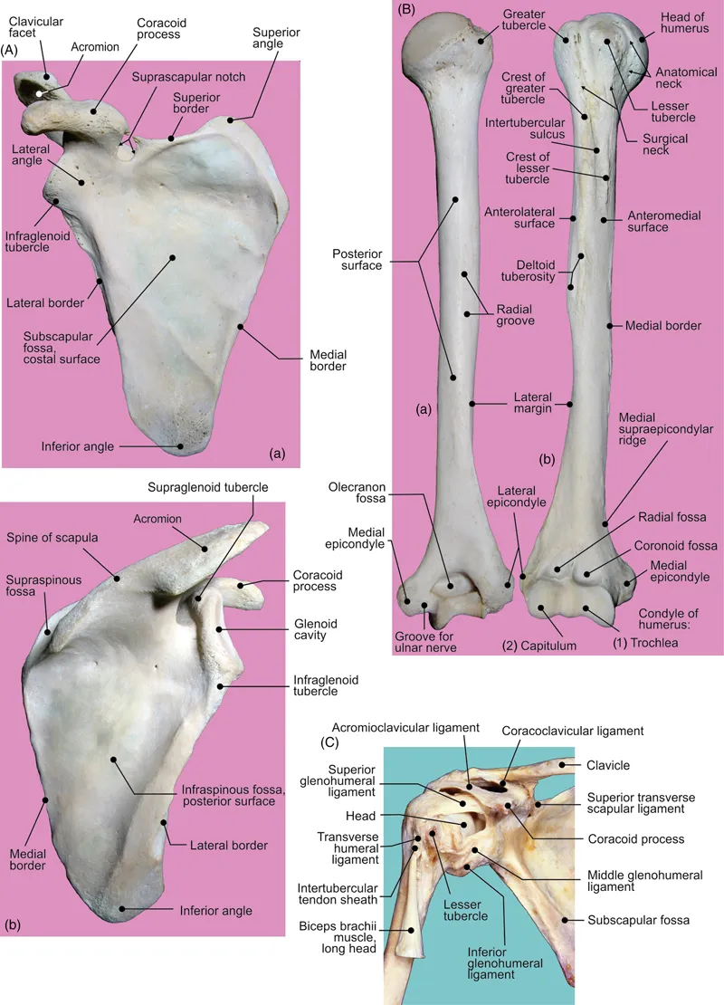
Figure 1.2 (A) Right scapula: (a) anterior and (b) posterior views. (B) Right humerus: (a) posterior and (b) anterior views. (C) Anterior view of the shoulder joint.
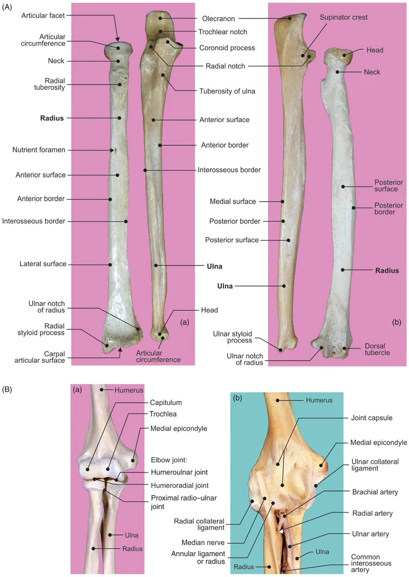
Figure 1.3 (A) Right forearm: (a) anterior and (b) posterior views of radius and ulna. (B) Elbow joint: anterior view (a) bones and (b) ligaments.
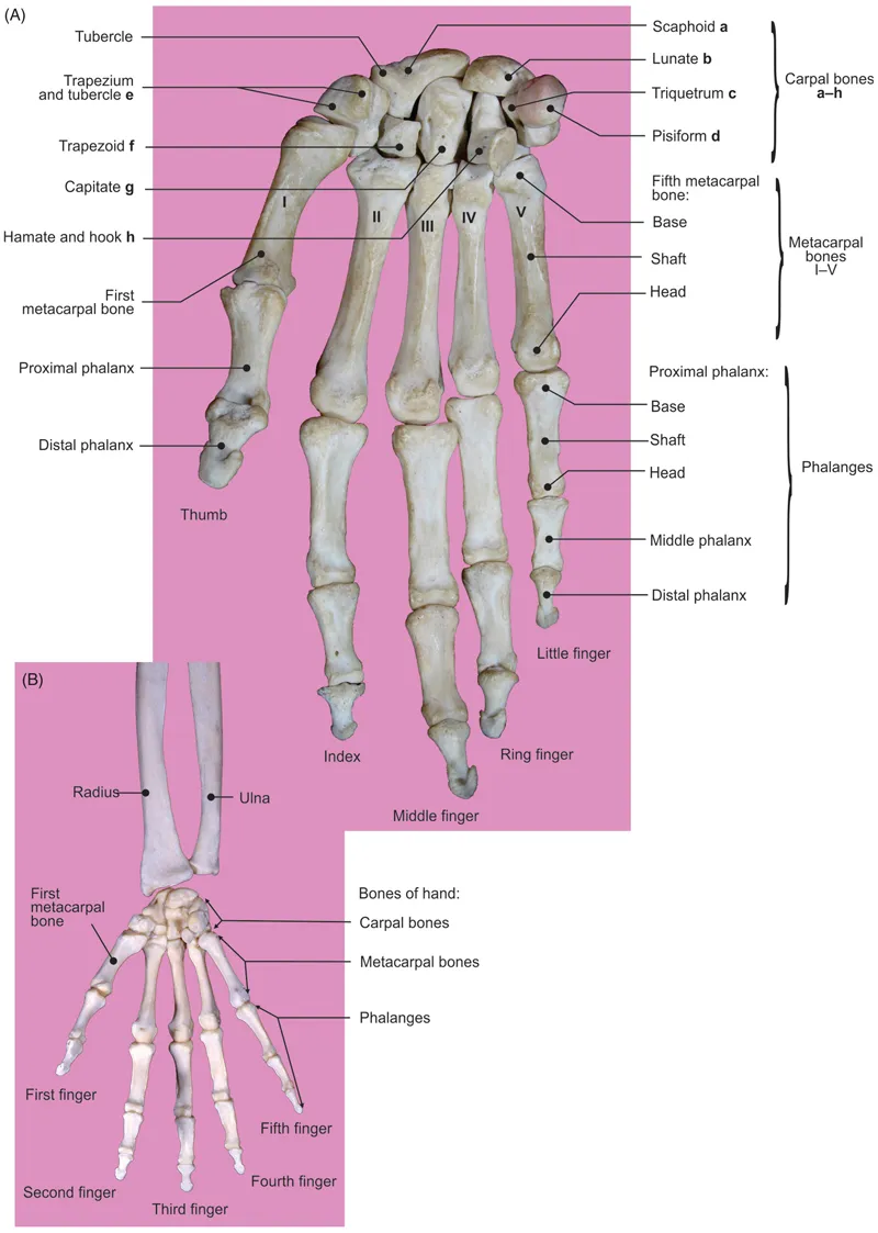
Figure 1.4 Palmar surfaces of hand skeleton.
(A) Bones of the hand. (B) Wrist joint.
(A) Bones of the hand. (B) Wrist joint.
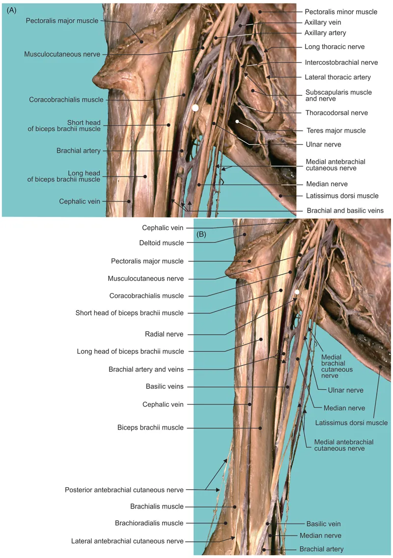
Figure 1.5 Anterior views of the (A) axillary and (B) brachial regions.
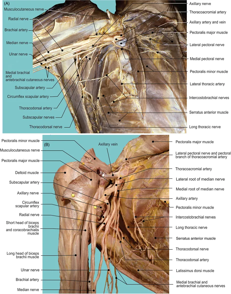
Figure 1.6 Anterior views of the (A) axillary and (B) brachial regions.
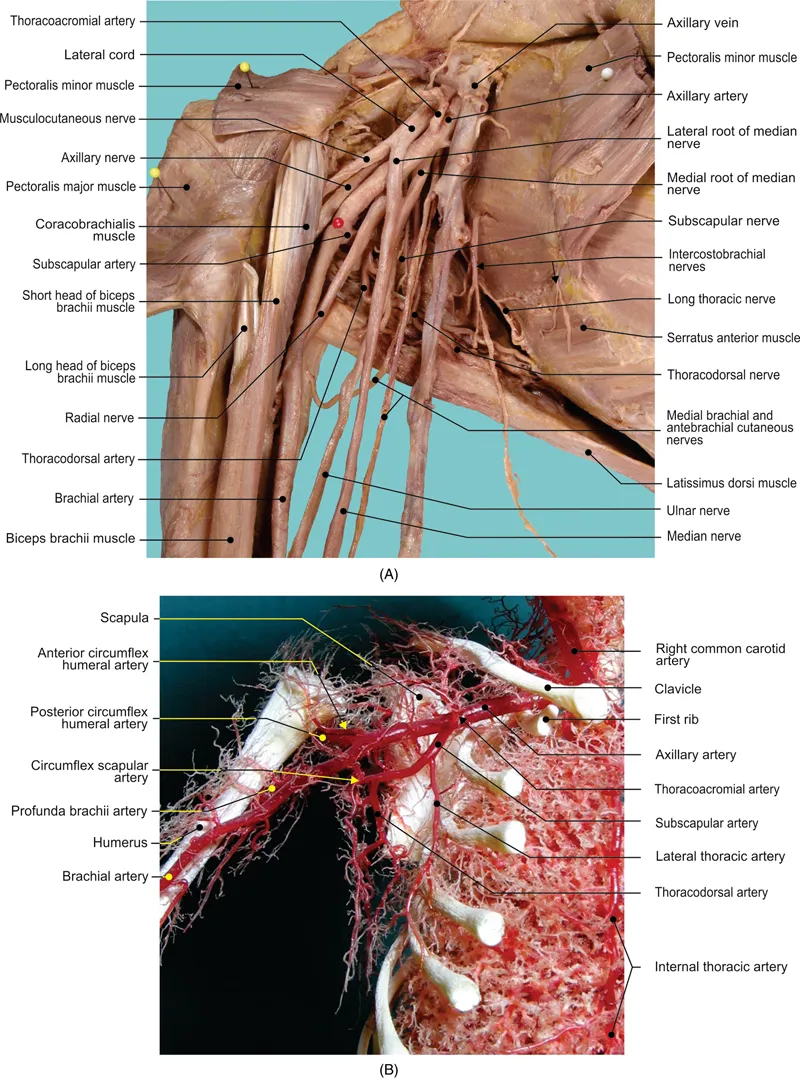
Figure 1.7 (A) Anterior view of the axillary fossa. (B) Anterior view of the fetal arterial distribution over rib cage and arm (corrosion cast).
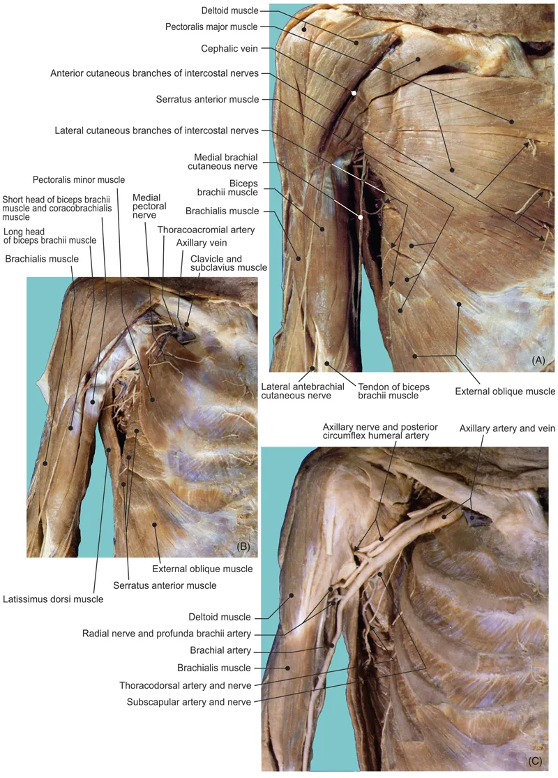
Figure 1.8 The pectoral region.
(A) Superficial layer. (B) Deep layer. (C) Axillary fossa after the removal of medial and lateral cords of the brachial plexus.
(A) Superficial layer. (B) Deep layer. (C) Axillary fossa after the removal of medial and lateral cords of the brachial plexus.
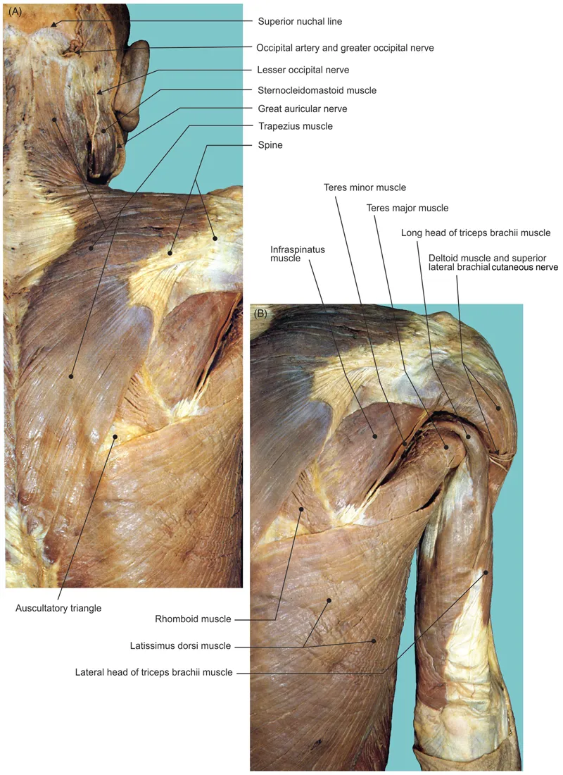
Figure 1.9 Superficial layer of the scapular region.
(A) Scapular and (B) posterior brachial regions.
(A) Scapular and (B) posterior brachial regions.
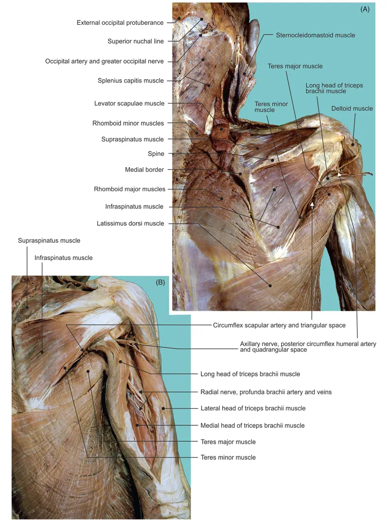
Figure 1.10 (A) Middl...
Table of contents
- Cover
- Title page
- Table of Contents
- Copyright
- Preface
- Chapter 1: Upper Limb and Vascularization
- Chapter 2: Lower Limb and Vascularization
- Chapter 3: Thorax and Vascularization
- Chapter 4: Abdomen and Vascularization
- Chapter 5: Pelvis and Perineum with 5–6-Month-Old Fetal Specimens
- Chapter 6: Head and Neck Regions and Vascularization
- Chapter 7: Cranial Central Nervous System and Spinal Cord
- Chapter 8: Vascularization of Head and Neck and the Cranial Central Nervous System
- References
- Index
Frequently asked questions
Yes, you can cancel anytime from the Subscription tab in your account settings on the Perlego website. Your subscription will stay active until the end of your current billing period. Learn how to cancel your subscription
No, books cannot be downloaded as external files, such as PDFs, for use outside of Perlego. However, you can download books within the Perlego app for offline reading on mobile or tablet. Learn how to download books offline
Perlego offers two plans: Essential and Complete
- Essential is ideal for learners and professionals who enjoy exploring a wide range of subjects. Access the Essential Library with 800,000+ trusted titles and best-sellers across business, personal growth, and the humanities. Includes unlimited reading time and Standard Read Aloud voice.
- Complete: Perfect for advanced learners and researchers needing full, unrestricted access. Unlock 1.5M+ books across hundreds of subjects, including academic and specialized titles. The Complete Plan also includes advanced features like Premium Read Aloud and Research Assistant.
We are an online textbook subscription service, where you can get access to an entire online library for less than the price of a single book per month. With over 1.5 million books across 990+ topics, we’ve got you covered! Learn about our mission
Look out for the read-aloud symbol on your next book to see if you can listen to it. The read-aloud tool reads text aloud for you, highlighting the text as it is being read. You can pause it, speed it up and slow it down. Learn more about Read Aloud
Yes! You can use the Perlego app on both iOS and Android devices to read anytime, anywhere — even offline. Perfect for commutes or when you’re on the go.
Please note we cannot support devices running on iOS 13 and Android 7 or earlier. Learn more about using the app
Please note we cannot support devices running on iOS 13 and Android 7 or earlier. Learn more about using the app
Yes, you can access Atlas of the Human Body by Branislav Vidic,Milan Milisavljevic in PDF and/or ePUB format, as well as other popular books in Biological Sciences & Medical Theory, Practice & Reference. We have over 1.5 million books available in our catalogue for you to explore.