- 352 pages
- English
- ePUB (mobile friendly)
- Available on iOS & Android
eBook - ePub
Physiology of the Cladocera
About this book
The Physiology of Cladocera is a much-needed summary of foundational information on these increasingly important model organisms. This unique and valuable summary is based on the world's literature, including Russian research not widely available until now. It offers systematically arranged data on the physiology of Cladocera, assisting with explanation of their life and distribution, as well as discussion on directions of future research. Special expert contributions in genetics, immunology, and cytology round out the physiological chapters and provide comprehensive insight into the state of knowledge of Cladocera and its underlying mechanisms.
Cladocera crustaceans make up a significant part of the natural communities and biological productivity of fresh waters. In recent decades, they have become globally studied for many purposes, including systematics, genetic, molecular, ecological and evolutionary biology studies. They are also used as "sentinel" organisms for assessing water quality and the environment. In addition, the genome of Daphnia (a genus within Cladocera) was recently sequenced and published, giving this system a much wider exposure. It has also led to a rapidly growing awareness of the importance of understanding physiological processes as they relate to evolutionary and ecological genomics and ecogenomic toxicology.
Despite the increasing use of Cladocera in research and study, physiological background information on these creatures is fragmentary. Hundreds of unconnected publications have been accumulated on their physiology, and a synthesis and general representation of the literature has been much needed for the many researchers working with this organism. The Physiology of Cladocera stands alone as a valuable and comprehensive offering in this area for many researchers and students.
- Collects and synthesizes from the worldwide literature the state of knowledge of cladoceran physiology
- Forward-looking perspective incorporates information from the emerging technological worlds of genomics, cytology, chemical communication, and immunology
- Provides foundational information on Cladocera physiology for researchers in various fields, including conservation and evolutionary biology, genomics, ecology, ecotoxicology, and comparative physiology
Trusted by 375,005 students
Access to over 1.5 million titles for a fair monthly price.
Study more efficiently using our study tools.
Information
Chapter 1
General
N.N. Smirnov
Some Cladocera species are dominant in the aquatic fauna. Some species are confined to narrow ecological niches, potentially as a result of their physiological adaptations. Specific studies require the precise identification of the species investigated. For this purpose, the keys to global faunas now available for most groups of Cladocera are indicated. The external and internal body structure is briefly described and size and weight characteristics are listed.
Keywords
Cladocera; species identification; keys; external structure; internal structure; size; weight
1.1 Systematic Position
It is now thought that there are over 700 species of the order Cladocera in the world fauna, many of which develop populations in enormous quantities and thus play a big role in the life of the biosphere. New species are still being described.
The Cladocera belong to the subclass Phyllopoda of the class Crustacea. Most Cladocera belong to the orders Anomopoda and Ctenopoda. Anomopoda principally comprise the families Daphniidae (e.g. the genera Daphnia, Ceriodaphnia, Simocephalus, and Scapholeberis), Moinidae (e.g. Moina), Ilyocryptidae (Ilyocryptus), Macrothricidae (e.g. Macrothrix and Streblocerus), Acantholeberidae, Ophryoxidae, Eurycercidae (Eurycercus), Chydoridae (e.g. Chydorus and Pleuroxus), Bosminidae (Bosmina, Bosminopsis); and Ctenopoda comprise the families Sididae (e.g. Sida, Pseudosida, and Diaphanosoma) and Holopedidae (Holopedium). Others belong to the order Onychopoda (Polyphemus, as well as marine and brackish water species) and the order Haplopoda with the family Leptodoridae (Leptodora).
As physiological studies should be accompanied by the reliable identification of the subjects being investigated, keys to the worldwide fauna of Cladocera are indicated: “Guides to the identification of the macroinvertebrates of the continental waters of the World,” issues 1, Macrothricidae (Smirnov, 1992); 3, Ctenopoda (Korovchinsky, 1992), 11, Chydorinae (Smirnov, 1996); 17, Simocephalus (Orlova-Bienkowskaja, 2001), 13, The predatory Cladocera (Rivier, 1998); 21, Daphnia (Benzie, 2005); and 22, Ilyocryptidae (Kotov and Štifter, 2006). There are also newer, general worldwide resources for Ctenopods, created by Korovchinsky (2004); Leydigia (Chydoridae), by Kotov (2009); and Eurycercus (Bekker, et al., 2012); as well as recent regional keys.
As investigations into Cladocera are actively developing, the aforementioned summaries are rapidly becoming incomplete, and more recent literature should also be taken into consideration.
1.2 General Morphological Background
As animal functions are linked to their form, some comments on the body structure and organs of Cladocera are provided here. Most of the animals attributed to the order Cladocera have the same principal structure, with various modifications present in different species. Investigations into comparative and functional morphology (such as, e.g. those by Fryer, 1968, 1974, 1991, etc.) have revealed exciting data on particular species, permitting a better understanding of their lifestyles.
Cladocerans have inherited from their ancestors a weakly segmented body covered with a chitinous, mostly bivalved, shell and bearing few pairs of appendages—antennules, antennae (biramous, with the single exception of female Holopedium), mandibles, maxillulae, maxillae (may be completely reduced), mandibles, and five or six pairs of thoracic limbs (Figs. 1.1–1.4).
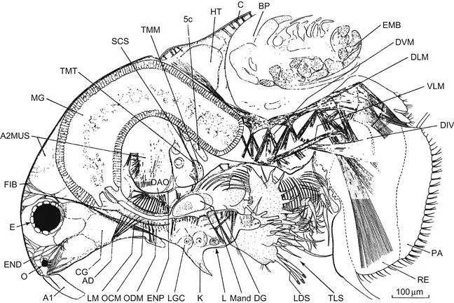
Figure 1.1 General anatomy of Acantholeberis curvirostris.
A1, antennule; A2MUS, antennary muscles; AD, apodeme; BP, brood pouch; C, carapace; CG, cerebral ganglion; DAO, dilator muscle of atrium oris; DG, duct of labral glands; DIV, diverticulum; DLM, dorsal longitudinal muscles; DVM, dorsoventral trunk muscles; E, compound eye; EMB, embryo; END, endoskeleton; ENP, endoskeletal plate; FIB, fibrils; HT, heart; K, keel of labrum; L, labrum; LDS, long distal setae of outer distal lobe of trunk limb 1; LGC, labral gland cells; LM, levator muscle of labrum; Mand, mandible; MG, mid-gut; O, ocellus; OCM, esophageal constrictor muscles; ODM, esophageal dilator muscles; PA, postabdominal lamella; RE, rectum; SUS, suspensory ligament; TLS, trunk limbs; TMM, 5c, transverse muscle of mandible; TMT, transverse mandibular tendon; VLM, ventral longitudinal trunk muscles. Source: Fryer (1974).
A1, antennule; A2MUS, antennary muscles; AD, apodeme; BP, brood pouch; C, carapace; CG, cerebral ganglion; DAO, dilator muscle of atrium oris; DG, duct of labral glands; DIV, diverticulum; DLM, dorsal longitudinal muscles; DVM, dorsoventral trunk muscles; E, compound eye; EMB, embryo; END, endoskeleton; ENP, endoskeletal plate; FIB, fibrils; HT, heart; K, keel of labrum; L, labrum; LDS, long distal setae of outer distal lobe of trunk limb 1; LGC, labral gland cells; LM, levator muscle of labrum; Mand, mandible; MG, mid-gut; O, ocellus; OCM, esophageal constrictor muscles; ODM, esophageal dilator muscles; PA, postabdominal lamella; RE, rectum; SUS, suspensory ligament; TLS, trunk limbs; TMM, 5c, transverse muscle of mandible; TMT, transverse mandibular tendon; VLM, ventral longitudinal trunk muscles. Source: Fryer (1974).
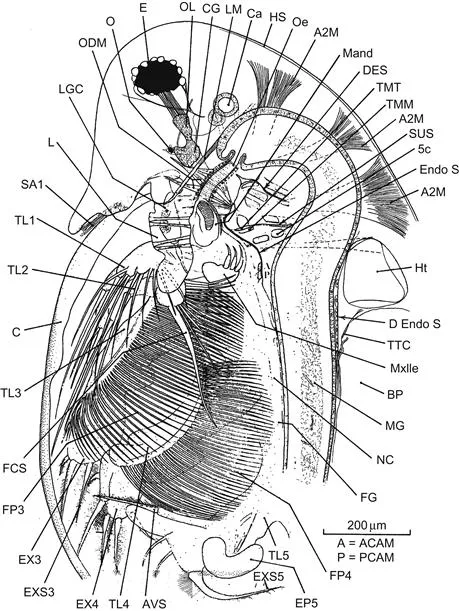
Figure 1.2 General anatomy of Daphnia longispina.
A, anterior carapax adductor muscle; A2M, antennal muscles; AVS, anterior vertical seta of trunk limb 5; Ca, cecum; D Endo S, dorsal endoskeletal sheet; DES, dorsal extension of ventral endoskeletal sheet; Endo S, endoskeletal sheet; EP5, epipodite of trunk limb 5; EX3, 4, exopod of trunk limbs 3, 4; EXS5, exopod seta 5; FCS, filter-cleaning spine of trunk limb 2; FG, food grove; FP3, gnathobasic filter plate of trunk limb 3; FP4, gnathobasic filter plate of trunk limb 4; Ht, heart; HS, head shield; LGC, labral gland cells; Mxlle, maxillule; NC, nerve cord; Oe, esophagus; OL, optic lobe of cerebral ganglion; P, posterior carapax adductor muscle; SA1, sensory seta of antennule; TL1, 2, 3, 4, 5, trunk limbs 1, 2, 3, 4, 5; TTC, thickened trunk cuticle. For other abbreviations, see Figure 1.1. Source: Fryer (1991).
A, anterior carapax adductor muscle; A2M, antennal muscles; AVS, anterior vertical seta of trunk limb 5; Ca, cecum; D Endo S, dorsal endoskeletal sheet; DES, dorsal extension of ventral endoskeletal sheet; Endo S, endoskeletal sheet; EP5, epipodite of trunk limb 5; EX3, 4, exopod of trunk limbs 3, 4; EXS5, exopod seta 5; FCS, filter-cleaning spine of trunk limb 2; FG, food grove; FP3, gnathobasic filter plate of trunk limb 3; FP4, gnathobasic filter plate of trunk limb 4; Ht, heart; HS, head shield; LGC, labral gland cells; Mxlle, maxillule; NC, nerve cord; Oe, esophagus; OL, optic lobe of cerebral ganglion; P, posterior carapax adductor muscle; SA1, sensory seta of antennule; TL1, 2, 3, 4, 5, trunk limbs 1, 2, 3, 4, 5; TTC, thickened trunk cuticle. For other abbreviations, see Figure 1.1. Source: Fryer (1991).
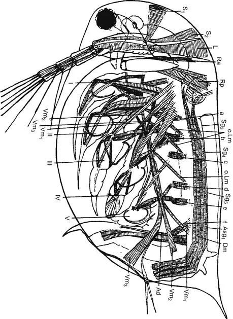
Figure 1.3 Muscles of Daphnia magna. Source: Binder (1931).
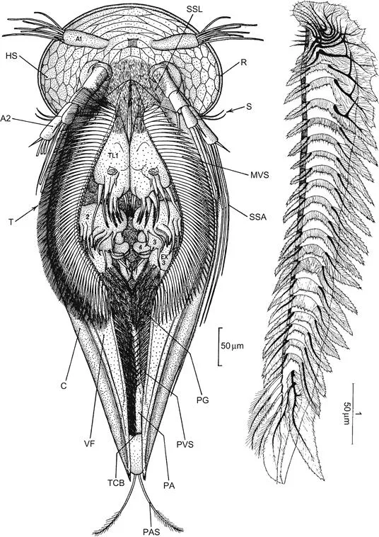
Figure 1.4 Modification of the ventral side for movement over flat surfaces.
Right, edge of valve of Scapholeberis mucronata. Left, Graptoleberis testudinaria. A1, antennule; A2, antenna; C, carapace; EX 3, exopod of trunk limb 3; HS, cuticle of head; MVS, medium ventral setae; PA, postabdomen; PAS, Postabdominal seta; PG, posterior gap; PVS, posterior ventral setae; R, thickened rim of head shield; S, Sensory setae of AII; SSA, Swimming setae of AII; SSL, setules of sealing seta of trunk limb 1; TCB, transverse chitinous bar; TL1, trunk limb 1; VF, ventral flange. Sources: right, Dumont and Pensaert (1983); left, Fryer (1968).
Right, edge of valve of Scapholeberis mucronata. Left, Graptoleberis testudinaria. A1, antennule; A2, antenna; C, carapace; EX 3, exopod of trunk limb 3; HS, cuticle of head; MVS, medium ventral setae; PA, postabdomen; PAS, Postabdominal seta; PG, posterior gap; PVS, posterior ventral setae; R, thickened rim of head shield; S, Sensory setae of AII; SSA, Swimming setae of AII; SSL, setules of sealing seta of trunk limb 1; TCB, transverse chitinous bar; TL1, trunk limb 1; VF, ventral flange. Sources: right, Dumont and Pensaert (1983); left, Fryer (1968).
Cladocerans are mostly oval in shape, compressed from the sides, but many are spherical. In the case of Graptoleberis, there is a curious and unique combination of lateral ...
Table of contents
- Cover image
- Title page
- Table of Contents
- Front-matter
- Copyright
- Preface
- Contributors
- Acknowledgments
- Chapter 1. General
- Chapter 2. Methods
- Chapter 3. Chemical Composition
- Chapter 4. Nutrition
- Chapter 5. Respiration
- Chapter 6. Circulation
- Chapter 7. Excretion
- Chapter 8. Osmotic Regulation
- Chapter 9. Cell and Tissue Metabolism
- Chapter 10. Growth and Molting
- Chapter 11. Reproduction
- Chapter 12. Locomotion
- Chapter 13. Nervous System and Sense Organs
- Chapter 14. Behavior
- Chapter 15. Ecophysiology
- Chapter 16. A Cytological Perspective
- Chapter 17. Immunology and Immunity
- Chapter 18. The Genomics of Cladoceran Physiology
- Conclusions: Special Traits of Cladoceran Physiology
- References
- Index of Latin Names of Cladocera
- Index of Chemical Substances
- Subject Index
Frequently asked questions
Yes, you can cancel anytime from the Subscription tab in your account settings on the Perlego website. Your subscription will stay active until the end of your current billing period. Learn how to cancel your subscription
No, books cannot be downloaded as external files, such as PDFs, for use outside of Perlego. However, you can download books within the Perlego app for offline reading on mobile or tablet. Learn how to download books offline
Perlego offers two plans: Essential and Complete
- Essential is ideal for learners and professionals who enjoy exploring a wide range of subjects. Access the Essential Library with 800,000+ trusted titles and best-sellers across business, personal growth, and the humanities. Includes unlimited reading time and Standard Read Aloud voice.
- Complete: Perfect for advanced learners and researchers needing full, unrestricted access. Unlock 1.5M+ books across hundreds of subjects, including academic and specialized titles. The Complete Plan also includes advanced features like Premium Read Aloud and Research Assistant.
We are an online textbook subscription service, where you can get access to an entire online library for less than the price of a single book per month. With over 1.5 million books across 990+ topics, we’ve got you covered! Learn about our mission
Look out for the read-aloud symbol on your next book to see if you can listen to it. The read-aloud tool reads text aloud for you, highlighting the text as it is being read. You can pause it, speed it up and slow it down. Learn more about Read Aloud
Yes! You can use the Perlego app on both iOS and Android devices to read anytime, anywhere — even offline. Perfect for commutes or when you’re on the go.
Please note we cannot support devices running on iOS 13 and Android 7 or earlier. Learn more about using the app
Please note we cannot support devices running on iOS 13 and Android 7 or earlier. Learn more about using the app
Yes, you can access Physiology of the Cladocera by Nikolai N. Smirnov in PDF and/or ePUB format, as well as other popular books in Technology & Engineering & Physiology. We have over 1.5 million books available in our catalogue for you to explore.