eBook - ePub
Sleep and Affect
Assessment, Theory, and Clinical Implications
- 532 pages
- English
- ePUB (mobile friendly)
- Available on iOS & Android
eBook - ePub
About this book
Sleep and Affect: Assessment, Theory, and Clinical Implications synthesizes affective neuroscience research as it relates to sleep psychology and medicine. Evidence is provided that normal sleep plays an emotional regulatory role in healthy humans. The book investigates interactions of sleep with both negative and positive emotions, along with their clinical implications. Sleep research is discussed from a neurobiological, cognitive, and behavioral approach. Sleep and emotions are explored across the spectrum of mental health from normal mood and sleep to the pathological extremes. The book, additionally, offers researchers a guide to methods and research design for studying sleep and affect.
This book will be of use to sleep researchers, affective neuroscientists, and clinical psychologists in order to better understand the impact of emotion on sleep as well as the effect of sleep on physical and mental well-being.
- Contains neurobiological, cognitive, and behavioral approaches
- Explains methods for examining sleep and affect
- Summarizes research on sleep and specific affect states
- Translates research for clinical use in treating disorders
Trusted by 375,005 students
Access to over 1.5 million titles for a fair monthly price.
Study more efficiently using our study tools.
Information
Topic
PsicologiaPart 1
Definitions
Chapter 1
Neurophysiology of Sleep and Circadian Rhythms‡
Jeff Dyche*; Katherine C. Couturier†; M. Kate Hall* * Department of Psychology, James Madison University, Harrisonburg, Virginia, USA
† Naval Submarine Medical Research Laboratory, Groton, Connecticut, USA
† Naval Submarine Medical Research Laboratory, Groton, Connecticut, USA
Abstract
This chapter will outline the biological machinery currently known to underpin the stages of consciousness and the rhythms that modulate them. We will briefly outline the basic functions of our central nervous system and how most researchers divide and categorize the major workings of our brain with an emphasis on the basic behaviors of sleep and alertness and how these two entities are not categorical. During wakefulness, we are kept in an alert state by the workings of some of the most primitive divisions of our brain. However, there are times when a person might be awake but their descriptions of their state varies as a function of what time of day it is and how long they have been awake. Therefore, wake and sleep states are more nuanced; the neural explanations of this will be discussed in detail.
Keywords
NREM sleep
REM
Circadian rhythm
Wakefulness
Sleep deprivation
Neurobiology
During the past half century, behavioral scientists have generally recognized three stages of consciousness: Non-Rapid Eye Movement (NREM) sleep, rapid eye movement (REM) sleep, and wakefulness (National Sleep Foundation, 2014). These three stages are choreographed in predictable processes across a 24-h period, which is the circadian rhythm of the sleep/wake cycle. This cycle depends on multifaceted switching machinery, all of which are modulated by neural mechanisms. The past 50 years have also brought huge advances in neuroscience methodology. Some obvious examples include expensive neuroimaging devices such as functional magnetic resonance imaging (fMRI) and positron emission tomography (PET) scans that have added knowledge on brain functioning and behavior that was heretofore impervious to human understanding (Chee et al., 2006; Drummond & Brown, 2001; Germain, Nofzinger, Kupfer, & Buysse, 2004; Maquet et al., 1996; Ruottinen et al., 2000). But even with additional progress in more traditional electrophysiological and neuroanatomical techniques, sleep remains one of the great mysteries of modern behavioral neuroscience. We spend about a third of our lives asleep yet the exact biological mechanism underlying sleep is not completely described in the literature. Still, with the 1990s being dubbed the “decade of the brain,” neuroscience research flourished, and with that momentum continuing as we near the middle of the second decade of the twenty-first century, scientists have been making enormous progress in understanding the mechanisms that control sleep and wakefulness.
In this chapter, we focus on four main areas of the central nervous system: the brainstem, spinal cord, medulla oblongata, and pons. Specifically, nerve cells that form the brainstem, the oldest part of our brain, are located just on top of the spinal cord and extend to the near middle of our brain, looking somewhat like the stem of an ice cream cone with a large overflowing scoop. The spinal cord, the major communication passageway for the motor and sensory portions of our brain, progresses from the inferior portions in the lower back to superior regions near the neck, and then it begins to widen, swell if you will, as it eases into the enigmatic confines of our cranium. This “swelling” of nerve cells is the beginning of the brainstem, is called the medulla oblongata, and is located at the bottom (inferior region) of the brainstem. As you might guess, the medulla isn’t where conscious thought occurs, but it is a very important center of behavior as it regulates functions such as breathing, swallowing, and vomiting—automatic behaviors that we don’t have to (or sometimes don’t want to) think about. Only 1.5 in. in length, this oblong material is conical in shape. The bottom smaller portion is basically continuous with our spinal cord while the top wider portion is connected to the bridge of the brainstem called the pons (literally “bridge” in Latin, as it connects the lower and upper regions of the brainstem). However, as primitive and as small as the medulla is, it also plays an important role in the functioning of sleep. Animal models have demonstrated that if we lesion the medial (middle) portion of the medulla, REM sleep is altered. When that lesion extends into the pons, REM sleep may be completely eliminated. If that part of a person’s brain was damaged, what are the behavioral ramifications of such a sleep change? How is NREM sleep implicated? And are circadian rhythms modulated by the same or different mechanisms? What are some of the myths and misconceptions of fatigue? These questions and more will be discussed in this chapter.
If I didn’t wake up, I’d still be sleeping.
Yogi Berra
Overview
Normal sleep is divided into two basic groups. One is Rapid Eye Movement sleep or “REM,” famous for dream mentation and muscle paralysis and the other is the less celebrated yet equally important “non-REM” sleep or “NREM.” NREM sleep is further divided into increasingly deeper stages of sleep: stage N1, stage N2, and stage N3. The latter is also referred to as the deepest stage of sleep where the brain produces large, synchronous “delta” waves sometimes referred to as “slow wave sleep” (Siegel, 2002). While REM sleep is not formally sub-divided, research has demonstrated that REM sleep is comprised of phasic and tonic components (Carskadon & Dement, 2011). Phasic REM sleep is a sympathetically driven state characterized by the stereotypical REMs with some distal twitching of the face and limbs. The tonic portion of REM is more parasympathetically enervated and involves little or no eye movements or twitching of distal muscle groups. Most REM sleep that occurs early in the night is tonic and later episodes are more phasic. The individual role of each subdivision is not yet understood. Of course a paramount feature of REM, whether tonic or phasic, is paralysis of the more proximal skeletal muscles.
Sleep is a state that may not be described as being truly unconscious as even in the deepest levels of sleep there is some monitoring of the external environment. Most, for example, can awaken at the sound of a baby crying or of a loud shout of the sleeping person’s first name, so the brain must be aware albeit at a reduced level. Indeed, sleep researchers typically define sleep in a multistaged fashion in that there is (1) a reduction in awareness of the environment, (2) lowered motility and muscular activity, (3) partial suppression of voluntary behavior, and, to distinguish sleep from coma or a true loss of consciousness, it is (4) reversible (Everson, 2009).
One of the most important points when discussing the physiology of sleep is that sleep is a state where the brain is very active. To fall asleep is not the central nervous system slowing across a period of time, perhaps in reaction to a long day’s work of cogitating and muscle contractions that lead to a passive quiescence where consciousness is lost due to the brain slowing inexorably under the workload. No, it doesn’t work that way. In fact, there are certain regions of the brain that work very rigorously to generate sleep (Lu, Greco, Shiromani, & Saper, 2000; McGinty & Sterman, 1968; Nauta, 1946; Sherin et al., 1996; Siegel, 2002; von Economo, 1930). The absence of these regions in laboratory animals leads to profound insomnia and, in certain cases, death (Lu et al., 2000; Nauta, 1946). Similarly, destroying other regions of the brain in animals leads to coma-like states (Lindsley, Schreiner, Knowles, & Magoun, 1950). We will outline those structures that not only generate the major sleep categories, NREM and REM, but also what brain structures are vital for wakefulness. You will notice an important relationship between the sleep generating structures, the waking generating circuitry and the wake reducing circuitry.
Sleeping is no mean art: for its sake one must stay awake all day.
Friedrich Nietzsche
Wakefulness
In the early twentieth century, most scientists who studied the brain believed that wakefulness was maintained due to sensory input to the brain. If that input was removed, then loss of consciousness, or sleep, would occur. This is, of course, a passive view of how sleep is generated. In 1916, a Vienna psychiatrist named Baron Constantine von Economo had been evaluating patients with an interesting form of brain infection he termed “encephalitis lethargica” that profoundly impacted the normal sleep-wake cycle (von Economo, 1930). Unlike normal sleepers who sleep 7-8 h each day, most of his patients slept excessively long (up to 20 h a day, an amount normally seen only in bats, the longest sleeping animal known). Autopsies on such patients revealed that these hypersomnolent individuals had lesions in the most rostral region (the superior portions) of the reticular formation (RF; Figure 1.1). This was the first clue that the midbrain portion of the brainstem contained some wake-promoting circuits. Moreover, a few of his patients actually experienced an opposite phenomenon as they were profoundly insomniac and slept just a few hours a day (i.e., as much as a typical horse, who are very short sleepers). These patients also had lesions but away from the brainstem proper in the anterior portions of the hypothalamus and in the adjacent basal forebrain (BF), suggesting to von Economo that these regions were involved in the induction of sleep.
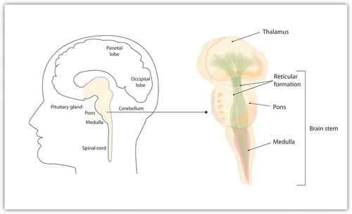
Figure 1.1 The reticular formation runs up the brainstem. This structure was damaged in von Economo's patients. wikicommons.
Of course nowadays, based on animal literature and a few pathological conditions in humans (e.g., encephalitis lethargica), it is known that the waking state depends on the active working neurons within the RF region of the brainstem (Moruzzi & Magoun, 1949; Siegel, 2002). The RF, in turn, communicates with the higher functions of the brain via an intricate system of neurons that project from the RF called the ascending reticular activating system (ARAS). The ARAS initially moves to the switchboard of the sensory system, the thalamus, and from there it makes its way through various synapses in all lobes of the cerebrum, without which consciousness is not possible without at least partial regeneration of pathways via a plasticity driven recovery of function. This interesting discovery of how the ARAS functioned was first demonstrated on cats by Moruzzi and Magoun in 1949 and is considered to be one of the most important concepts in the sleep-wake field (Siegel, 2002).
Incidentally, numerous psychological and physiological discoveries in the wake of Moruzzi and Magoun’s discovery contributed to much more than just sleep science knowledge. Indeed, many psychologists with neurobiology backgrounds and neurobiologists with psychology backgrounds began collaborating with anatomists and physicians and the new interdisciplinary field of neuroscience was born. Therefore, sleep science might lay at least partial claim to being the progeny of one of the largest scientific disciplines in the world based on the total membership of the Society for Neuroscience (SfN, 2013).
To return to the anatomy and function of the ARAS—the sine qua non for arousal (and therefore consciousness)—it is further divided into two pathways in order to maintain normal alertness. The first dorsal (lower) branch consists of cholinergic neurons extending from the ...
Table of contents
- Cover image
- Title page
- Table of Contents
- Copyright
- Foreword: Interrelationships between Sleep and Affect
- Preface
- Acknowledgements
- Contributors
- Part 1: Definitions
- Part 2: Methods
- Part 3: Evidence Regarding Sleep and Specific Types of Affect
- Part 4: Future Directions
- Index
Frequently asked questions
Yes, you can cancel anytime from the Subscription tab in your account settings on the Perlego website. Your subscription will stay active until the end of your current billing period. Learn how to cancel your subscription
No, books cannot be downloaded as external files, such as PDFs, for use outside of Perlego. However, you can download books within the Perlego app for offline reading on mobile or tablet. Learn how to download books offline
Perlego offers two plans: Essential and Complete
- Essential is ideal for learners and professionals who enjoy exploring a wide range of subjects. Access the Essential Library with 800,000+ trusted titles and best-sellers across business, personal growth, and the humanities. Includes unlimited reading time and Standard Read Aloud voice.
- Complete: Perfect for advanced learners and researchers needing full, unrestricted access. Unlock 1.5M+ books across hundreds of subjects, including academic and specialized titles. The Complete Plan also includes advanced features like Premium Read Aloud and Research Assistant.
We are an online textbook subscription service, where you can get access to an entire online library for less than the price of a single book per month. With over 1.5 million books across 990+ topics, we’ve got you covered! Learn about our mission
Look out for the read-aloud symbol on your next book to see if you can listen to it. The read-aloud tool reads text aloud for you, highlighting the text as it is being read. You can pause it, speed it up and slow it down. Learn more about Read Aloud
Yes! You can use the Perlego app on both iOS and Android devices to read anytime, anywhere — even offline. Perfect for commutes or when you’re on the go.
Please note we cannot support devices running on iOS 13 and Android 7 or earlier. Learn more about using the app
Please note we cannot support devices running on iOS 13 and Android 7 or earlier. Learn more about using the app
Yes, you can access Sleep and Affect by Kimberly Babson,Matthew Feldner in PDF and/or ePUB format, as well as other popular books in Psicologia & Psicologia cognitiva e cognizione. We have over 1.5 million books available in our catalogue for you to explore.