- 282 pages
- English
- ePUB (mobile friendly)
- Available on iOS & Android
eBook - ePub
About this book
Receptors in the Evolution and Development of the Brain: Matter into Mind presents the key role of receptors and their cognate ligands in wiring the mammalian brain from an evolutionary developmental biology perspective. It examines receptor function in the evolution and development of the nervous system in the large vertebrate brain, and discusses rapid eye movement sleep and apoptosis as mechanisms to destroy miswired neurons. Possible links between trophic deficits and connectional diseases including Alzheimer's, Parkinson's, and ALS are also discussed. This book is extremely useful to those with an interest in the molecular and cellular neurosciences, including those in cognitive and clinical branches of this subject, and anyone interested in how the incredibly complex human brain can build itself.
- Provides an understanding of the key role receptors play in brain development and the selection process necessary to construct a large brain
- Traces the evolution of receptors from the most primitive organisms to humans
- Emphasizes the roles that REM sleep and apoptosis play in this selection via trophic factors and receptors
- Describes the role that trophic factor-receptor interactions play throughout life and how trophic deficits can lead to connectional diseases, including Alzheimer's, Parkinson's and ALS
- Provides a potential mechanism whereby neuronal stem cells can cure these diseases
Trusted by 375,005 students
Access to over 1.5 million titles for a fair monthly price.
Study more efficiently using our study tools.
Information
Chapter 1
Classes of receptors, their signaling pathways, and their synthesis and transport
Abstract
This chapter provides basic information on how a few embryonic cells can form a big brain that contains over 1011 neurons and about the same number of glial cells, and that is able of doing amazing things. Receptors will be defined as will their binding partners, ligands/trophic factors. Receptors and trophic factors are key elements in the evolution and development of the big brain. The different families of signaling plasma membrane and cytoplasmic/nuclear receptors will be described as will nutrient receptors. The types of signaling mechanisms employed by both plasma membrane and cytoplasmic/nuclear receptors are discussed, as is the mechanism of synthesis of secretory and integral membrane proteins including receptors and many trophic factors.
Keywords
Calnexin; calreticulin; SMADS; translocon; axin; disheveled; ADAM-10
The human brain is the most complex organ ever assembled. It is composed of at least 100 billion neurons and about the same number of glial cells, which communicate through a network of trillions of chemical synapses and electrical channels called gap junctions, as well as other cell types such as endothelial cells that are present in much smaller numbers. This conglomerate of connections can solve incredibly difficult mathematical problems, compose symphonies, communicate with other brains either by speech or writing, build rockets to reach the moon, hit a baseball moving at 100 mph, and produce incredibly beautiful paintings.
Perhaps the most amazing thing about the human brain is that it can build itself from a small group of undefined cells generated early in development. The purpose of this book is to outline a plausible strategy for the development of the human brain. Central to this strategy is the use of two classes of molecules, receptors, and ligands—a scientific term for molecules to which receptors bind with great specificity and selectivity. Once a receptor binds to its ligand, it changes its shape, thereby sending a signal to the cell interior. This fundamental mechanism has evolved from the most ancient unicellular organisms, Archaea, and has been critical in the evolution of multicellular organisms and in the origin of the brain, leading to the development of the most complex assemblage of all, the human brain.
This book is entitled The Key Role of Receptors in the Evolution and Development of the Brain: Matter into Mind. To understand what this key role is, one must understand the unique properties of receptors. In this chapter the reader will be acquainted with these properties before moving into the central areas of the book, the evolution of receptors and organisms and the concomitant evolution of neurons leading to the most complex system that we know of: the mammalian brain. (Humans are thought to have the highest intelligence. However, one can argue that whales and elephants, which have brains containing more neurons and glia than humans, are also more intelligent. Perhaps their greater intellectual ability makes them unwilling to destroy our planet.)
Receptors are proteins found at the outer membrane of the cell, the plasma membrane, or within the cell either in the cytoplasm or the nucleus. An individual receptor has several key characteristics. It is able to recognize and tightly bind a very specific molecule including small molecules, another protein, a lipid, or a sugar. This selective tight binding is a key feature of the receptor–ligand interaction. A receptor can bind to a specific molecule when the latter’s concentration is 10–12 molar or lower. At the same time it will not recognize and bind to a very similar molecule that is present at a million-fold higher concentration.
Upon binding to its specific ligand, the receptor changes its shape—the scientific word is conformation. This in turn causes information to be transferred to other molecules in the cytoplasm of the cell, leading to an almost infinite variety of changes in the behavior of the cell. These include alteration in the direction and/or the speed of movement, increases in the type and the amount of various proteins produced by the cell, and an alteration of the electrical excitability of a neuron. In the most extreme cases, receptor–ligand binding leads to the suicide of the cell, also called apoptosis. This topic will be discussed in detail in Chapter 9, The importance of rapid eye movement sleep and other forms of sleep in selecting the appropriate neuronal circuitry; programmed cell death/apoptosis.
While there was pharmacological and physiological evidence that specific receptors existed for quite some time, the first isolation of a receptor, the nicotinic acetylcholine receptor (NAchR), occurred in 1972. The NAchR from the eel electric organ, a modified muscle that contains many NAchRs, was used. The isolation was based on the coupling of a small molecule, ɑ-bungarotoxin, which specifically bound to this receptor, to a resin poured into a column. Only the NAchR specifically bound to the immobilized ɑ-bungarotoxin. After washing several times the receptor was eluted with a mild detergent, which preserved its structure. It was then incorporated into an artificial membrane and shown to have the pharmacological properties identical to the NAchR. This method has been used with modifications to purify many important receptors.
1.1 Types of Receptors
As expected for a class of molecules that carries out a myriad of functions and can bind to a huge number of divergent molecules, there are many types of receptors. In fact, receptors comprise the most abundant group of proteins in the human genome. However, they can be organized into a relatively small number of classes, suggesting that the members of each group evolved from a single receptor.
G Protein Linked Receptors
The most numerous class of receptors is called the G protein coupled receptors (GPCRs), referring to the ability of these plasma membrane receptors to bind to a cytoplasmic protein called the G protein after binding to an extracellular ligand. The G protein subsequently transfers information into the cell. A large percentage of drugs are directed toward the GPCRs.
All of the GPCRs contain seven α-helical segments or protein domains composed of hydrophobic (“water hating”) or neutral amino acids that traverse the plasma membrane and form a hydrophilic (“water loving”) channel in the middle. The α-helix, whose structure was first solved by the Nobel Prize winning chemist Linus Pauling, is ideal to pass through the very hydrophobic environment of the plasma membrane. The α-helix, when composed of hydrophobic amino acids, can neutralize all of its charged peptide bonds by hydrogen bonding to another amino acid 3.6 amino acids away in the α-helix. Fig. 1.1A. It requires 20–21 either hydrophobic or neutral amino acids to completely traverse the 35-angstrom hydrophobic environment of the bilayer portion of the plasma membrane.

(A) α-Helix. The stick figure (left) shows a right-handed α-helix with the N-terminus at the bottom and side chains R represented by the β-carbon. Hydrogen bonds between backbone atoms are indicated by blue lines. In this orientation, the carbonyl oxygens point upward, the amide protons point downward, and the R groups trail toward the N-terminus. Space filling models (middle) show a polyalanine α-helix. The end-on views show how the backbone atoms fill the center of the helix. A space filling model (right) of α-helix 5 from bacterial rhodopsin shows the side chains. Some key dimensions are 0.15 nm rise per residue, 0.55 nm per turn, and diameter of approximately 1.0 nm. (B) Stick figure and space filling models of an antiparallel β-sheet. The arrows indicate the polarity of each chain. With the polypeptide extended in this way, the amide protons and carbonyl oxygens lie in the plane of the sheet, where they make hydrogen bonds (blue lines) with the neighboring strands. The amino acid side chains alternate pointing upward and downward from the plane of the sheet. Some key dimensions are 0.35 nm rise per residue in a β-strand and 0.45 nm separation between strands. (C) Stick figure and space filling models of a parallel β-sheet. All strands have the same orientation (arrows). The orientations of the hydrogen bonds are somewhat less favorable than in an antiparallel sheet. (D and E) Stick figures of two types of reverse turns found between strands of antiparallel β-sheets. (F) Stick figure of an omega loop. Pollard TD, Earnshaw WC, Lippincott-Schwartz J, Johnson GT. Cell Biology. 3rd ed. Philadelphia, PA: Elsevier; 2016 [Chapters 1, 3, 13–16, 21–24, 27, 30, 33 and 34]. Fig. 3.8, p. 38. With permission.
The amino acids comprising bacteriorhodopsin like those in the GPCRs, contain seven transmembrane α-helices that cross the plasma membrane (Fig. 1.2). Bacteriorhodopsin was the first membrane protein whose three-dimensional structure was determined.
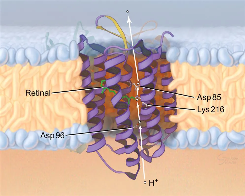
Numerous atomic structures, fast spectroscopic measurements of reaction intermediates, and analysis of a wide array of mutations revealed the pathway for protons through the middle of the bundle of seven α-helices. A cytoplasmic proton binds successively to Asp96, the Schiff base linking retinal to lysine 216 (Lys216), Asp85, and Glu204 before releasing outside the cell. Absorption of light by retinal drives conformational changes in the protein that favor the transfer of the proton across the membrane up its concentration gradient. Pollard TD, Earnshaw WC, Lippincott-Schwartz J, Johnson GT. Cell Biology. 3rd ed. Philadelphia, PA: Elsevier; 2016 [Chapters 1, 3, 13–16, 21–24, 27, 30, 33, and 34]. Fig. 14.3, p. 38. With permission.
Another protein domain whose structure allows it to traverse the plasma membrane is the β-pleated sheet (Fig. 1.1B–F). However this structure is primarily used by protists, which include bacteria and other single-celled organisms. β-pleated sheets also can form the cross β-pleated sheets, which are the insoluble structures called amyloid fibers.
As mentioned previously, the 35-angstrom portion of the plasma membrane as well as that of almost all cellular membranes is a bilayer composed of phospholipids interspersed with cholesterol molecules and glycolipids, which contain complex sugars. The lipids that form the bilayer are also asymmetrically oriented with some phospholipids only on one side of the bilayer. Also some glycolipids form clusters on the extracellular side of the bilayer (Fig. 1.3). Integral membrane proteins including GPCRs completely traverse the bilayer; some proteins only extend through one layer of the bilayer. Others are present only on the membrane surface attached through electrostatic interaction (Fig. 1.3).
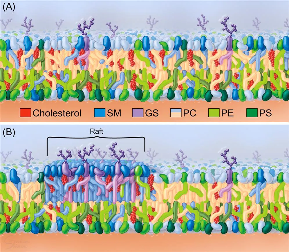
(A) Sphingomyelin (SM) and cholesterol form a small cluster in the external leaflet. GS, glycosphingolipid; PC, phosphatidylcholine; PE, phosphatidylethanolamine; PS, phosphatidylserine. PS is enriched in the inner leaflet. (B) Lipid raft in the outer leaflet ...
Table of contents
- Cover image
- Title page
- Table of Contents
- Copyright
- Acknowledgments
- Introduction
- Chapter 1. Classes of receptors, their signaling pathways, and their synthesis and transport
- Chapter 2. The RNA world: receptors and their cognate ligands in Archaea, Bacteria, and Choanoflagellates
- Chapter 3. Dictyostelium discoideum, sponges, comb jellies, and hydra: the earliest animals
- Chapter 4. The growth cone and the synapse
- Chapter 5. Neuronal cell biology
- Chapter 6. Glial cells, the myelinated axon, and the blood-brain barrier
- Chapter 7. A testable theory on the wiring of the brain
- Chapter 8. Development of the cerebral cortex
- Chapter 9. The importance of sleep in selecting neuronal circuitry; programmed cell death/apoptosis
- Chapter 10. BDNF and endocannabinoids in brain development: neuronal commitment, migration, and synaptogenesis
- Chapter 11. Axon and dendrite guidance molecules and the extracellular matrix in brain development
- Chapter 12. Key receptors involved in laminar and terminal specification and synapse construction
- Chapter 13. Steroid hormones, glucocorticoids, and the hypothalamus
- Chapter 14. Receptors and the development of the enteric nervous system
- Chapter 15. Synaptic pruning and trophic factor interactions during development
- Chapter 16. Receptor-mediated mechanisms for drugs of abuse and brain development
- Chapter 17. Role of trophic factors and receptors in developmental disorders
- Chapter 18. Neuronal survival and connectional neurodegenerative diseases
- Chapter 19. Neuronal stem cells
- Chapter 20. Summary and conclusions
- Index
Frequently asked questions
Yes, you can cancel anytime from the Subscription tab in your account settings on the Perlego website. Your subscription will stay active until the end of your current billing period. Learn how to cancel your subscription
No, books cannot be downloaded as external files, such as PDFs, for use outside of Perlego. However, you can download books within the Perlego app for offline reading on mobile or tablet. Learn how to download books offline
Perlego offers two plans: Essential and Complete
- Essential is ideal for learners and professionals who enjoy exploring a wide range of subjects. Access the Essential Library with 800,000+ trusted titles and best-sellers across business, personal growth, and the humanities. Includes unlimited reading time and Standard Read Aloud voice.
- Complete: Perfect for advanced learners and researchers needing full, unrestricted access. Unlock 1.5M+ books across hundreds of subjects, including academic and specialized titles. The Complete Plan also includes advanced features like Premium Read Aloud and Research Assistant.
We are an online textbook subscription service, where you can get access to an entire online library for less than the price of a single book per month. With over 1.5 million books across 990+ topics, we’ve got you covered! Learn about our mission
Look out for the read-aloud symbol on your next book to see if you can listen to it. The read-aloud tool reads text aloud for you, highlighting the text as it is being read. You can pause it, speed it up and slow it down. Learn more about Read Aloud
Yes! You can use the Perlego app on both iOS and Android devices to read anytime, anywhere — even offline. Perfect for commutes or when you’re on the go.
Please note we cannot support devices running on iOS 13 and Android 7 or earlier. Learn more about using the app
Please note we cannot support devices running on iOS 13 and Android 7 or earlier. Learn more about using the app
Yes, you can access Receptors in the Evolution and Development of the Brain by Richard E. Fine in PDF and/or ePUB format, as well as other popular books in Biological Sciences & Neuroscience. We have over 1.5 million books available in our catalogue for you to explore.