- 532 pages
- English
- ePUB (mobile friendly)
- Available on iOS & Android
eBook - ePub
Imaging of the Human Brain in Health and Disease
About this book
Brain imaging technology remains at the forefront of advances in both our understanding of the brain and our ability to diagnose and treat brain disease and disorders. Imaging of the Human Brain in Health and Disease examines the localization of neurotransmitter receptors in the nervous system of normal, healthy humans and compares that with humans who are suffering from various neurologic diseases.
Opening chapters introduce the basic science of imaging neurotransmitters, including sigma, acetylcholine, opioid, and dopamine receptors. Imaging the healthy and diseased brain includes brain imaging of anger, pain, autism, the release of dopamine, the impact of cannabinoids, and Alzheimer's disease.
This book is a valuable companion to a wide range of scholars, students, and researchers in neuroscience, clinical neurology, and psychiatry, and provides a detailed introduction to the application of advanced imaging to the treatment of brain disorders and disease.
- A focused introduction to imaging healthy and diseased brains
- Focuses on the primary neurotransmitter release
- Includes sigma, acetylcholine, opioid, and dopamine receptors
- Presents the imaging of healthy and diseased brains via anger, pain, autism, and Alzheimer's disease
Trusted by 375,005 students
Access to over 1.5 million titles for a fair monthly price.
Study more efficiently using our study tools.
Information
Chapter One
Neuroimaging of Addiction
Nora D. Volkow1, Gene-Jack Wang2, Joanna S. Fowler2, Dardo Tomasi3 and Ruben Baler1, 1National Institute on Drug Abuse, Bethesda, MD, USA, 2Brookhaven National Laboratory, Upton, NY, USA, 3National Institute on Alcohol Abuse and Alcoholism, Bethesda, MD, USA
Abstract
Modern imaging techniques have allowed researchers to noninvasively peer into the human brain and investigate, among many other things, the acute effects and long-term consequences of drug abuse. Here, we review the most commonly used and some emerging imaging techniques in addiction research, explain how the various techniques generate their characteristic images, and describe the rational that researchers use to interpret them. In addition, examples of seminal imaging findings are highlighted that illustrate the contribution of each imaging modality to the expansion in our understanding of the neurobiological bases of drug abuse and addiction, and how they can be parlayed in the future into clinical and therapeutic applications.
Keywords
Addiction; MRI; Neuroimaging; NMR; Prefrontal Cortex; Diffusion Tensor Imaging; Dopamine; Methamphetamine; μ-Opioid; Nicotine
Acknowledgments
Some of the work described in this article was performed at the Brookhaven National Laboratory under contract DEAC02-98CH10886 with the US Department of Energy and was supported by its Office of Biological and Environmental Research and by the National Institute on Drug Abuse (K24-DA16170 and Ko5-DA020001) and NIH GCRC (MO1RR10710).
1 Introduction
Scientific advances over the past 20 to 30 years have established drug addiction as a chronic brain disease (Leshner, 1997). Key evidence supporting this concept was produced by brain imaging studies of drug abusers obtained during or following various periods of drug exposure. These studies have provided information on drugs’ neurobiological effects, helped explain the causes and mechanisms of vulnerability to drug abuse, and yielded important insights into abusers’ subjective experiences and behaviors, including their difficulty to attain a sustained, relapse-free recovery. Clinicians may be able, in the not too distant future, to use brain imaging to evaluate the level and pattern of brain dysfunction in their addicted patients, helping them to tailor their treatments and to monitor their response to therapy.
The seven primary brain imaging techniques - structural magnetic resonance imaging (MRI), functional MRI, resting functional MRI, Diffusion Tensor Imaging (DTI), magnetic resonance spectroscopy (MRS), positron emission tomography (PET), and single photon emission computed tomography (SPECT) - reveal different aspects of brain structure and/or function (Bandettini, 2009; Detre and Floyd, 2001; Duyn and Koretsky, 2011; Johansen-Berg and Rushworth, 2009; Sharma and Ebadi, 2008). Individually, the techniques yield highly complementary information about brain anatomy and tissue composition; biochemical, physiological, and functional processes; neurotransmitter levels; energy utilization and blood flow; and drug distribution and kinetics. Together, and in combination with other research techniques they contribute to continuously improve our understanding of drug abuse and addiction.
2.1 Magnetic Resonance-Based Imaging Techniques
2.1.1 Structural Magnetic Resonance Imaging
Structural magnetic resonance imaging (sMRI) translates the local differences in water content into different shades of gray that serve to outline the shapes and sizes of the brain’s various subregions. An MRI scanner delivers a specific radiofrequency that excites hydrogen atoms in the water molecule, which return some of this energy in the form of a characteristic nuclear magnetic resonance signal. Not all protons “resonate” in that way, but enough do such that the resulting computer-generated image constitutes a highly detailed map of the brain’s tissues and structures. Thus, this tool can be used to discover the presence of abnormal tissue through the changes in tissue density or composition. Scientists examining an sMRI can readily distinguish between gray and white matter and other types of tissue—both normal, such as blood vessels, and abnormal, such as tumors—by their different shading and contrast with surrounding areas.
Such measurements can help scientists and doctors to home in on the regions that are most heavily affected by drugs. Importantly, these initial observations often guide additional investigations, using other research tools and techniques, to determine the reasons for the structural changes as well as their experiential and behavioral consequences. As explained below, sMRI studies have provided detailed evidence that chronic drug exposure can lead to both increases and reductions in the volume of specific brain regions.
Drug Exposure can Trigger Abnormalities in Prefrontal Cortex and Other Brain Regions
Numerous sMRI studies have documented that addictive drugs can cause volume and tissue composition changes in the prefrontal cortex (PFC), a brain region that supports logical thinking, goal-directed behaviors, planning, and self-control. These changes in turn are likely to be associated with drug abusers’ cognitive and decision-making deficiencies. Related to this finding, another sMRI study found that individuals with a history of abusing multiple substances have smaller prefrontal lobes than did matched controls (Liu et al., 1998).
These findings add to the growing evidence associating prefrontal abnormalities with the abuse of various substances (Goldstein and Volkow, 2002; Stapleton et al., 1995; Volkow et al., 1991). For example, using sMRI, Schlaepfer and colleagues found that chronic substance abusers’ frontal lobe tissues contained a lower proportion of white matter than those of matched controls did (Schlaepfer et al, 2006). Interestingly, similar deficits in white matter content have been found in individuals with other psychiatric disorders that tend to cooccur with substance abuse.
Pertaining to the abuse of stimulants, Kim and colleagues (Kim et al., 2006) documented a reduction in the gray-matter density in the right middle frontal cortex of abstinent methamphetamine abusers (Figure 1). A lower density correlated with a worse performance on a test that measures a person’s ability to switch mental gears (Wisconsin Card Sorting Task). Gray matter was closer to normal in individuals who had been abstinent for >6 months than in others with a shorter period of abstinence.
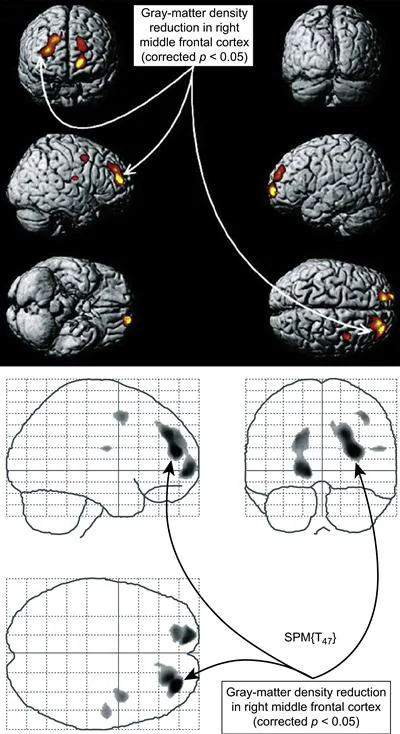
Figure 1 MRI: methamphetamine reduces gray matter. The yellow and red area in the central brain view indicates a reduced gray-matter density in the right middle frontal cortex. The same deficit is shown from other perspectives in the flanking views. Reprinted with permission from Kim et al. (2006).
In another sMRI study, cocaine abusers who had been abstinent for 20 days exhibited a reduced gray-matter density in the regions of the frontal cortex. Interestingly, no differences were found with respect to white matter density (Matochik et al., 2003). With regards to other brain regions, several sMRI studies have shown an enlargement of the brain’s basal ganglia in cocaine-dependent (Jacobsen et al., 2001) and methamphetamine-dependent (Chang et al., 2005; Jernigan et al., 2005) subjects compared with healthy subjects. This is similar to other observations made in schizophrenic subjects who were treated with typical antipsychotics (Gur et al., 1998).
The fact that stimulant drugs, such as cocaine or methamphetamine, and typical antipsychotics that occupy receptors for dopamine in the basal ganglia appear to cause an enlargement of the basal ganglia and are related to psychosis, support the hypothesis that the hyperstimulation of dopamine in basal ganglia structures is involved in psychosis. Finally, an automated morphometric analysis of MR images also showed that a group of chronic methamphetamine abusers had severe gray-matter deficits in cingulate, limbic, and paralimbic cortices. They also had smaller hippocampi than did nondrug abusers of drugs.
The hippocampus is a key site for memory storage, and the volume decrements correlated with a poorer performance on a word recall test (Thompson et al., 2004). Furthermore, sMRI studies have also reported amygdala volume reductions in cocaine addicts (Makris et al., 2004).
Alcohol abuse provides a case study on the utility of MRI to evaluate the structural damage that can result from the chronic use of a psychoactive substance. Investigators using sMRI have reported diminished cortical gray matter, most prominently in the PFC, in alcoholic patients in treatment (Pfefferbaum et al., 1998). In another study, researchers found that alcohol-dependent individuals had reduced whole brain, prefrontal cortical, and parietal cortical gray matter compared with controls (Fein et al., 2002). Two additional studies have shown alcoholics’ frontal cortex and other structures beginning to recover their normal volumes within weeks of stopping drinking (Bendszus et al., 2001; O’Neill et al., 2001; Pfefferbaum et al., 1995) [see Mann et al. (Mann et al., 2001), for a comprehensive review on the brain imaging of alcoholism].
Another MRI study indicated that the amygdala, a brain structure that helps shape our emotional responses to experiences, is relatively smaller in children of alcoholics (Hill et al., 2001; Wrase et al., 2008), a finding that might be a clue to brain-dependent vulnerabilities to alcohol abuse disorders.
2.1.2 Functional MRI
Like sMRI, functional MRI (fMRI) produces images by applying a magnetic field and detecting the radiofrequency energy from the excited protons in water molecules. However, fMRI is an ultrafast technique that can image the whole brain in a second and has the ability to detect changes in the ratio of oxygenated to deoxygenated hemoglobin in the capillary bed of the brain by contrasting task and baseline conditions. Since neurons use oxygen as the main fuel source, this measure turns out to be a reliable proxy for brain activity. In an fMRI image, differences in oxygen content appear as variations in the signal intensity, which is referred to as blood oxygen level-dependent (BOLD) contrast.
In fMRI studies, researchers compare multiple images, which may be of single or different individuals. Images of a single individual taken under varying conditions—for example, at rest and then working on a cognitive task, such as a puzzle, or before and after taking a drug—enable researchers to map which brain regions were activated during the performance of that task or in response to experiences or chemical exposures.
Studies of individuals from different groups—for example, drug-addicted and nonaddicted—can reveal differences in the brain regions that the two groups tap into in order to perform identical tasks or respond to stimuli or exposures. In turn, the differences in brain activity patterns revealed by fMRI provide valuable information on a wide range of issues. For example, studies have correlated regional brain patterns in response to taking a drug with a vulnerability to drug abuse, addictive symptoms and behaviors, and long-term cognitive capacity.
Increasingly, fMRI is being used to investigate the pattern of interactions associated with a given task and how these differed as a function of performance and intersubject variability. This change in the emphasis, from the identification of specific brain region toward the identification of networks (regions working together) reflect the understanding that any given process in the brain results from the complex inter...
Table of contents
- Cover image
- Title page
- Table of Contents
- Copyright
- List of Contributors
- Chapter One. Neuroimaging of Addiction
- Chapter Two. Brain PET Imaging in the Cannabinoid System
- Chapter Three. Brain Imaging of Cannabinoid Receptors
- Chapter Four. Human Brain Imaging of Opioid Receptors: Application to CNS Biomarker and Drug Development
- Chapter Five. Brain Imaging of Sigma Receptors
- Chapter Six. Human Brain Imaging of Acetylcholine Receptors
- Chapter Seven. Human Brain Imaging of Adenosine Receptors
- Chapter Eight. Human Brain Imaging of Dopamine D1 Receptors
- Chapter Nine. Human Brain Imaging of Dopamine Transporters
- Chapter Ten. Imaging of Dopamine and Serotonin Receptors and Transporters
- Chapter Eleven. Imaging the Dopamine D3 Receptor In Vivo
- Chapter Twelve. Dopamine Receptors and Dopamine Release
- Chapter Thirteen. Dopamine Receptor Imaging in Schizophrenia: Focus on Genetic Vulnerability
- Chapter Fourteen. Human Brain Imaging in Tardive Dyskinesia
- Chapter Fifteen. Human Brain Imaging of Autism Spectrum Disorders
- Chapter Sixteen. Radiotracers Used to Image the Brains of Patients with Alzheimer’s Disease
- Chapter Seventeen. Human Brain Imaging of Anger
- Chapter Eighteen. Imaging Pain in the Human Brain
- Chapter Nineteen. Imaging of Neurochemical Transmission in the Central Nervous System
- Chapter Twenty. Characterizing Recovery of the Human Brain following Stroke: Evidence from fMRI Studies
- Index
Frequently asked questions
Yes, you can cancel anytime from the Subscription tab in your account settings on the Perlego website. Your subscription will stay active until the end of your current billing period. Learn how to cancel your subscription
No, books cannot be downloaded as external files, such as PDFs, for use outside of Perlego. However, you can download books within the Perlego app for offline reading on mobile or tablet. Learn how to download books offline
Perlego offers two plans: Essential and Complete
- Essential is ideal for learners and professionals who enjoy exploring a wide range of subjects. Access the Essential Library with 800,000+ trusted titles and best-sellers across business, personal growth, and the humanities. Includes unlimited reading time and Standard Read Aloud voice.
- Complete: Perfect for advanced learners and researchers needing full, unrestricted access. Unlock 1.5M+ books across hundreds of subjects, including academic and specialized titles. The Complete Plan also includes advanced features like Premium Read Aloud and Research Assistant.
We are an online textbook subscription service, where you can get access to an entire online library for less than the price of a single book per month. With over 1.5 million books across 990+ topics, we’ve got you covered! Learn about our mission
Look out for the read-aloud symbol on your next book to see if you can listen to it. The read-aloud tool reads text aloud for you, highlighting the text as it is being read. You can pause it, speed it up and slow it down. Learn more about Read Aloud
Yes! You can use the Perlego app on both iOS and Android devices to read anytime, anywhere — even offline. Perfect for commutes or when you’re on the go.
Please note we cannot support devices running on iOS 13 and Android 7 or earlier. Learn more about using the app
Please note we cannot support devices running on iOS 13 and Android 7 or earlier. Learn more about using the app
Yes, you can access Imaging of the Human Brain in Health and Disease by Philip Seeman,Bertha Madras in PDF and/or ePUB format, as well as other popular books in Biological Sciences & Neurology. We have over 1.5 million books available in our catalogue for you to explore.