- 410 pages
- English
- ePUB (mobile friendly)
- Available on iOS & Android
eBook - ePub
About this book
Biomaterials: A Systems Approach to Engineering Concepts provides readers with a systems approach to biomaterials and materials engineering. By focusing on the mechanical needs of implants, disease states, and current clinical needs, readers are encouraged to design materials and systems targeted at specific conditions, and to identify the impact of their proposed solutions.
This inventive text is a useful resource for researchers, students, and course providers of biomaterials and biomedical engineering.
- Provides a fully comprehensive treatment relating to the construction and use of materials in medicine
- Presents perspectives of disease states to encourage the design of materials and systems targeted at specific conditions
- Defines current issues experienced by clinics to enable optimized engineering solutions
Trusted by 375,005 students
Access to over 1 million titles for a fair monthly price.
Study more efficiently using our study tools.
Information
Chapter 1
Cell Biology
Abstract
The goal of this chapter is to present the reader with a working knowledge of cell types and distinctions, compositional make up of cells, and descriptions of activities that cells perform. The rationale for understanding cell anatomy and physiology is due to the fact that disease can arise from cell dysfunction, and any synthetic intervention leads to cells that ultimately encounter these synthetic, hybrid, and transplanted materials. It is important to create relevant classifications of specialized cell types and to distinguish between functional physiology and pathology or disease. Ultimately bioengineered solutions using materials require some haptic interface with biological tissue and by colocation, cells as well. There are multiple complete books that are dedicated to this one topic. Those are terrific references for this chapter. There are great image resources on the web that can supplement the bulk of the text. The presentation of this chapter coupled with Chapter 2, Cell Expression: Proteins and Their Characterization, tied to proteins and amino acids should help engineers reading this book appreciate the more detailed nuances of normal cell physiology, and allow readers to possess a deeper language linked with cell biology in the context of biomaterials used to replace tissue and organ function, and to seamlessly interface with viable tissues.
Keywords
Cell nomenclature; classifications; cell function; mitosis; motility; communication; trafficking; apoptosis; nucleus; mitochondria
Learning Objectives
By reading this chapter, the reader should be able to
1. Recognize the various naming conventions for cells including functions, tissues from which they are derived, morphological features, and what they ultimately produce as proteins.
2. Describe features and subunits of typical eukaryotic cells which include mitochondria, the nucleus, the cytosol, the lipid membrane, and other structural features that are tied to the cytoskeleton and to the glycocalyx.
3. Understand functions of cells, which include cell division, protein expression, extracellular sensing and communicating within the environment, assembly into larger tissue constructs, and cell death, whether by apoptosis, cell rupture, or some other mechanism.
4. Recognize cell types contained within blood, tissues, and organs.
1.1 Introduction
The genesis of living matter is regulated and organized by cellular production, encoded by different genetic sequences by cells in the presence of local tissues and fluids. There are a number of different classifications for cells in terms of their shape, function, location, and origin and these differences are noted in terms of their activities. The goal here is not necessarily to create a stand-alone cell biology textbook within this treatise. Other books are both more comprehensive and objectively, just better. There are some exemplary images and micrographs to represent detailed structural features of different cell types, organelles, and ensembles thereof. The goal here is to classify cells based on their attributes, functions, and influences sufficiently for bioengineers, and other interested scientists and engineers who have a working knowledge that is useful. A second goal is for readers to have sufficient background so that they can effectively communicate with collaborating life scientists using a more common language. Here the focus is also to identify both morphological features and chemical characteristics of normal eukaryotic cells, followed by a broader description of cellular physiology including the mechanics of cellular function.
1.2 Cell Composition and Make-Up
Normal eukaryotic cells are highly organized and regulated structures and are shown schematically in Fig. 1.1. Eukaryotic (mammalian) cells are distinguished by a lipid bilayer that separates them from their environment. They contain a series of organelles (machinery) to drive cellular function. One such organelle is the nucleus that houses the genetic encoding (DNA, chromatin) and is contained by a separate lipid membrane isolating the DNA from the other organelles contained by the larger cell membrane. Examples of organelle machinery include the mitochondria that convert stored chemical energy channeled through adenosine triphosphate (ATP) into useable energy for cellular function, the endoplasmic reticulum (ER) that is involved in protein synthesis, and the Golgi apparatus that is involved in sorting and protein separations. Each distinct region is partitioned as a separate entity and identified as a different spatial location of the cell through microscopy. The cytosol makes up the fluid fractions of the cell outside of these functional domains with the proteins shuttled from one domain to the next as part of the production cycle. Also contained with the cell are filamentous proteins that compose the cytoskeleton. As a result, the cell has a mechanical structure with mechanical properties such as stiffness and recovery due to the equilibrium structure of the cytoskeleton. Pushing on a cell takes force to sustain the compression and cell retraction results upon unloading from the perturbed state. Contained both inside and outside of the cell and bound to the lipid bilayer are the so-called receptor molecules. Receptor proteins protrude either into the ECM or into the cell and are used to probe the external environment, communicate with neighboring cells, and respond through what is called receptor-binding links to trigger specific physiologic consequences.

1.2.1 The Nucleus
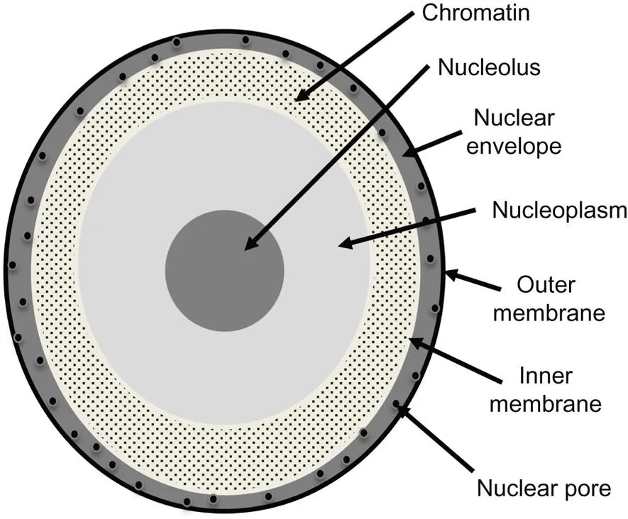
The nucleus of a cell is also bound by an internal lipid bilayer that separates the genetic encoding sequences (transcription) from the rest of the cytoplasm, as shown in Fig. 1.2. The nucleus is often the largest and most easily identifiable organelle in a cell. Functions within the nucleus include controlling gene expression and mediating the replication of DNA during the cell cycle. Mutations in cellular DNA that lead to protein defects and the failure to control DNA replication usually trigger either apoptotic cell death during the normal cellular function or the formation of a cancer cell that grows in an uncontrolled fashion into a larger tumor mass.
1.2.2 The Endoplasmic Reticulum, ER
The ER houses a network of cisternae (sac-like structures) fixtured and shaped by the cytoskeleton, as shown in Fig. 1.3. A lipid bilayer isolates the cisternal space (or lumen) from the fluid region of the cell identified as the cytosol. The ER is involved in protein synthesis as the ribosomes are contained within a morphologically distinct region called the rough ER. The specific role of each ER is cell dependent, as not all cells produce proteins that are commonly transported (exocytosed) out of the cell. The ER ultimately sorts proteins that are conveyed out of the ER usually through a series of vesicles, lipid bilayers surrounding the secreted proteins. Ultimately, proteins are internally digested, internally sequestered, routed to the cell membrane, or exocytosed outside of the cell.
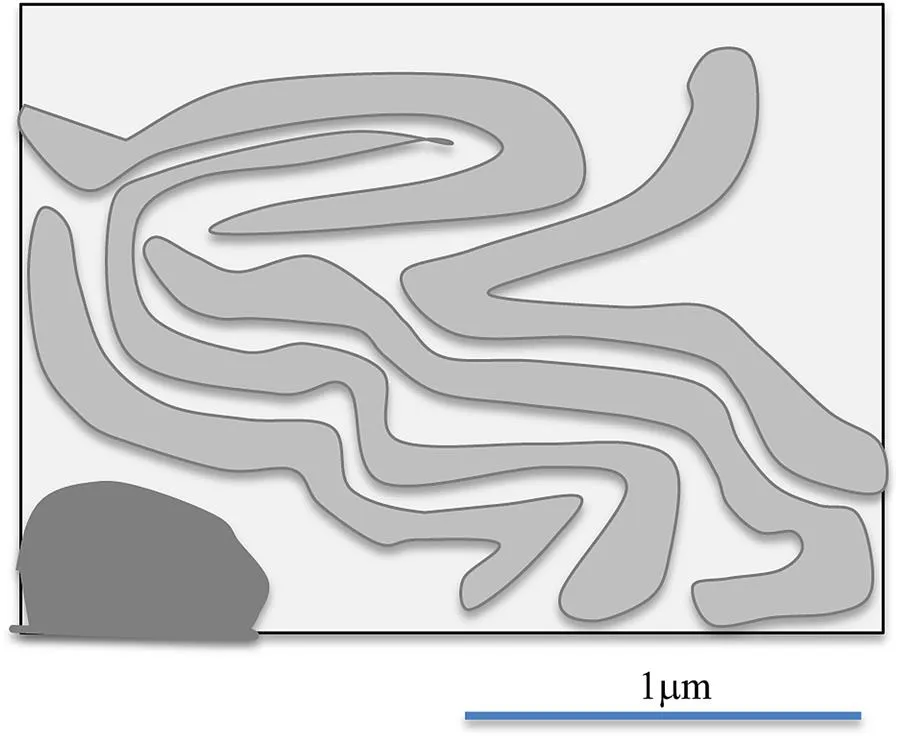
Protein folding occurs in the ER that is regulated by amino acid sequencing, the local charge, the presence of chaperone molecules to control local charge and ionic strength to drive the appropriate conformational shape. Defects linked with protein synthesis (primary amino acid structure) and folding (tertiary and quaternary structure) are often conveyed for digestion, while correct shapes are expressed appropriately. Inappropriately folded or amyloid protein structures that are expressed are the current focus of a range of age-related diseases called amyloid diseases including Alzheimer’s disease, Parkinson’s disease, Huntington’s disease, and a host of tissue amyloidosis related diseases that are often encountered in the last hours of life [1].
1.2.3 Mitochondria
Mitochondria drive energy conversion in cells due to their production of ATP, used as a primary cellular source of chemical energy, as shown conceptually in Fig. 1.4. Individual cells can have multiple mitochondria which are also involved in a range of other cellular functions associated with the cell cycle, cell signaling, and apoptosis.

1.2.4 The Golgi Apparatus
The Golgi apparatus is an organelle downstream from the ER. Vesicles containing proteins are fused to the input region of the Golgi that causes the vesicle to break down and helps to convey the now liberated proteins into the entering lumen of the Golgi, as shown in Figs. 1.5 and 1.6. Once separated from the vesicles, many proteins are grafted with hydrophilic phosphates (a process called phosphorylation) and carboxylic acids (glycosylation). Specific glycosylation or phosphorylation steps are kinds of labeling exercises that facilitate protein separations and to redirect proteins destined for different fates. There is often a small change in the molecular weight of the protein as a result of the grafting, which are discussed in Chapter 2, Cell Expression: Proteins and Their Characterization.
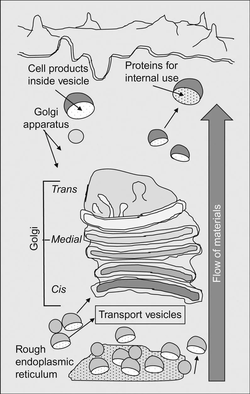
Table of contents
- Cover image
- Title page
- Table of Contents
- Copyright
- Dedication
- Author Bio
- Preface
- Acknowledgments
- Chapter 1. Cell Biology
- Chapter 2. Cell Expression: Proteins and Their Characterization
- Chapter 3. Bones and Mineralized Tissues
- Chapter 4. Connective and Soft Tissues
- Chapter 5. Property Assessments of Tissues
- Chapter 6. Environmental Effects on Natural Tissues
- Chapter 7. Metallic Biomaterials
- Chapter 8. Ceramic Biomaterials
- Chapter 9. Polymeric Biomaterials
- Chapter 10. Nanomaterials and Phase Contrast Imaging Agents
- Chapter 11. Orthopedics
- Chapter 12. Neural Interventions
- Chapter 13. Cardiovascular Interventions
- Chapter 14. Artificial Organs
- Chapter 15. Special Topics: Assays Applied to Both Health and Sports
- Postface
- Index
Frequently asked questions
Yes, you can cancel anytime from the Subscription tab in your account settings on the Perlego website. Your subscription will stay active until the end of your current billing period. Learn how to cancel your subscription
No, books cannot be downloaded as external files, such as PDFs, for use outside of Perlego. However, you can download books within the Perlego app for offline reading on mobile or tablet. Learn how to download books offline
Perlego offers two plans: Essential and Complete
- Essential is ideal for learners and professionals who enjoy exploring a wide range of subjects. Access the Essential Library with 800,000+ trusted titles and best-sellers across business, personal growth, and the humanities. Includes unlimited reading time and Standard Read Aloud voice.
- Complete: Perfect for advanced learners and researchers needing full, unrestricted access. Unlock 1.4M+ books across hundreds of subjects, including academic and specialized titles. The Complete Plan also includes advanced features like Premium Read Aloud and Research Assistant.
We are an online textbook subscription service, where you can get access to an entire online library for less than the price of a single book per month. With over 1 million books across 990+ topics, we’ve got you covered! Learn about our mission
Look out for the read-aloud symbol on your next book to see if you can listen to it. The read-aloud tool reads text aloud for you, highlighting the text as it is being read. You can pause it, speed it up and slow it down. Learn more about Read Aloud
Yes! You can use the Perlego app on both iOS and Android devices to read anytime, anywhere — even offline. Perfect for commutes or when you’re on the go.
Please note we cannot support devices running on iOS 13 and Android 7 or earlier. Learn more about using the app
Please note we cannot support devices running on iOS 13 and Android 7 or earlier. Learn more about using the app
Yes, you can access Biomaterials by Brian J. Love in PDF and/or ePUB format, as well as other popular books in Technology & Engineering & Biotechnology in Medicine. We have over one million books available in our catalogue for you to explore.