eBook - ePub
The Choroid Plexus and Cerebrospinal Fluid
Emerging Roles in CNS Development, Maintenance, and Disease Progression
- 210 pages
- English
- ePUB (mobile friendly)
- Available on iOS & Android
eBook - ePub
The Choroid Plexus and Cerebrospinal Fluid
Emerging Roles in CNS Development, Maintenance, and Disease Progression
About this book
The Choroid Plexus and Cerebrospinal Fluid: Emerging Roles in CNS Development, Maintenance, and Disease Progression combines new and established work to allow for cross-disciplinary discussion and showcase newfound excitement surrounding the choroid plexus and cerebrospinal fluid (CSF). This book is of great utility to neuroscientists interested in biological questions about cancer, multiple sclerosis, Alzheimer's, choroid plexus, or CSF research, and especially for researchers looking to expand their research into later stages of their disease of interest, such as metastasis. No other resource is currently available which addresses these issues in this fashion. The focus on the choroid plexus provides a practical resource on modeling clinical issues influenced by this brain region for researchers from students to principal investigators.
- Presents recent progress made in the research of choroid plexus and cerebrospinal fluid across multi-disciplinary fields, including neuroscience, cancer biology, and immunology
- Includes numerous illustrations of light, fluorescent, and electron micrographs
- Provides data analysis boxes in each chapter to help with data interpretation and offer guidelines on how best to represent results
- Includes chapters written by prominent researchers in the field
Trusted by 375,005 students
Access to over 1.5 million titles for a fair monthly price.
Study more efficiently using our study tools.
Information
Chapter 1
Introduction to the Ventricular System and Choroid Plexus
Tatsuhiro Fujii*
Joshua Youssefzadeh*,†
Michael Novel**
Josh Neman*
* Department of Neurosurgery, Keck School of Medicine, University of Southern California, Los Angeles, CA, USA
† Department of Health Promotion and Disease Prevention, Keck School of Medicine of USC, Los Angeles, CA, USA
** Department of History, University of California at Los Angeles, Los Angeles, CA, USA
Abstract
Despite our ever-increasing understanding of the central nervous system and intricacies that allow human behavior and functioning, certain areas of the brain have received much less attention. One such region of the brain is the choroid plexus, a highly vascularized structure situated in each of the ventricles and crucial in maintaining homeostasis through cerebrospinal fluid secretion. We begin this introductory chapter of the choroid plexus by reviewing the concepts of early central nervous system development with a particular focus on the formation of the ventricular system. We also discuss the formation of the choroid plexus as well as the physiology behind cerebrospinal fluid production. Finally, we review the vascular supply to the choroid plexus and some of the pathologies that can occur should this be compromised.
Keywords
development
cerebral spinal fluid
vascular supply
embryonic cerebral spinal fluid
lateral ventricle formation
Development of the ventricular system
Within the first 4 weeks of human development, the formation of the central nervous system (CNS) has begun to take shape in the form of the neural tube. From within this enclosed cavity emerge the future ventricles of the brain as well as the central canal of the spinal cord. As the primitive neural tube continues to enlarge in way of rapid cell division, the appearance of the pontine flexure and diencephalic–telencephalic sulcus gives rise to five distinctive vesicles namely, the telencephalon, diencephalon, mesencephalon, metencephalon, and myelencephalon. Differing rates of cell division of each vesicle results in the transformation of a cylindrical neural tube into a more complex, folded structure. This in turn influences the size and shape of the cavities of each of the five divisions, which gives rise to their respective parts of the ventricular system. As the cerebral hemispheres continue to expand, so do the lateral ventricles, which are in close association. The lateral ventricles communicate with the single and narrow third ventricle, through the interventricular foramina of Monro. As the cells of mesencephalon continue to divide, the ventricular cavity is reduced in size giving rise to a narrowed cerebral aqueduct, which connects the third and fourth ventricle. With the closures of the rostral and caudal neuropores early in embryogenesis, the neural tube space gives rise to an enclosed ventricular system. By the third month of fetal development, foramina appear within the roof of the fourth ventricle that allows communication between the once closed ventricular system and the surrounding subarachnoid space. As the layer of connective tissue and ependymal cells that line the fourth ventricle, begin to break down, this gives rise to the formation of the three openings: a single, medial foramen of Magendie and two, lateral foramina of Luschka (Fig. 1.1).
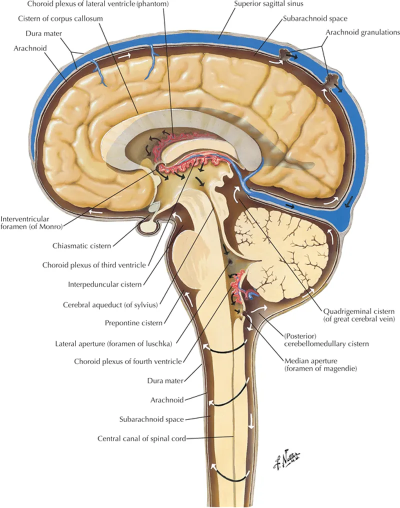
Figure 1.1 CSF flow through the ventricular system.
CSF produced from the choroid plexus flow from the lateral ventricles to the third ventricle through the interventricular foramina of Monro. From the third ventricle, CSF flows through the cerebral aqueduct and into the fourth ventricle. From here, CSF can continue further into the central canal of the spinal cord or into the subarachnoid space through the foramen of Magendie and foramina of Luschka. Netter medical illustration used with permission of Elsevier Inc. Copyright 2016. All rights reserved. www.netterimages.com.
CSF produced from the choroid plexus flow from the lateral ventricles to the third ventricle through the interventricular foramina of Monro. From the third ventricle, CSF flows through the cerebral aqueduct and into the fourth ventricle. From here, CSF can continue further into the central canal of the spinal cord or into the subarachnoid space through the foramen of Magendie and foramina of Luschka. Netter medical illustration used with permission of Elsevier Inc. Copyright 2016. All rights reserved. www.netterimages.com.
Despite our increasing understanding of the development of the CNS, the function and purpose of the ventricular system is yet to be fully comprehended. Following the development of the embryonic forebrain, midbrain, and hindbrain ventricle formation, these ventricles expand at a much more rapid rate than brain tissue, thus making ventricle volume notably faster in growth.1 Research on the molecular and cellular mechanism gives more insight into the brain ventricular system. Formation of the ventricles is dependent upon the neuroepithelium.2 The surrounding neuroepithelium gives position and shape to the developing embryonic brain ventricular system. The neuroepithelium is arranged along the anteroposterior axis. With this pattern of placement, correct positioning of the ventricles is allowed and morphogenesis of the brain tissue is directed downstream. The arrangement of neuroepithelium occurs before and during neurulation.2 During this period, embryonic brain tissue is subdivided into various gene expression domains. Patterning genes are responsible for the positioning of brain ventricles, including the characteristic as well as conserved constrictions and bends within each region of the brain. The patterning genes may be responsible prox...
Table of contents
- Cover
- Title page
- Table of Contents
- Copyright
- Dedication
- List of Contributors
- About the Editors
- Preface
- Acknowledgment
- List of Abbreviations
- Chapter 1: Introduction to the Ventricular System and Choroid Plexus
- Chapter 2: Development of Brain Ventricles and Choroid Plexus
- Chapter 3: Choroid Plexus: Structure and Function
- Chapter 4: Toward an Artificial Choroid Plexus, Concept and Clinical Implications
- Chapter 5: Choroid Plexus Tumors
- Chapter 6: Role of Blood–Brain Barrier, Choroid Plexus, and Cerebral Spinal Fluid in Extravasation and Colonization of Brain Metastases
- Chapter 7: The Role of the Choroid Plexus in the Pathogenesis of Multiple Sclerosis
- Chapter 8: The Choroid Plexus and Cerebrospinal Fluid System: Roles in Neurodegenerative Diseases
- Chapter 9: Delivery Considerations for Targeting the Choroid Plexus–Cerebrospinal Fluid Route
- Glossary
- Subject Index
Frequently asked questions
Yes, you can cancel anytime from the Subscription tab in your account settings on the Perlego website. Your subscription will stay active until the end of your current billing period. Learn how to cancel your subscription
No, books cannot be downloaded as external files, such as PDFs, for use outside of Perlego. However, you can download books within the Perlego app for offline reading on mobile or tablet. Learn how to download books offline
Perlego offers two plans: Essential and Complete
- Essential is ideal for learners and professionals who enjoy exploring a wide range of subjects. Access the Essential Library with 800,000+ trusted titles and best-sellers across business, personal growth, and the humanities. Includes unlimited reading time and Standard Read Aloud voice.
- Complete: Perfect for advanced learners and researchers needing full, unrestricted access. Unlock 1.5M+ books across hundreds of subjects, including academic and specialized titles. The Complete Plan also includes advanced features like Premium Read Aloud and Research Assistant.
We are an online textbook subscription service, where you can get access to an entire online library for less than the price of a single book per month. With over 1.5 million books across 990+ topics, we’ve got you covered! Learn about our mission
Look out for the read-aloud symbol on your next book to see if you can listen to it. The read-aloud tool reads text aloud for you, highlighting the text as it is being read. You can pause it, speed it up and slow it down. Learn more about Read Aloud
Yes! You can use the Perlego app on both iOS and Android devices to read anytime, anywhere — even offline. Perfect for commutes or when you’re on the go.
Please note we cannot support devices running on iOS 13 and Android 7 or earlier. Learn more about using the app
Please note we cannot support devices running on iOS 13 and Android 7 or earlier. Learn more about using the app
Yes, you can access The Choroid Plexus and Cerebrospinal Fluid by Josh Neman,Thomas C. Chen in PDF and/or ePUB format, as well as other popular books in Biological Sciences & Neuroscience. We have over 1.5 million books available in our catalogue for you to explore.