- 194 pages
- English
- ePUB (mobile friendly)
- Available on iOS & Android
eBook - ePub
Implantable Electronic Medical Devices
About this book
Implantable Electronic Medical Devices provides a thorough review of the application of implantable devices, illustrating the techniques currently being used together with overviews of the latest commercially available medical devices. This book provides an overview of the design of medical devices and is a reference on existing medical devices.
The book groups devices with similar functionality into distinct chapters, looking at the latest design ideas and techniques in each area, including retinal implants, glucose biosensors, cochlear implants, pacemakers, electrical stimulation therapy devices, and much more. Implantable Electronic Medical Devices equips the reader with essential background knowledge on the application of existing medical devices as well as providing an introduction to the latest techniques being used.
- A catalogue of existing implantable electronic medical devices
- Up-to-date information on the design of implantable electronic medical devices
- Background information and reviews on the application and design of up-to-date implantable electronic medical devices
Trusted by 375,005 students
Access to over 1.5 million titles for a fair monthly price.
Study more efficiently using our study tools.
Information
Chapter 1
Retinal Implants
This chapter introduces the main anatomical features of the eye, the processes involved in vision and eye disorders, and diseases such as retinitis pigmentosa and age-related macular degeneration which can affect normal vision. The use of microelectrodes and microphotodiodes in retinal, subretinal, and suprachoroidal implants is introduced with an emphasis on the Argus II Retinal Prosthesis (Second Sight Medical Products); the Artificial Silicon Retina (Optobionics); Alpha-IMS (Retina Implant AG); Wide-View BVA and High-Acuity BVA (Bionic Vision Australia); and Boston Retinal Implant Project (Bionic Eye Technologies, Inc. and Visus Technologies, Inc.).
Keywords
Retina; retinitis pigmentosa; AMD; microphotodiodes; microelectrodes; epiretinal; subretinal; suprachoroidal
1.1 Introduction
Figure 1.1 shows the main anatomical features of the eye. In normal sight, light enters the eye through the pupil and is focused onto the retina at the back of the eye, stimulating photocells that translate the light into electrical signals. These electrical signals travel down the optic nerve to the visual centers in the brain where they are decoded and perceived as images. Progressive diseases of the eye that result in partial or total loss of vision include glaucoma, retinitis pigmentosa, and macular degeneration.

Glaucoma results from an increase in the internal pressure of the eye, the effects of which are irreversible, eventually leading to loss of sight. However, if detected early, the onset of the disease can be managed with medical treatment or laser surgery. Measuring the intraocular pressure of the eye can help in detecting the early stages of the disease (see Chapter 2).
Retinitis pigmentosa is a genetic disorder resulting in the degeneration of the photoreceptor cells in the retina, leading to partial or complete loss of sight. Currently there is no cure, although gene therapy in which a virus is used to deliver sight-restoring therapeutic genes to the photoreceptors at the back of the eye may offer an alternative form of treatment in the future.
Age-related macular degeneration (AMD) is another disease of the retina, but it only affects a small area of the retina known as the macula which contains a small population of cone-type photoreceptor cells that are more responsive to bright light levels required for reading and viewing objects close up and in greater detail. The onset of AMD occurs in the later stages of life and only leads to a partial degeneration of sight.
Retinal implants are used to help people with degenerative retinal diseases such as retinitis pigmentosa and AMD where the optic nerve and the visual centers in the brain are still functioning but the patient has lost light or sight perception due to degeneration of the outer layer of the retinal photoreceptor cells. However, the cells in the inner retinal layer are relatively intact compared to the outer cells and it is the inner cells which form a neuronal ganglion interface to the optical nerve. Retinal implants will not benefit people who have been blind from birth because their optical visual neuronal circuits and visual processing centers in the brain have not been developed or conditioned to perceive vision.
1.2 The Retina
Light entering the eye through the lens is focused onto the retina which consists of a thin layer of transparent neural tissue located at the back of the eye. Near the center of the retina is a region known as the macula which has a high concentration of neural cells responsible for seeing detailed colors and represents the center of vision. At the center of the macula is a small depression or dimple known as the fovea which represents the absolute center of vision and highest color resolution attainable, providing the clearest and sharpest images. Subsequently, the eye continuously moves (saccades) such that the lens focuses images of interest onto the fovea for the highest image of color resolution.
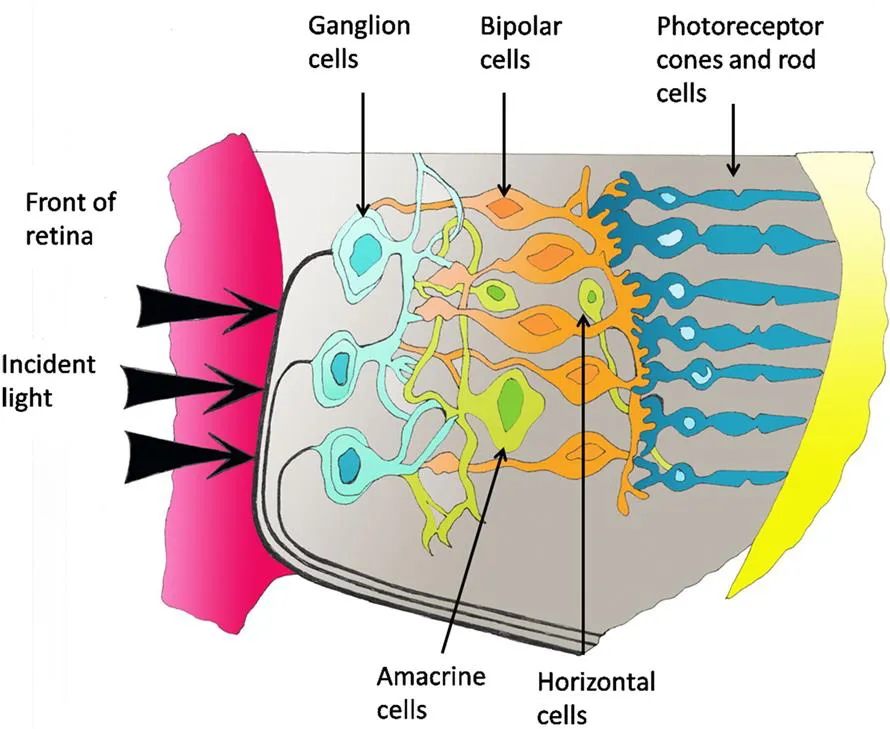
The retina is made up of three main functional neural cell layers: photoreceptor cells, bipolar cells, and ganglion cells. Interspersed between the layers are the horizontal and amacrine neural cells as shown in Figure 1.2. The photoreceptor cells at the back of the retina transduce photon light energy into graded neural signals which are transmitted and processed via the bipolar and ganglion cell layers. It is the axons of the ganglion cells which together collectively form the optic nerve which leads to the visual processing centers in the brain.
1.3 Photoreceptor Cells
There are two types of photoreceptor cells: rods, which have the ability to detect color but are sensitive to low light levels (scotopic vision), and cones, which in bright light are sensitive to colors (photopic vision) in the visible spectrum. The rods and cones are made up of four segments (Figure 1.3): the outer segment, inner segment, cell body (nucleus), and synaptic terminals.
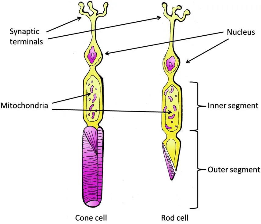
The outer segment in rods and cones consists of the outer membrane folding in on itself and stacking up to form disks. In the case of rods, the in-folded membranes become detached and the disks float inside the outer segment. Located on the disks are light-sensitive pigment proteins, rhodopsin in rods, and iodopsin in cones. The inner segment contains mitochondria which provide the energy required for chemical reactions and the cell body which contains the cell nucleus and other cell organelles essential to maintain cell functionality. The synaptic terminals provide for the transmission of glutamate neurotransmitters between neural cell synaptic bodies.
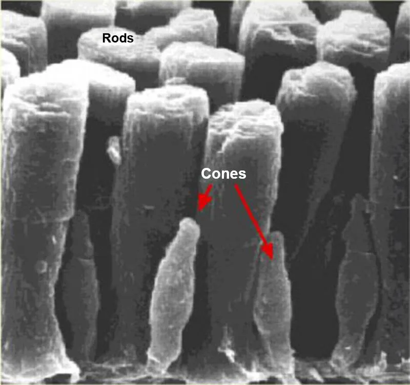
In rods, the outer segment is cylindrical, whereas for cones, the outer segment is conical in shape (Figure 1.4). Typical outside diameters for the inner and outer segments are 2 µm for rods and 6 µm for cones. The rods also contain a greater number of light-sensitive disks in the outer segment compared to cones, resulting in a greater sensitivity to light. There are typically 120 million rods compared to 6 million cones in the retina.
In rods, all the disks contain the same light-sensitive pigment, rhodopsin, which exhibits a peak absorption of light energy at a wavelength of 500 nm which lies within the blue-green region of the visual light spectrum. In cones, the light-sensitive iodopsin pigment occurs in three varieties due to differences in their amino acid sequence, each with different peak absorption wavelengths in the red (560 nm), blue (420 nm), and green (530 nm) regions of the visible light spectrum, respectively.
Although each cone contains three different opsin pigment types, there are three different types of defined cones: short-wave (blue light), medium-wave (green light), and long-wave (red light), each with a predominant opsin variety in the cone. The superimposition of the light absorption response of each opsin pigment will result in a peak response around the area of the defined cone color type. For example, the peak response of a long-wave cone will be shifted due to the superimposition of the individual blue and green opsin spectrum absorption responses, toward the yellow-green region of the visible spectrum as shown in Figure 1.5.

Figure 1.6 shows a rod photoreceptor cell with sodium- and potassium-specific ion channels in the outer membrane. In the absence of light, there will be a continuous flow of positively charged sodium ions into the cell and potassium ions out of the cel...
Table of contents
- Cover image
- Title page
- Table of Contents
- Copyright
- Preface
- Chapter 1. Retinal Implants
- Chapter 2. Smart Contact Lens
- Chapter 3. Phrenic Nerve Stimulation
- Chapter 4. Glucose Biosensors
- Chapter 5. Cochlear Implants
- Chapter 6. Pacemakers and Implantable Cardioverter Defibrillators
- Chapter 7. Bladder Implants
- Chapter 8. Electrical Stimulation Therapy for Pain Relief and Management
- Chapter 9. Electrical Stimulation Therapy for Parkinson’s Disease and Dystonia
- Chapter 10. Electrical Stimulation Therapy for Epilepsy
- Chapter 11. Peripheral Nerve Stimulation
- Chapter 12. Lower Esophagus Stimulator
- Chapter 13. Vagal Blocking Therapy
- Chapter 14. Implantable Drug Delivery Systems
- Chapter 15. Wireless Endoscopy Capsules
- Index
Frequently asked questions
Yes, you can cancel anytime from the Subscription tab in your account settings on the Perlego website. Your subscription will stay active until the end of your current billing period. Learn how to cancel your subscription
No, books cannot be downloaded as external files, such as PDFs, for use outside of Perlego. However, you can download books within the Perlego app for offline reading on mobile or tablet. Learn how to download books offline
Perlego offers two plans: Essential and Complete
- Essential is ideal for learners and professionals who enjoy exploring a wide range of subjects. Access the Essential Library with 800,000+ trusted titles and best-sellers across business, personal growth, and the humanities. Includes unlimited reading time and Standard Read Aloud voice.
- Complete: Perfect for advanced learners and researchers needing full, unrestricted access. Unlock 1.5M+ books across hundreds of subjects, including academic and specialized titles. The Complete Plan also includes advanced features like Premium Read Aloud and Research Assistant.
We are an online textbook subscription service, where you can get access to an entire online library for less than the price of a single book per month. With over 1.5 million books across 990+ topics, we’ve got you covered! Learn about our mission
Look out for the read-aloud symbol on your next book to see if you can listen to it. The read-aloud tool reads text aloud for you, highlighting the text as it is being read. You can pause it, speed it up and slow it down. Learn more about Read Aloud
Yes! You can use the Perlego app on both iOS and Android devices to read anytime, anywhere — even offline. Perfect for commutes or when you’re on the go.
Please note we cannot support devices running on iOS 13 and Android 7 or earlier. Learn more about using the app
Please note we cannot support devices running on iOS 13 and Android 7 or earlier. Learn more about using the app
Yes, you can access Implantable Electronic Medical Devices by Dennis Fitzpatrick in PDF and/or ePUB format, as well as other popular books in Technology & Engineering & Biomedical Science. We have over 1.5 million books available in our catalogue for you to explore.