eBook - ePub
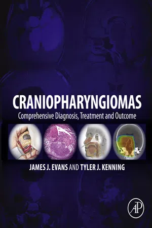
Craniopharyngiomas
Comprehensive Diagnosis, Treatment and Outcome
- 612 pages
- English
- ePUB (mobile friendly)
- Available on iOS & Android
eBook - ePub
About this book
Craniopharyngiomas: A Comprehensive Guide to Diagnosis, Treatment and Outcome is a comprehensive guide to the science, diagnosis and treatment of craniopharyngiomas, rare brain tumors that grow near the pituitary gland. Even though these tumors are generally benign, due to the location, prognosis was often bleak. This reference provides a resource for specialists requiring a comprehensive overview of this rare form of tumor, outlining new diagnostic, imaging and surgical techniques, including endoscopic endonasal cranial base surgery, which enable successful neurosurgical intervention. With a better prognosis, patients require multidisciplinary management from neurology, otolaryngology, radiation oncology, endocrinology, and neuropathology.
- The only comprehensive guide to the science, diagnosis and treatment of craniopharyngiomas
- Includes multidisciplinary management from neurology, otolaryngology, radiation oncology, endocrinology, and neuropathology
- Detailed coverage of the impacts of new diagnostic, imaging and neurosurgical techniques including endoscopic endonasal cranial base surgery
Information
Section III
Surgical Approaches
Chapter 7
Surgical Approaches
Introduction and Outcomes
Paolo Cappabianca∗, Luigi Maria Cavallo∗, Domenico Solari∗, Alberto Di Somma∗, and Maria Laura Del Basso De Caro† ∗Department of Neurosciences and Reproductive and Odontostomatological Sciences, Division of Neurosurgery, Università degli Studi di Napoli Federico II, Naples, Italy †Department of Advanced Biomedical Sciences, Division of Pathology, Università degli Studi di Napoli Federico II, Naples, Italy
Abstract
Craniopharyngiomas account for 1.2 to 4.6% of all intracranial tumors, and were first described in 1857 by Friedrich Albert von Zenker. They exist as two subtypes, namely adamantinomatous and papillary, with the former occurring in children and adults, but the form is seen almost exclusively in adults. Craniopharyngiomas involve the sella and parasellar areas and can recur even after radical resection.
Over the years, several authors have defined classifications for craniopharyngiomas in terms of their growth path and the surgical route used. Fronto-temporal and transsphenoidal approaches are used, with the aid of the microscope and endoscope. Radiation treatment is usually reserved for residual or recurrent craniopharyngiomas.
The outcome of primary surgery for craniopharyngiomas is affected by different factors such as age, clinical conditions, and previous treatment. Visual and endocrinological complications may persist.
Keywords
adamantinomatous; craniopharyngioma; endoscope; fronto-temporal approach; microscope; papillary; radiation treatment; transsphenoidal approach; tumor“The most forbidding of the intracranial tumors.”
Harvey Cushing, 1932
Introduction
Craniopharyngiomas are disembryogenetic tumors arising from the squamous epithelial remnants of Rathke’s pouch and, for this reason, can arise from its whole pathway, from the rhinopharynx to the hypothalamus (Jane and Laws, 2006; Karavitaki, 2014). They tend to involve a great number of vital neurovascular structures, including the optic nerves, internal carotid arteries (ICAs), the third ventricle – above all of its floor – the hypothalamus, and the pituitary gland, causing a variety of symptoms.
Common clinical presentations include visual dysfunction, with symptoms of chiasmatic and/or retrochiasmatic compression, hypothalamic dysfunction, such as behavioral changes, alterations of eating patterns, apathy, or even stupor, and finally pituitary dysfunction, often presenting as panhypopituitarism (Karavitaki, 2014).
These lesions account for 1.2 to 4.6% of all intracranial tumors (Central Brain Tumor Registry of the United States) and exhibit a bimodal distribution, with one peak occurring during childhood (5–14 years of age) and another in late adulthood (50–74 years of age) (Bunin et al., 1998).
Description of a craniopharyngioma was first presented by the German pathologist, Friedrich Albert von Zenker (Zenker, 1857), who in 1857 depicted a cystic suprasellar mass containing cholesterol crystals and squamous epithelium. In 1904, Jakob Erdheim accurately defined the histopathological features of a craniopharyngioma (Erdheim, 1904); according also to previous reports by Mott and Barret (Mott and Barret, 1899), he confirmed that these tumors arise from squamous cell rests originating from a partially involuted hypophyseal-pharyngeal duct.
Rupert Boyce and Cecil Beadles (Boyce and Beadles, 1893) described a 35-year-old blind patient who became comatose and died as a result of a large, partially ossified, cystic lesion – suspected to be a craniopharyngioma – causing brainstem, optic chiasm, and optic tract compression. Subsequently, Babinski (Babinski, 1900) and Frohlich (Frohlich, 1901) reported similar clinical signs in patients with cystic pituitary masses without acromegaly.
From a neuropathological standpoint, two subtypes of craniopharyngioma have been recognized, namely adamantinomatous and papillary. The adamantinomatous form occurs in children (5–10 years) and adults (50–60 years), while the papillary form is seen almost exclusively in adults; generally, the adamantinomatous form is the more common (ratio: 9/1) (Yasargil et al., 1990; Paulus et al., 1997). Regarding macroscopic features, the adamantinomatous subtype shows an irregular interface, adhesions to surrounding structures, and cystic contents have dark “motor oil” fluid, with cholesterol crystals inside; calcification may occur in the majority of cases. In contrast, the papillary form shows no adherence to surrounding structures and/or calcifications, while the cystic contents are often clear (Crotty et al., 1995). The two distinct craniopharyngioma types have different biological behaviors, and immunohistochemical examinations appear to be relevant in order to analyze this issue; as a matter of fact, the adamantinomatous subtype demonstrates positivity for CK7, CK8, CK14 (Kurosaki et al., 2001; Tateyama et al., 2001; Xin et al., 2002), and beta-catenin (Sekine et al., 2002; Hofmann et al., 2006; Holsken et al., 2013). In particular, clusters of cells with nuclear beta-catenin are predominantly represented in invasive tumors. This pattern is suggestive of an involvement of the beta-catenin signaling in the migratory behavior of these tumors; the cellular clusters with nuclear beta-catenin may coordinate the infiltration of the tumor into surrounding tissues, thus explaining the greater aggressiveness of the adamantinomatous tumor histotype (Hussain et al., 2013; Larkin and Ansorge, 2013).
Craniopharyngiomas involve the sella and parasellar areas, but in contrast to pituitary tumors, they often adhere to the neurovascular structures of the suprasellar space, such as perforating vessels from the anterior and posterior cerebral arteries and/or ICAs, the optic chiasm and pathways, the hypothalamus, and third ventricular regions. These close anatomi...
Table of contents
- Cover image
- Title page
- Table of Contents
- Copyright
- Dedications
- List of Contributors
- Foreword
- Preface
- Section I. Introduction
- Section II. Medical Evaluation
- Section III. Surgical Approaches
- Section IV. Radiotherapy
- Section V. Intracavitary Therapy
- Section VI. Management of Recurrence
- Section VII. Special Considerations
- Section VIII. Controversies
- Index
Trusted by 375,005 students
Access to over 1.5 million titles for a fair monthly price.
Study more efficiently using our study tools.
Frequently asked questions
Yes, you can cancel anytime from the Subscription tab in your account settings on the Perlego website. Your subscription will stay active until the end of your current billing period. Learn how to cancel your subscription
No, books cannot be downloaded as external files, such as PDFs, for use outside of Perlego. However, you can download books within the Perlego app for offline reading on mobile or tablet. Learn how to download books offline
Perlego offers two plans: Essential and Complete
- Essential is ideal for learners and professionals who enjoy exploring a wide range of subjects. Access the Essential Library with 800,000+ trusted titles and best-sellers across business, personal growth, and the humanities. Includes unlimited reading time and Standard Read Aloud voice.
- Complete: Perfect for advanced learners and researchers needing full, unrestricted access. Unlock 1.5M+ books across hundreds of subjects, including academic and specialized titles. The Complete Plan also includes advanced features like Premium Read Aloud and Research Assistant.
We are an online textbook subscription service, where you can get access to an entire online library for less than the price of a single book per month. With over 1.5 million books across 990+ topics, we’ve got you covered! Learn about our mission
Look out for the read-aloud symbol on your next book to see if you can listen to it. The read-aloud tool reads text aloud for you, highlighting the text as it is being read. You can pause it, speed it up and slow it down. Learn more about Read Aloud
Yes! You can use the Perlego app on both iOS and Android devices to read anytime, anywhere — even offline. Perfect for commutes or when you’re on the go.
Please note we cannot support devices running on iOS 13 and Android 7 or earlier. Learn more about using the app
Please note we cannot support devices running on iOS 13 and Android 7 or earlier. Learn more about using the app
Yes, you can access Craniopharyngiomas by James J. Evans,Tyler J. Kenning in PDF and/or ePUB format, as well as other popular books in Medicine & Neurology. We have over 1.5 million books available in our catalogue for you to explore.