eBook - ePub
Lung Epithelial Biology in the Pathogenesis of Pulmonary Disease
- 276 pages
- English
- ePUB (mobile friendly)
- Available on iOS & Android
eBook - ePub
Lung Epithelial Biology in the Pathogenesis of Pulmonary Disease
About this book
Lung Epithelial Biology in the Pathogenesis of Pulmonary Disease provides a one-stop resource capturing developments in lung epithelial biology related to basic physiology, pathophysiology, and links to human disease. The book provides access to knowledge of molecular and cellular aspects of lung homeostasis and repair, including the molecular basis of lung epithelial intercellular communication and lung epithelial channels and transporters.
Also included is coverage of lung epithelial biology as it relates to fluid balance, basic ion/fluid molecular processes, and human disease. Useful to physician and clinical scientists, the contents of this book compile the important and most current findings about the role of epithelial cells in lung disease. Medical and graduate students, postdoctoral and clinical fellows, as well as clinicians interested in the mechanistic basis for lung disease will benefit from the books examination of principles of lung epithelium functions in physiological condition.
- Provides a single source of information on lung epithelial junctions and transporters
- Discusses of the role of the epithelium in lung homeostasis and disease
- Includes capsule summaries of main conclusions as well as highlights of future directions in the field
- Covers the mechanistic basis for lung disease for a range of audiences
Trusted by 375,005 students
Access to over 1.5 million titles for a fair monthly price.
Study more efficiently using our study tools.
Information
Topic
MedicineChapter 1
Junctional Interplay in Lung Epithelial Barrier Function
Michael Koval
Abstract
Lung epithelial cells are oriented in order to properly function. Top-bottom orientation, called the apical–basolateral polarity axis, is determined by several cues in the cell microenvironment. Particularly critical are contacts between cells that establish a demarcation zone that defines the border between the apical and basolateral elements of the plasma membrane. This contact zone, called the apical junctional complex (AJC), is an area where the polarity complex proteins interlink with tight junctions, adherens junctions, and gap junctions. Each of these junctions has unique composition and function, moreover, their organization and function is coordinated. Junctions also serve as signaling platforms that regulate the traffic of transcription factors between the plasma membrane and nucleus. This chapter focuses on AJC components that have been best characterized for lung epithelial cells: adherens junctions, that serve as initiators of junction formation and sites that coordinate the actin cytoskeleton, tight junctions that regulate paracellular permeability of small solutes and ions through cell contact sites and gap junctions, communicating junctions that enable cytosolic molecules and ions to transfer from one cell to another.
Keywords
Apical junctional complex; apical; basolateral; epithelia; alveolus; airway; claudin; connexin; cytoskeleton; pulmonary edema; acute respiratory distress syndrome
1.1 Introduction
Epithelial cells line the entire airspace in order to maintain a functional barrier between the outside environment of the atmosphere and inner fluid-filled tissues such as the interstitium and circulatory system. While different zones of the respiratory system have different function and phenotype, lung epithelial cells in general share common features with each other as well as with epithelia present in other tissues. Of particular note are intercellular junctions that enable the individual cells of an epithelial monolayer to coherently function as a unified tissue barrier.
This chapter focuses on the composition, structure, and function of different classes of intercellular junctions found in lung epithelia: adherens junctions, tight junctions, and gap junctions. In addition to general principles, specific roles for these intercellular structures in lung function will be described.
1.2 Epithelial Cell Polarity Complex
Cells define their basal, or bottom, surface through direct contact with the extracellular matrix mediated by integrin-family transmembrane proteins [1–3]. Integrins provide an organizing point where actin cytoskeletal filaments can attach and be interlinked with other structures in the cell [4]. Among these actin tethered structures is the apical junctional complex (AJC), a multicomponent structure where neighboring cells are in direct contact.
The AJC has several distinct components that serve different functions (Fig. 1.1) [5]. The most apical of these is the polarity complex, which is initiated by transport of a Crumbs-related (Crb) family transmembrane protein to the plasma membrane [6]. There are three mammalian Crb isoforms, of which Crb3 is the most broadly expressed [7]. Crb3 nucleates formation of the AJC by recruiting several cytosolic scaffold proteins, including Protein Associated with Lin Seven (PALS1) and PALS1 associated TJ protein (PATJ) [8]. The Crb3–PALS1–PATJ complex associates with other signaling complexes, including the Par3–Par6-atypical protein kinase C complex, which acts to reinforce polarity by phosphorylating Crb3 and Par3 as well as lateral proteins (e.g., Lgl) and downstream kinases such as MAP kinase [9–11].
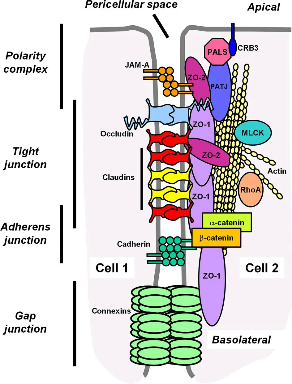
Shown are major functional zones of the Apical Junctional Complex (AJC), including the polarity complex, tight junctions, adherens junctions and gap junctions. A common theme for the structure of the AJC are layers of transmembrane proteins complexed to scaffold proteins, such as ZO-1 and ZO-2, that crosslink them to cortical actin cytoskeletal filaments. Transcellular stability of the junctions arises from interactions of proteins across the pericellular space. Cross regulation of scaffold proteins with different classes of junction proteins also provides structural cues that organize the AJC. Additional scaffold proteins are present in AJCs, but for simplicity are not shown here. Of particular relevance to the regulation of paracellular permeability are head-to-head interactions between claudins on adjacent cells that form paracellular channels. Gap junctions form channels that provide a pathway for transfer of cytosolic molecules from one cell to another. Modified from Koval M. Structure and function of epithelial and endothelial barriers. In: Muro S, editor. Drug delivery across physiological barriers. Singapore: Pan Stanford Publishing; 2016. p. 3–40 with permission.
An essential role for Crb3 in epithelial development was demonstrated by the observation that Crb3-deficient mice die shortly after birth and have disrupted epithelial morphology [12]. Of particular note, the lung airspaces of neonatal Crb3-deficient mice are filled with debris and the polarity complex is disordered. The effect of Crb3-depletion on epithelia is not limited to the lung as other tissues are also affected.
A specific role for Crb3 in lung development was elucidated using mice with a loxP-flanked Crb3 gene combined with a Cre recombinase driven by a Sonic hedgehog promoter that is active at the earliest stages of lung development [13]. The developing trachea of these tissue-specific Crb3-deficient mice contained largely Keratin5-positive cells suggestive of basal epithelial cell hyperplasia. This was due in large part to the inability of Crb3-deficient cells to retain the Yap transcription factor as part of the AJC [13]. Instead, in the absence of Crb3, Yap translocated to the nucleus where it acted as a transcription factor to influence gene expression. This suggests that regulation of Yap is a key step in lung epithelial differentiation and underscores the role for Yap/Taz/Hippo pathway in regulating lung branching morphogenesis [14,15]. However, given the central role for Crb3 in assembling the AJC, the dysregulation of Yap is not necessarily immediately downstream of Crb3. Instead, YAP may more directly bind to other polarity complex proteins or more broadly to other AJC proteins. Nonetheless, this reflects a common theme with respect to junctions, in that they frequently act as sensors reacting to intercellular contact that regulate genes by sequestering transcription factors in properly assembled AJCs.
1.3 Adherens Junctions
1.3.1 Cadherins
Along with the polarity complex, adherens junctions represent another lynchpin that is necessary for establishment of the AJC [16,17]. Adherens junctions mediate cell–cell contact through transmembrane proteins known as cadherins that bind to each other across the cell junction with high affinity in a calcium-dependent manner [18,19]. Epithelial (E)-cadherin is the canonical classical cadherin most prominently expressed by epithelial cells [20], including lung epithelium [21]. However, there are several other classical cadherin homologues expressed by lung epithelia, including N-cadherin that complements the function of E-cadherin [22].
In addition, the nonclassical cadherins, such as desmocollin and desmoglein, are a structurally distinct subset of cadherin homologues. Nonclassical cadherins form desmosomes, junctional structures distinct from that AJC that mediate a very strong form of intercellular binding and interconnect with intermediate filaments to protect epithelia from mechanical stress [23]. Atypical cadherins are a third group of cadherin homologues that generally are not expressed by lung epithelia [20]. The most broadly expressed atypical cadherin is vascular endothelial (VE) cadherin that is predominantly localized to the circulatory system [24].
1.3.2 Scaffold-Cytoskeletal Interactions
Cadherins recruit cytoplasmic scaffold proteins to adherens junctions, including α-catenin, β-catenin, and p120 catenin [20,25]. It is well established that β-catenin binds directly to the cytoplasmic terminus of E-cadherin [26]. Adherens junction scaffold proteins also facilitate an interaction with the actin cytoskeleton that requires the junction to be under tension [27]. Thus, the polarity complex and adherens junctions act as two cytoskeleton attachment sites to provide loci that orient and stabilize other elements of the AJC.
That α-catenin has the ability to directly interact both with the cadherin/β-catenin complex as well as bind to actin filaments has been interpreted as implicating a role for α-catenin in cross-linking adherens junctions and the cytoskeleton. Experiments showing that cadherin/α-catenin fusion proteins interact with actin support this model [28,29].
However, an indirect role for α-catenin in regulating cytoskeleton/adherens junction interactions has also been proposed and tested. In this model, recruitment of α-catenin to adherens junctions is followed by interactions with formin proteins that dissociate α-catenin from β-catenin to then provide a platform that binds to the arp2/3 complex that subsequently initiates actin filament formation and bundling [25,30]. Although this is not concordant with evidence demonstrating that cadherin/α-catenin fusion proteins can cross-link adherens junctions with the cytoskeleton [28,29], one possibility is that cadherin/scaffold protein chimeras do not reflect a native conformation of α-catenin and instead enable a nonphysiologic interaction with actin to occur [31]. However, different α-catenin isoforms have been shown to differ in the capacity to directly cross-link adherens junctions to filamentous actin [32]. Other scaffold proteins, such as EPLIN, can directly cross-link actin to adherens junctions [33]. Thus, a direct cross-linking role for α-catenin is not necessarily required to tether cadherins to the cytoskeleton. In addition, other functions of α-catenin, such as recruitment of tight junction scaffold proteins [e.g., zonula occludens 1 (ZO-1)], also help organize the AJC [34].
1.3.3 Cross-talk With Wnt Signaling
A co...
Table of contents
- Cover image
- Title page
- Table of Contents
- Copyright
- List of Contributors
- Acknowledgments
- Introduction: The Lung Epithelium
- Chapter 1. Junctional Interplay in Lung Epithelial Barrier Function
- Chapter 2. Ion Transport and Lung Fluid Balance
- Chapter 3. Glucose Transport and Homeostasis in Lung Epithelia
- Chapter 4. Pulmonary Surfactant Trafficking and Homeostasis
- Chapter 5. Integrin Regulation of the Lung Epithelium
- Chapter 6. Epithelial Regeneration and Lung Stem Cells
- Chapter 7. The Function of Epithelial Cells in Pulmonary Fibrosis
- Chapter 8. The Role of Epithelial Cell Quality Control in Health and Disease of the Distal Lung
- Chapter 9. The Respiratory Epithelium in COPD
- Chapter 10. Acute Respiratory Distress Syndrome
- Chapter 11. Epithelial Barrier Dysfunction in Asthma
- Chapter 12. Cystic Fibrosis: An Overview of the Past, Present, and the Future
- Index
Frequently asked questions
Yes, you can cancel anytime from the Subscription tab in your account settings on the Perlego website. Your subscription will stay active until the end of your current billing period. Learn how to cancel your subscription
No, books cannot be downloaded as external files, such as PDFs, for use outside of Perlego. However, you can download books within the Perlego app for offline reading on mobile or tablet. Learn how to download books offline
Perlego offers two plans: Essential and Complete
- Essential is ideal for learners and professionals who enjoy exploring a wide range of subjects. Access the Essential Library with 800,000+ trusted titles and best-sellers across business, personal growth, and the humanities. Includes unlimited reading time and Standard Read Aloud voice.
- Complete: Perfect for advanced learners and researchers needing full, unrestricted access. Unlock 1.5M+ books across hundreds of subjects, including academic and specialized titles. The Complete Plan also includes advanced features like Premium Read Aloud and Research Assistant.
We are an online textbook subscription service, where you can get access to an entire online library for less than the price of a single book per month. With over 1.5 million books across 990+ topics, we’ve got you covered! Learn about our mission
Look out for the read-aloud symbol on your next book to see if you can listen to it. The read-aloud tool reads text aloud for you, highlighting the text as it is being read. You can pause it, speed it up and slow it down. Learn more about Read Aloud
Yes! You can use the Perlego app on both iOS and Android devices to read anytime, anywhere — even offline. Perfect for commutes or when you’re on the go.
Please note we cannot support devices running on iOS 13 and Android 7 or earlier. Learn more about using the app
Please note we cannot support devices running on iOS 13 and Android 7 or earlier. Learn more about using the app
Yes, you can access Lung Epithelial Biology in the Pathogenesis of Pulmonary Disease by Venkataramana K Sidhaye,Michael Koval in PDF and/or ePUB format, as well as other popular books in Medicine & Medical Theory, Practice & Reference. We have over 1.5 million books available in our catalogue for you to explore.