- 178 pages
- English
- ePUB (mobile friendly)
- Available on iOS & Android
eBook - ePub
Clinical Anatomy of the Cranial Nerves
About this book
Clinical Anatomy of the Cranial Nerves combines anatomical knowledge, pathology, clinical examination, and explanation of clinical findings, drawing together material typically scattered throughout anatomical textbooks. All of the pertinent anatomical topics are conveniently organized to instruct on anatomy, but also on how to examine the functioning of this anatomy in the patient. Providing a clear and succinct presentation of the underlying anatomy, with directly related applications of the anatomy to clinical examination, the book also provides unique images of anatomical structures of plastinated cadaveric dissections. These images are the only ones that exist in this form, and have been professionally produced in the Laboratory of Human Anatomy, University of Glasgow under the auspices of the author. These specimens offer a novel way of visualizing the cranial nerves and related important anatomical structures.
- Anatomy of cranial nerves described in text format with accompanying high-resolution images of professional, high-quality prosected cadaveric material, demonstrating exactly what the structures (and related ones) look like
- Succinct yet comprehensive format with quick and easy access to facts in clearly laid out key regions, common throughout the different cranial nerves
- Includes clinical examination and related pathologies, featuring diagnostic summaries of potential clinical presentations and clinically relevant questions on the anatomy of these nerves
Trusted by 375,005 students
Access to over 1.5 million titles for a fair monthly price.
Study more efficiently using our study tools.
Information
Chapter 1
Olfactory Nerve
The olfactory nerve is the first cranial nerve and conveys special sensory information related to smell. It is the shortest of the cranial nerves and passes from its receptors in the nasal mucosa to the forebrain. It enters the skull through the cribriform plate of the ethmoid bone. It then sends its impulses to be interpreted at various brain regions including the temporal lobe, amygdala, and entorhinal cortex. Simple bedside testing of the olfactory nerve can be done using vanilla essence or coffee extracts. The sense of smell can be altered due to a variety of conditions referred to as hyperosmia, hypoosmia, anosmia, and dysosmia. However, the most common pathology to affect the olfactory nerve is the common cold.
Keywords
Anosmia; dysosmia; hyperosmia; hypoosmia; olfactory nerve
The Anatomy—Summary
The first cranial nerve is the olfactory nerve. It is the shortest of all the cranial nerves and is one of the two nerves that do not join with the brainstem. It is the cranial nerve responsible for conduction of impulses related to the special sense of smell (i.e., special sensory). Specifically, it is a special visceral afferent nerve.
The Anatomy—in More Detail
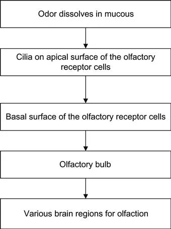
The cell bodies of the olfactory receptor neurons are located in the olfactory organ. These are found in the upper part of the nasal cavity, nasal septum, and on the inner aspect of the superior nasal concha. The olfactory receptor nerves have two surfaces: basal and apical, and these receive information from the odors which then dissolve in the mucous fluid to allow for electrical transmission of those impulses.
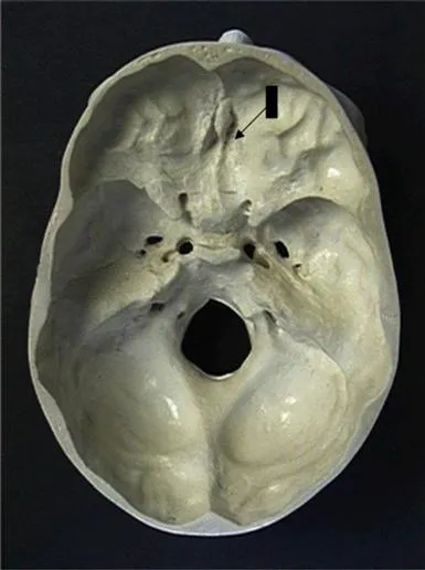
When the odor dissolves in the mucous, it is then detected by the olfactory nerves. Each olfactory nerve has two components: an apical and a basal division. It is the cilia on the apical portion that detects the dissolved “smell.” This then passes to the basal portion which constitutes the main processes of the olfactory nerve. The olfactory nerves at that point then enter the cranial cavity via the cribriform plate of the ethmoid bone (Figure 1.1). The special sense of smell is then transmitted toward the olfactory bulb where the cells then synapse. The transmission of the impulses carries posteriorly toward the brain via the olfactory tract. Specifically, the information passes to the piriform cortex of the anterior temporal lobe, anterior olfactory nucleus, amygdala, and entorhinal cortex.
The Components
The olfactory nerve is comprised of a single component allowing it to perform its role—special sensation (i.e., smell). The following diagram summarizes the pathway of innervation.
Special Sensory
• Smell from the nasal septum, superior concha, and the roof of the nasal cavity. It is a special visceral afferent nerve.
The Important Branches
Within the nasal cavity, there are two types of fibers—those of the trigeminal nerve (see Chapter 5) which responds to irritating substances and temperature and those of the olfactory nerve for olfaction.
The olfactory receptor neurons have two parts:
1. Olfactory neurons in the olfactory epithelium have central processes which pass to the olfactory bulb where they synapse.
2. The processes from the synapse pass via the olfactory tracts to the brain regions for olfaction.
The Clinical Application
Testing of the olfactory nerve is often missed in routine clinical examination. As it is involved in the special sensation of smell, testing of this nerve is undertaken by using a substance with a recognizable smell.
Testing at the Bedside
• Tell the patient that you want to test their sense of smell and ask permission to do so. Use vanilla essence, coffee, orange peel, or lemon juice.
• Then do the following:
1. Ask them to cover one nostril at a time and then close their eyes and present the testing substance to each nostril. The testing substance must not be visible to the patient.
2. Ask the patient to report if they smell anything. This allows identification of the ability to detect an odor. Asking them to identify what it is involves an “olfactory memory,” that is, higher cortical functioning, only if they recognize the substance. DO NOT touch the patient when doing ...
Table of contents
- Cover image
- Title page
- Table of Contents
- Copyright
- Preface
- Acknowledgments
- Introduction to the Nervous System
- Chapter 1. Olfactory Nerve
- Chapter 2. Optic Nerve
- Chapter 3. Oculomotor Nerve
- Chapter 4. Trochlear Nerve
- Chapter 5. Trigeminal Nerve
- Chapter 6. Abducent Nerve
- Chapter 7. Facial Nerve
- Chapter 8. Vestibulocochlear Nerve
- Chapter 9. Glossopharyngeal Nerve
- Chapter 10. Vagus Nerve
- Chapter 11. Spinal Accessory Nerve
- Chapter 12. Hypoglossal Nerve
- Summary Table of Cranial Nerves
- Index
Frequently asked questions
Yes, you can cancel anytime from the Subscription tab in your account settings on the Perlego website. Your subscription will stay active until the end of your current billing period. Learn how to cancel your subscription
No, books cannot be downloaded as external files, such as PDFs, for use outside of Perlego. However, you can download books within the Perlego app for offline reading on mobile or tablet. Learn how to download books offline
Perlego offers two plans: Essential and Complete
- Essential is ideal for learners and professionals who enjoy exploring a wide range of subjects. Access the Essential Library with 800,000+ trusted titles and best-sellers across business, personal growth, and the humanities. Includes unlimited reading time and Standard Read Aloud voice.
- Complete: Perfect for advanced learners and researchers needing full, unrestricted access. Unlock 1.5M+ books across hundreds of subjects, including academic and specialized titles. The Complete Plan also includes advanced features like Premium Read Aloud and Research Assistant.
We are an online textbook subscription service, where you can get access to an entire online library for less than the price of a single book per month. With over 1.5 million books across 990+ topics, we’ve got you covered! Learn about our mission
Look out for the read-aloud symbol on your next book to see if you can listen to it. The read-aloud tool reads text aloud for you, highlighting the text as it is being read. You can pause it, speed it up and slow it down. Learn more about Read Aloud
Yes! You can use the Perlego app on both iOS and Android devices to read anytime, anywhere — even offline. Perfect for commutes or when you’re on the go.
Please note we cannot support devices running on iOS 13 and Android 7 or earlier. Learn more about using the app
Please note we cannot support devices running on iOS 13 and Android 7 or earlier. Learn more about using the app
Yes, you can access Clinical Anatomy of the Cranial Nerves by Paul Rea in PDF and/or ePUB format, as well as other popular books in Biological Sciences & Neurology. We have over 1.5 million books available in our catalogue for you to explore.