- 512 pages
- English
- ePUB (mobile friendly)
- Available on iOS & Android
eBook - ePub
About this book
The Synapse summarizes recent advances in cellular and molecular mechanisms of synaptic transmission and provides new insights into neuronal plasticity and the cellular basis of neurological diseases.
- Part 1 provides an in-depth look at structural differences and distribution of various pre- and post-synaptic proteins found at glutamatergic synapses.
- Part 2 is dedicated to dendritic spines and their associated perisynaptic glia, which together constitute the tripartite synapse. The spines are portrayed as major sites for calcium sequestration and local protein synthesis.
- Part 3 highlights the important regional and cellular differences between glutamatergic transmission and that of neurotransmitters such as dopamine and acetylcholine that are commonly found in axon terminals without synaptic membrane specializations.
- Part 4 provides an overview of the synapse from the time of formation to degeneration under the powerful influence of aging or hormonal decline that leads to severe deficits in cognitive function.
Each chapter is illustrated with drawings and images derived from calcium imaging, electron microscopic immunolabeling, or electrophysiology. This book is a valuable reference for neuroscientists and clinical neurologists in both research and clinical settings.
- A comprehensive reference focused on the structure and function of the synapse
- Covers the links between the synapse and neural plasticity and the cellular basis of neurologic disease
- Detailed coverage of dendritic spines and associated perisynaptic glia—the tripartite synapse
- Includes in-depth coverage of synapse degeneration due to aging or hormonal decline related to severe cognitive impairment
Trusted by 375,005 students
Access to over 1.5 million titles for a fair monthly price.
Study more efficiently using our study tools.
Information
Topic
MedizinSubtopic
NeurologieChapter One
Structure and Complexity of the Synapse and Dendritic Spine
Michael G. Stewart1, Victor I. Popov2, Igor V. Kraev1, Nikolay Medvedev1 and Heather A. Davies1, 1Laboratory of Neurocytology, Deparment of Life Sciences, The Open University, Milton Keynes, UK, 2Institute of Cell Biophysics, Russian Academy of Sciences, Pushchino, Russia
Abstract
Synapses exist in different structural forms exhibiting a variety of morphological complexities, which are complimented by a myriad of dendritic spine shapes. This chapter shows how electron microscopy can demonstrate the incredible variety of synapse and spine relationships. It uses both two-dimensional images and three-dimensional reconstructions of ultrathin sections of synapses and spines to illustrate this great variety of morphological forms. The diversity of synapses, spines, and dendritic structures is also reflected in the nature of their relationships to each other, which far from being on a simple one-to-one basis, may be very complex either as multisynaptic boutons, or multi-innervated spines. We also examine those structures which can be visualized within both the synaptic bouton and dendritic spine, and the dendrite itself. All the images and reconstructions are from tissue in the mammalian hippocampus, and the region from which the images are taken is shown in Figure 1(A) and (B).
Keywords
Dendrites; Dendritic Spines; Synapses
1 Introduction
Synapses are the point at which an impulse is transmitted from one nerve cell to another. Some signalling is purely electrical, where transmission occurs via electrical coupling of ion movements at gap junctions. However, most transmission occurs at chemical synapses where an action potential causes release of neurotransmitter from the presynaptic component (the bouton), which acts on receptors on the postsynaptic cell. Knowledge of the structure of synapse and their postsynaptic contacts is the key to understanding synaptic function. In this chapter, discussion will be restricted to the hippocampus of central nervous system (Figure 1) and will focus on a description of synapses and their dendritic spines contacts.
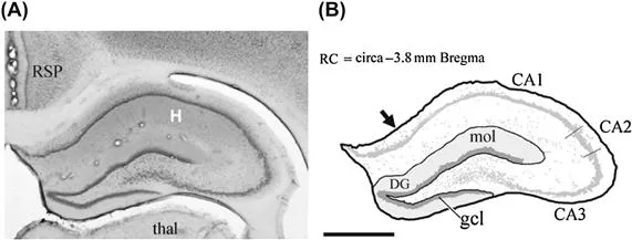
Figure 1 The hippocampus is shown in section of tissue (A) and reference diagram (B).
2.1 Synapses and Dendritic Spines
2.1.1 Synapses
Synapses are axonal swellings, found either at the end of an axon, or formed as en passant contacts—made on a dendritic component (spine or shaft) as an axon courses on its pathway. Synapses are not visible at light microscope level but in a Golgi impregnated granule cell in the dentate gyrus of the hippocampus (Figure 2(A)), the dendritic spines are visible as small “bumps” and more clearly in Figure 2(B), as twig-like structures. Camillo Golgi first recognized spines in 1873 using a silver nitrate impregnation method of neurons, and these were later described in detail by Santiago Ramon y Cajal in 1893 (DeFelipe, 2006). However, to see spines in detail, electron microscopy (EM) is necessary, and Figure 2(C) is an electron micrograph from a semi thin section (300 nm) of a portion of the granule cell in Figure 2(B) where the silver from the Golgi technique has been replaced by gold (gold toning). Spines can be seen extending from either side of the dendrite and can be up to 3 μm in length. A limitation, however, of the Golgi technique is that even with gold toning, it is difficult to see contacts between synapses and spines.
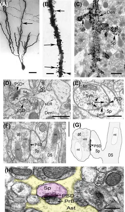
Figure 2 (A) Golgi impregnated granule cell in dentate gyrus of rat, arrow points to an apical dendritic branch, scale bar = 30 μm; (B) higher power image of dendrite shown by arrow in (A), scale bar = 10 μm. Note the presence of dendritic spines which cover the entire dendrite, some show by arrows; (C) electron micrograph of a portion of dendrite in (B) in which the gold has been replaced by silver. The dendritic spines (Sp) are now clearly visible but the synapses cannot be seen with this method of staining; (D) a small portion of dendrite as in (C) but with the tissue now stained with lead and uranium salts. Not only is the dendrite (den) and spine (sp neck) clearly visible but also the PSD (postsynaptic density), this is an asymmetric synapse (see below for definition). The dendrite contains smooth endoplasmic reticulum (sER) and there is a spine apparatus (SA) in the spine head, scale bar = 5 μm (E) Synapse at higher magnification with bouton containing vesicles (ves) and a mitochondrion (M). The bouton makes contact with a spine and the PSD is perforated (perf) (scale bar = 0.5 μm); (F) Electron micrograph of an asymmetric axo-spinous synaptic junction from stratum lacunosum-moleculare of dorsal anterior CA1 hippocampus. Spherical synaptic vesicles (ves) are present in the presynaptic axon terminal (also termed presynaptic bouton) (at). The PSD is indicated by the arrow. The post-synaptic spine head (Sp) arises from a dendritic shaft (DS). Mitochondria (mt) are visible in the axon terminal and the dendritic shaft. Scale bar: 0.5 μm (G) Schematic representation of the asymmetric axo-spinous synapse seen in H. The profiles of synaptic vesicles (ves) and mitochondria (mt) are illustrated. (H) A synapse with presynaptic bouton PrB makes contact with a dendritic spine (Sp) (purple color). The PSD is arrowed. Most notable is the fact that the synapse is surrounded by an astrocyte (Ast) (yellow color) scale bar = 0.5 μm. Originals except (F) and (G), with permission from Donohue et al., 2006.
To see synapses, EM with “normal” staining—using lead and uranium salts (Davies and Stewart, 1998)—is necessary. While confocal microscopy and newer methods including stimulated emission depletion microscopy (Wilt et al., 2009; Nagerl et al., 2008, 2010) enable quantification of factors such as Ca2+ entry into spines, they do not yet enable visualization of synaptic boutons or postsynaptic density (PSD) parameters.
2.1.2 Postsynaptic Density
Synapses can be identified at the electron microscope (EM) level by the presence of a PSD and presynaptic vesicles close to the presynaptic membrane, as in Figure 1.2(D–H). Synaptic vesicle distribution appears to be related to function activity with redistribution of synaptic vesicles in the synaptic bouton toward the synaptic active zone following long-term potentiation (LTP) (Applegate et al., 1987; Applegate and Landfield, 1988; Lushnikova et al., 2008). PSDs are found typically on dendritic spines which play primarily connective, electrical, and biochemical roles in neuronal physiology (DeFelipe, 2006; Harris, 1999; Popov et al., 2004, 2005; Segal, 2005). The PSD contains receptors, scaffold, and signalling proteins (Gu and Zeng, 2009). There is only a fine synaptic cleft (∼20 nm) separating the pre- and postsynaptic components. In Figure 1(D) and (F), the spine is attached to the dendrite by a thin spine (Tsp) neck and at the head of the spine is a PSD in apposition to a synaptic bouton, while at the side of the spine there is another synapse with a very fine PSD. The structure and form of spines is due a cytoskeleton, which is primarily made of filamentous actin (F-actin), but this cytoskeleton is not visible with normal staining in the electron microscope, immunocytochemistry would be required.
2.1.3 Three-Dimensional Reconstructions
To determine the structural organization of PSDs and dendritic spines most clearly, three-dimensional (3-D) reconstructions from ultrathin serial sections are necessary. Typically up to 150 serial ultrathin sections, from fixed and resin embedded material, are cut at ∼50–60 nm and imaged in an electron microscope at ∼10 K. The images are reconstructed in 3-D using software developed by Drs John Fiala and Kristen Harris (http://synapses.clm.utexas.edu/). The availability of these programmes has made the ability to carry out reconstructions of 2-D sections accessible to electron microscopists but the procedures require patience and experience. However, features of synapses and spines become readily apparent in 3-D reconstructions and these are described below.
2.1.4 Synapses and Spinules
Spinules are prominent structures arising from discontinuities in the PSD on the spine head, which evaginate into the presynaptic bouton. Figure 3(A) and (B) shows two images of a series of 100 sections that were used to reconstruct the spine and PSD which are presented in Figure 3(C).
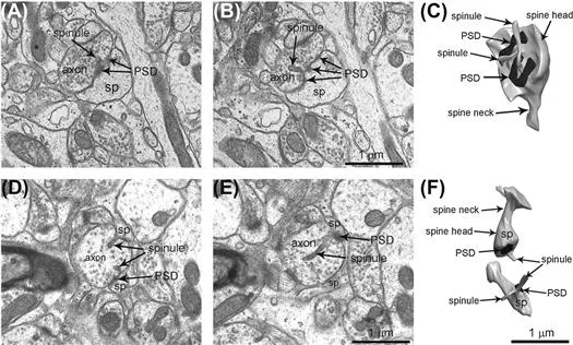
Figure 3 Spinules on Mushroom and Thin Spines. Two electron micrographs (from a series of 100) containing identified synapses and spines. Reconstructions of 100 serial sections of the synapse shown in (A) and (B), and presented in (C). This synaptic contact is on a mushroom spine and two spinules from the spine arise as discontinuities in the PSD on the spine head and evaginate into the presynaptic bouton. (D,E) Two serial electron micrographs through a thin spine from which two spinules project into an axon, as shown in the 3-D reconstruction in (F). Spinules from thin spine heads are less frequent than on mushroom spines (see below); abbreviations: sp, spine; PSD, postsynaptic density. Reproduced with permission from Medvedev et al., 2010.
This example is of a synaptic contact on a mushroom spine (see definitions below) and there are two spinules from this mushroom spine. Figure 3(D) and (E) are two serial electron micrographs from a Tsp in which two spinules project into an axon, and these are seen clearly in the 3-D reconstruction of the spine in Figure 3(F). Spinules from Tsp heads are less frequent than on mushr...
Table of contents
- Cover image
- Title page
- Table of Contents
- Copyright
- List of Contributors
- Chapter One. Structure and Complexity of the Synapse and Dendritic Spine
- Chapter Two. The Molecular Mechanisms Underlying Synaptic Transmission: A View of the Presynaptic Terminal
- Chapter Three. The First Hour in the Life of a Synapse: Contact Formation, Partner Selection, and Onset of Function
- Chapter Four. Structural and Functional Organization of the Postsynaptic Density
- Chapter Five. The Tripartite Synapse: A Role for Glial Cells in Modulating Synaptic Transmission
- Chapter Six. Local Protein Synthesis at Synapses
- Chapter Seven. Estrogen Effects on Hippocampal Synapses
- Chapter Eight. Trafficking of Glutamate Receptors and Associated Proteins in Synaptic Plasticity
- Chapter Nine. Structural Alterations of Synapses in Psychiatric and Neurodegenerative Disorders
- Chapter Ten. Synaptic Correlates of Aging and Cognitive Decline
- Chapter Eleven. Activity-Induced Fine Structural Changes of Synapses in Mammalian Central Nervous System
- Chapter Twelve. Activity-Mediated Structural Plasticity of Dendritic Spines
- Chapter Thirteen. Experience-Dependent Synaptic Plasticity in the Developing Cerebral Cortex
- Chapter Fourteen. Asynaptic and Synaptic Innervation by Acetylcholine Neurons of the Central Nervous System
- Chapter Fifteen. Prefrontal Cortical Dopamine Transmission: Ultrastructural Studies and Their Functional Implications
- Index
Frequently asked questions
Yes, you can cancel anytime from the Subscription tab in your account settings on the Perlego website. Your subscription will stay active until the end of your current billing period. Learn how to cancel your subscription
No, books cannot be downloaded as external files, such as PDFs, for use outside of Perlego. However, you can download books within the Perlego app for offline reading on mobile or tablet. Learn how to download books offline
We are an online textbook subscription service, where you can get access to an entire online library for less than the price of a single book per month. With over 1.5 million books across 990+ topics, we’ve got you covered! Learn about our mission
Look out for the read-aloud symbol on your next book to see if you can listen to it. The read-aloud tool reads text aloud for you, highlighting the text as it is being read. You can pause it, speed it up and slow it down. Learn more about Read Aloud
Yes! You can use the Perlego app on both iOS and Android devices to read anytime, anywhere — even offline. Perfect for commutes or when you’re on the go.
Please note we cannot support devices running on iOS 13 and Android 7 or earlier. Learn more about using the app
Please note we cannot support devices running on iOS 13 and Android 7 or earlier. Learn more about using the app
Yes, you can access The Synapse by Virginia M. Pickel,Menahem Segal in PDF and/or ePUB format, as well as other popular books in Medizin & Neurologie. We have over 1.5 million books available in our catalogue for you to explore.