- 214 pages
- English
- ePUB (mobile friendly)
- Available on iOS & Android
eBook - ePub
Neurology for Nurses
About this book
Neurology for Nurses is an attempt to make neurology as clear as possible, using the nursing model. The first portion of this book provides a diagram of the planes of the body that considers the nervous system anatomically, which is referenced throughout the book. The different orientations and planes of the body include the anterior (ventral) surface, posterior (dorsal) surface, lateral, medial, sagittal (median) section, Coronal (frontal) section, and transverse. Other than detailed descriptions of the anatomy and functions of nerves and the nervous system, this book provides diagnostic evaluation of diseases and clinical conditions, such as multiple sclerosis, cerebrovascular accidents, brain tumors, head injury, epilepsy, Parkinson's disease, and meningitis. This book includes as well discussions on neurological examinations, investigations, and observations. The topic on nursing care for unconscious patients is also provided. This text is aimed primarily at nursing students in training, but will also benefits those taking a post-basic nursing course in neurology.
Trusted by 375,005 students
Access to over 1.5 million titles for a fair monthly price.
Study more efficiently using our study tools.
Information
1
The planes of the body
When considering the nervous system anatomically it is necessary to divide the body diagrammatically along different planes in order to look at the various parts from different directions. The following are the different orientations and planes used in this book.
| Anterior (ventral) surface | - towards or at the front |
| Posterior (dorsal) surface | - towards or at the back |
| Lateral | - away from the midline, on the side. |
| Medial | - towards the midline. |
| Sagittal (median) section | - dividing the body from top to bottom into a right and left part. |
| Coronal (frontal) section | - dividing the front from the back, i.e. vertically at right angles to the sagittal section. |
| Transverse | - dividing the upper part of the body from the lower. A horizontal section through the body. |
2
The neurone
Publisher Summary
This chapter discusses the structure and functions of neurons. The neuron or nerve cell is the functional unit of the nervous system. It consists of a cell body and a number of nerve fibers. The function of the neuron is to carry information in the form of nerve impulses from one point to another. As with any other cell in the body, cytoplasm forms the major part, with a nucleus at its center. Surrounding the nucleus are the Nissl granules that contain RNA and are thought to produce protein for the cells. The cells come in a variety of shapes and, compared with other cells in the body, are relatively large. The red blood cell is about 7–5 μm in diameter, whereas the neuron may vary from 10–100 μm. The nerve cell body is essential for the life of the nerve and if it is destroyed, it cannot regenerate. Attached to the cell body are the nerve fibers. These are of two types that include (1) the dendrites, which are short, branching fibers receiving information, of which there may be one or more, and (2) the axon, which may be short or more than a meter long and of varying thickness. The axon transmits information away from the nerve cell. It may give off branches along its length, and at its far end has several small branches. Within these nerve endings are vesicles containing chemical transmitter substances, necessary to produce another impulse in the connecting neuron, or to act on the muscle cell.
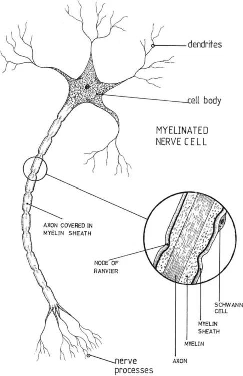
The neurone or nerve cell is the functional unit of the nervous system. It consists of a cell body and a number of nerve fibres. The function of the neurone is to carry information in the form of nerve impulses from one point to another.
As with any other cell in the body, cytoplasm forms the major part, with a nucleus at its centre. Surroundingthe nucleus are the Nissl granules which contain RNA and are thought to produce protein for the cells. The cells comein a variety of shapes, and compared with other cells in the body are relatively large. For example the red blood cell is about 7·5 microns in diameter whereas the neurone may vary from 10–100 microns. The nerve cell body isessential for the life of the nerve and if it is destroyed it cannot regenerate.
Attached to the cell body are the nerve fibres. These are of two types: the dendrites, which are short, branching fibres receiving information, of which there may be one or more, and the axon which may be short o more than a metre long and of varying thickness. The axon transmits information away from the nerve cell. It may giveoff branches along its length, and at its far end has several small branches. Within these nerve endings are vesiclescontaining chemical transmitter substances, necessary to produce another impulse in the connecting neurone, or to acton the muscle cell.
Those axons found in the peripheral nervous system and in reflex arcs, those connecting the spinal cord and cerebellum, and the motor fibres to the muscles, are all surrounded by a fatty sheath, the myelin sheath, and are therefore called myelinated nerves. This sheath is wound around the axon in a spiral fashion and is much thicker than the axon itself. It acts as an insulator preventing impulses jumping across to the neighbouring nerves. The myelin sheath is interrupted at intervals along its length, allowing the outer membrane, the neurilemma, to dip down and touch the axon. These dips are called the nodes of Ranvier. The neurilemma and Schwann cell are present in non-myelinated neurones as well. They are both necessary for the regeneration of the nerve fibre. Surrounding the whole is connective tissue, which binds the neurones together to form a nerve. Non-myelinated nerves are found in the autonomic nervous system.
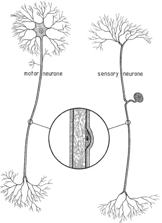
The motor neurone has its cell body at one end of the axon. Ittransmits information from the brain and spinal cord to the muscles and glands. Sensory neurones have the cell body to one side, with dendrites and axon coming off a short process. They receive information from receptors and sendit to the spinal cord and brain. Association neurones link one neurone to another. In the nervous system there are also cells which do not receive or transmit information; they are the connective tissue of the nervous system and support, feed and repair the neurones. In the brain and spinal cord they are called neuroglia, and in the peripheral nervous system, the Schwann cells.
The synapse
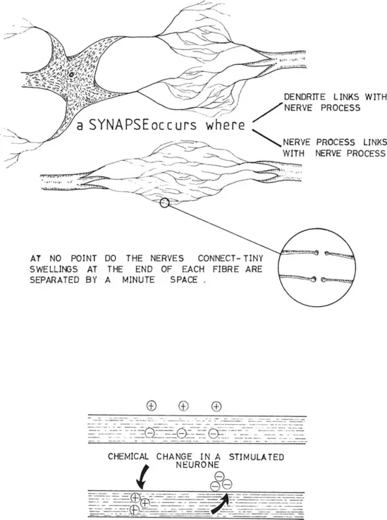
The nerve endings of the axon, with their vesicles, will connect with the dendrites or cell body of another neurone, or with a muscle cell or gland. The junction of one neurone with another it is called a synapse. Several neurones may be involved in the synapse and any one neurone is likely to link up with many others. Under the electron microscope it can be seen that there is a minute gap between the synaptic knobs of the axon and the dendrites or cell body with which they are linking.
The nerve impulse
As has already been said the function of a neurone is to carry nerve impulses. These have been likened to a ‘wave of negativity’ along the axon of a neurone. Information is picked up by receptor cells. This may be in a chemical form as in the eyes (see p. 63), or a physical form from pressure on the skin or sound waves on the ear drums, or it may start off as an electrical stimulus from another nerve. Sometimes the receptor is part of the neurone, sometimes it links with it. In either case the information is transformed into an electrical impulse, whatever causes the original stimulation.
When a neurone is at rest the cytoplasm in the axon is rich in potassium, but because it also has a large number of protein molecules and some chloride the membrane is negative on the inside. The tissue fluid bathing the outside of the membrane is rich in sodium, with some chloride. It is therefore p...
Table of contents
- Cover image
- Title page
- Table of Contents
- Copyright
- PREFACE
- Inside Front Cover
- Chapter 1: The planes of the body
- Chapter 2: The neurone
- Chapter 3: Introduction to the central nervous system
- Chapter 4: The forebrain
- Chapter 5: The thalamus
- Chapter 6: The basal nuclei
- Chapter 7: The hypothalamus
- Chapter 8: The neuro-hypophysis (posterior pituitary)
- Chapter 9: The brain stem
- Chapter 10: The cerebellum
- Chapter 11: The meninges
- Chapter 12: The ventricles
- Chapter 13: Cerebrospinal fluid
- Chapter 14: Arterial supply of the brain
- Chapter 15: Venous drainage of the brain
- Chapter 16: The spinal cord
- Chapter 17: Ascending sensory tracts
- Chapter 18: Descending motor tracts
- Chapter 19: The reflex arc
- Chapter 20: An introduction to the peripheral nervous system
- Chapter 21: The spinal nerves
- Chapter 22: The cranial nerves
- Chapter 23: The nerves of the upper limb
- Chapter 24: The nerves of the lower limb
- Chapter 25: Sight
- Chapter 26: Hearing
- Chapter 27: Balance
- Chapter 28: Smell and taste
- Chapter 29: Touch
- Chapter 30: The autonomic nervous system
- Chapter 31: Neurological examination
- Chapter 32: Neurological investigations
- Chapter 33: Neurological observations
- Chapter 34: Nursing care of the unconscious patient
- Chapter 35: The paralysed patient
- Chapter 36: Multiple sclerosis
- Chapter 37: Cerebrovascular accident
- Chapter 38: Brain tumours
- Chapter 39: Head injury
- Chapter 40: Epilepsy
- Chapter 41: Parkinson’s disease
- Chapter 42: Meningitis
- Chapter 43: Encephalitis
- Chapter 44: Subarachnoid haemorrhage
- Chapter 45: Lesions of the spinal cord
- Chapter 46: Anterior poliomyelitis
- Chapter 47: Spina bifida
- Chapter 48: Hydrocephalus
- Chapter 49: Peripheral neuritis
- Chapter 50: The trigeminal and facial nerves
- Chapter 51: Menières disease
- Chapter 52: Headaches and migraine
Frequently asked questions
Yes, you can cancel anytime from the Subscription tab in your account settings on the Perlego website. Your subscription will stay active until the end of your current billing period. Learn how to cancel your subscription
No, books cannot be downloaded as external files, such as PDFs, for use outside of Perlego. However, you can download books within the Perlego app for offline reading on mobile or tablet. Learn how to download books offline
Perlego offers two plans: Essential and Complete
- Essential is ideal for learners and professionals who enjoy exploring a wide range of subjects. Access the Essential Library with 800,000+ trusted titles and best-sellers across business, personal growth, and the humanities. Includes unlimited reading time and Standard Read Aloud voice.
- Complete: Perfect for advanced learners and researchers needing full, unrestricted access. Unlock 1.5M+ books across hundreds of subjects, including academic and specialized titles. The Complete Plan also includes advanced features like Premium Read Aloud and Research Assistant.
We are an online textbook subscription service, where you can get access to an entire online library for less than the price of a single book per month. With over 1.5 million books across 990+ topics, we’ve got you covered! Learn about our mission
Look out for the read-aloud symbol on your next book to see if you can listen to it. The read-aloud tool reads text aloud for you, highlighting the text as it is being read. You can pause it, speed it up and slow it down. Learn more about Read Aloud
Yes! You can use the Perlego app on both iOS and Android devices to read anytime, anywhere — even offline. Perfect for commutes or when you’re on the go.
Please note we cannot support devices running on iOS 13 and Android 7 or earlier. Learn more about using the app
Please note we cannot support devices running on iOS 13 and Android 7 or earlier. Learn more about using the app
Yes, you can access Neurology for Nurses by J Bickerton,J. Victor Small in PDF and/or ePUB format, as well as other popular books in Biological Sciences & Family Medicine & General Practice. We have over 1.5 million books available in our catalogue for you to explore.