- 276 pages
- English
- ePUB (mobile friendly)
- Available on iOS & Android
eBook - ePub
About this book
Ocular transporters and receptors contains detailed descriptions of major transporters and receptors expressed in the eye, with special emphasis on their role in drug delivery. The complex anatomy and the existence of multiple barriers in the eye pose a considerable challenge to successful drug delivery to the eye. Hence ocular transporters and receptors are important targets for drug delivery. A significant advancement has been made in the field of ocular transport research and their role in drug delivery. In this book the cutting edge research being carried out in this field is compiled and summarized. The book focuses on key areas, including the anatomy and physiology of the eye, biology of ocular transporters and receptors, techniques in characterization of transporters and receptors, transporters and receptors in the anterior and posterior segment in the eye, the role of ocular transporters and receptors in drug delivery, and transporter-metabolism interplay in the eye.
- Highly focused on ocular transporters
- Most up-to-date research compilation
- Detailed description of role of transporters and receptors in ocular drug discovery and delivery
Trusted by 375,005 students
Access to over 1.5 million titles for a fair monthly price.
Study more efficiently using our study tools.
Information
1
Eye: anatomy, physiology and barriers to drug delivery
Kishore Cholkar, Supriya Reddy Dasari, Dhananjay Pal and Ashim K. Mitra, Division of Pharmaceutical Sciences, School of Pharmacy, University of Missouri-Kansas City, Kansas City, MO, USA
Abstract:
This chapter describes current knowledge on the ocular structure, function and barrier properties to drug delivery. A broad description of different ocular parts and their histology is provided. Different routes of drug administration such as topical, oral, intravenous, subconjunctival, periocular and intravitreal routes are employed to treat ocular ailments. Critical anatomical and physiological factors that regulate drug absorption are discussed. Of the drug administration routes mentioned, topical for anterior ocular tissues, and periocular and intravitreal for back of the eye tissues are mostly preferred and recommended. Ocular drug absorption is impeded by static, dynamic and metabolic barriers. Static ocular barriers include corneal epithelium, blood aqueous barrier, sclera, retinal pigment epithelium and blood capillary endothelial cells, whereas the dynamic ocular barriers include tear drainage, conjunctival blood and lymph clearance, and choroidal blood and lymphatic circulations. These barriers are segregated and described as anterior segment and posterior segment barriers. As a barrier, the role of ocular efflux transporters, which reduces intracellular xenobiotic concentration, is briefly described.
Key words
anatomy
eye
barriers
static
dynamic
1.1 Introduction
The eyes are one of the most important and complex sensory organs; they act as a gateway to collect external images and transmit them to the brain as signals through the optic nerve. By this process they maintain a connection between the body and our surroundings. Various diseases, such as inflammations or bacterial and viral infections, affect the function of the eye. Most of the diseases affecting anterior eye tissues can be easily treated with high doses of drugs. However, diseases affecting the posterior tissues of the eye, are difficult to reach and treat. Age-related macular degeneration, macular edema, glaucoma, diabetic macular edema, proliferative vitreoretinopathy, cytomegalovirus retinitis, endophthalmitis and diabetic vitreoretinopathies are some of the common posterior eye diseases that may lead to vision loss if not treated.
The complex anatomy, physiology and biochemistry of the eye render this organ highly impervious to drugs/treatment. To provide an effective treatment for diseases affecting both anterior and posterior ocular tissues, a close examination of ocular anatomy, physiology and barriers is of great importance. This helps to understand the challenges associated with drug delivery to eye tissues. In this chapter we provide a detailed description of ocular anatomy and physiology, and the barriers that pose a challenge to drug delivery.
1.2 Anatomy and physiology of the eye
The eye can be broadly divided into two segments; the anterior segment and the posterior segment (Figure 1.1). The anterior segment consists of cornea, conjunctiva, aqueous humor, iris, ciliary body and crystalline lens. These occupy approximately one-third of the front of the eye. The remaining portion, i.e. posterior segment, comprises sclera, choroid, Bruch’s membrane, retinal pigment epithelium (RPE), neural retina and vitreous humor. A detailed description of the anatomy and physiology of the eye is presented below.
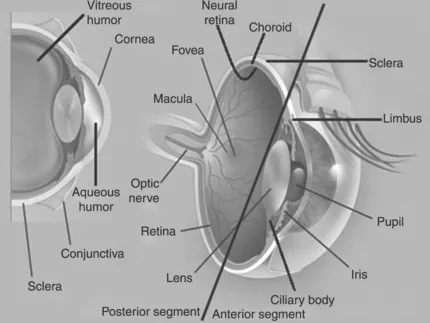
Figure 1.1 Structure of human eye
1.2.1 Anterior segment
Cornea
The cornea is thin, transparent, smooth, avascular, highly innervated and the most sensitive tissue in the body. It is convex, aspherical in shape and directly exposed to the external environment (Figure 1.1). The cornea is continuous with the white part of the eye, called the sclera, and the semi-transparent tissue, called the conjunctiva. The border of the cornea, where it continues with the sclera, is called the limbus (Figure 1.1). Limbus is highly vascularized and contains a reservoir of pluripotent stem cells [1]. The corneal surface exposed to the external environment is suffused by the tear film and its inner surface is directly in contact with fluid called aqueous humor. The thickness of the cornea gradually increases from center to periphery [2]. This is observed with corneal curvature, which is greatest at the center and smallest at the limbus.
Corneal contour, surface smoothness, transparency and refractive index determine the optical properties of the cornea. The corneal stroma is embedded with a relatively homogeneous and uniform arrangement of collagen fibers (diameter 25–35 nm) [3]. Such an arrangement is thought to be responsible for preventing and cancelling scattered light interference from the incident ray of light on collagen. The precise arrangement and function of fibers allow the light rays to pass through cornea without any interference. Corneal smoothness is maintained by the corneal epithelium and tear film. Any deviation from normal architecture of corneal collagen fibers and absence of tear film coverage causes dry eye and scattering of incident light rays, and leads to loss of corneal contours, transparency and smoothness.
As mentioned previously, cornea is highly innervated with nerve endings, with a density that is 300–400 times greater, relative to skin [4]. Sensory nerves, long ciliary nerves and sympathetic autonomic nerve fibers innervate the corneal tissue. The ciliary nerves of the ophthalmic trigeminal nerve supply sensory nerves to the cornea. The prelimbal nerve ring is provided by long ciliary nerves, which lose their myelin covering (for certain short distances) upon entering the cornea, and terminate at wing cells after penetrating the Bowman’s layer. These nerve fibers travel deeper into the stroma radially and anteriorly to form the subepithelial plexus [5]. Any damage or loss of corneal epithelium exposes nerve endings to the external environment, causing severe ocular pain [6].
Normal cornea does not have a supply of blood vessels. So this tissue is considered as one of the avascular tissues in the body, along with cartilage and lens. Despite being avascular, the epithelial and endothelial cells of cornea are metabolically active and are actively involved in wound healing. Both cell layers receive blood components and other requirements from the blood vessels of the internal and external carotid arteries that form an arcade around the cornea in the limbal region [2]. Aqueous humor supplies glucose and small amount of oxygen required by the cornea. Most of the oxygen supply to the cornea comes from exposure to air, where oxygen absorbed into the tear layer diffuses to corneal epithelial cells. This exposure of suffused tear layer on the corneal surface is necessary for oxygen supply, and maintenance of smoothness and integrity.
Histological section shows that the cornea is composed of six different layers namely, corneal epithelium, Bowman’s layer, stroma, Dua’s layer Descemet’s membrane and endothelium (Figure 1.2). Corneal epithelium is made of five to six layers of stratified and squamous non-keratinized epithelial cells. The different epithelial layers of cornea include two to three layers of superficial and wing cells and a single layer of basal cells. Multilayered corneal epithelial cells are made of cuboidal basal cells with tight junction complexes, which prevent tears from entering intercellular spaces. These cells upon differentiation gradually flatten as they move towards the corneal surface. Electron microscopy shows that the surface of the superficial epithelial cells is irregular with ridge-like folding of plasmalemma, termed microplicae (Figure 1.3) [7]. This folding increases the surface area of contact. The microplicae are covered by a very fine, closely apposed and charged glycocalyceal layer, which helps to spread tear fluid uniformly on the corneal surface with each blink (Figure 1.3) [7]. Superficial epith...
Table of contents
- Cover image
- Title page
- Table of Contents
- Copyright
- List of figures and tables
- About the authors
- Chapter 1: Eye: anatomy, physiology and barriers to drug delivery
- Chapter 2: Biology of ocular transporters: efflux and influx transporters in the eye
- Chapter 3: Characterization of ocular transporters
- Chapter 4: Transporters and receptors in the anterior segment of the eye
- Chapter 5: Transporters and receptors in the posterior segment of the eye
- Chapter 6: Transporters in drug discovery and delivery: a new paradigm in ocular drug design
- Chapter 7: Transporter–metabolism interplay in the eye
- Index
Frequently asked questions
Yes, you can cancel anytime from the Subscription tab in your account settings on the Perlego website. Your subscription will stay active until the end of your current billing period. Learn how to cancel your subscription
No, books cannot be downloaded as external files, such as PDFs, for use outside of Perlego. However, you can download books within the Perlego app for offline reading on mobile or tablet. Learn how to download books offline
We are an online textbook subscription service, where you can get access to an entire online library for less than the price of a single book per month. With over 1.5 million books across 990+ topics, we’ve got you covered! Learn about our mission
Look out for the read-aloud symbol on your next book to see if you can listen to it. The read-aloud tool reads text aloud for you, highlighting the text as it is being read. You can pause it, speed it up and slow it down. Learn more about Read Aloud
Yes! You can use the Perlego app on both iOS and Android devices to read anytime, anywhere — even offline. Perfect for commutes or when you’re on the go.
Please note we cannot support devices running on iOS 13 and Android 7 or earlier. Learn more about using the app
Please note we cannot support devices running on iOS 13 and Android 7 or earlier. Learn more about using the app
Yes, you can access Ocular Transporters and Receptors by Ashim K Mitra in PDF and/or ePUB format, as well as other popular books in Biological Sciences & Pharmacology. We have over 1.5 million books available in our catalogue for you to explore.