eBook - ePub
A Combined MRI and Histology Atlas of the Rhesus Monkey Brain in Stereotaxic Coordinates
- 402 pages
- English
- ePUB (mobile friendly)
- Available on iOS & Android
eBook - ePub
A Combined MRI and Histology Atlas of the Rhesus Monkey Brain in Stereotaxic Coordinates
About this book
A Combined MRI and Histology Atlas of the Rhesus Monkey Brain in Stereotaxic Coordinates, Second Edition maps the detailed architectonic subdivisions of the cortical and subcortical areas in the macaque monkey brain using high-resolution magnetic resonance (MR) images and the corresponding histology sections in the same animal. This edition of the atlas is unlike anything else available as it includes the detailed cyto- and chemoarchitectonic delineations of the brain areas in all three planes of sections (horizontal, coronal, and sagittal) that are derived from the same animal. This is a significant progress because in functional imaging studies, such as fMRI, both the horizontal and sagittal planes of sections are often the preferred planes given that multiple functionally active regions can be visualized simultaneously in a single horizontal or sagittal section. This combined MRI and histology atlas is designed to provide an easy-to-use reference for anatomical and physiological studies in macaque monkeys, and in functional-imaging studies in human and non-human primates using fMRI and PET.
- The first rhesus monkey brain atlas with horizontal, coronal, and sagittal planes of sections, derived from the same animal
- Shows the first detailed delineations of the cortical and subcortical areas in horizontal, coronal, and sagittal plane of sections in the same animal using different staining methods
- Horizonal series illustrates the dorsoventral extent of the left hemisphere in 47 horizontal MRI and photomicrographic sections matched with 47 detailed diagrams (Chapter 3)
- Coronal series presents the full rostrocaudal extent of the right hemisphere in 76 coronal MRI and photomicrographic sections, with 76 corresponding drawings (Chapter 4)
- Sagittal series shows the complete mediolateral extent of the left hemisphere in 30 sagittal MRI sections, with 30 corresponding drawings (Chapter 5). The sagittal series also illustrates the location of different fiber tracts in the white matter
- Individual variability - provides selected cortical and subcortical areas in three-dimensional MRI (horizontal, coronal, and sagittal MRI planes). For comparison, it also provides similar areas in coronal MRI section in six other monkeys. (Chapter 6)
- Vasculature - indicates the corresponding location of all major blood vessels in horizontal, coronal, and sagittal series of sections
- Provides updated information on the cortical and subcortical areas, such as architectonic areas and nomenclature, with references, in chapter 2
- Provides the sterotaxic grid derived from the in-vivo MR image
Trusted by 375,005 students
Access to over 1.5 million titles for a fair monthly price.
Study more efficiently using our study tools.
Information
Chapter 1
Introduction, Methods and Presentation of Data
Introduction
To understand the anatomical localization of functional activity in different cortical areas, we need an accurate map of the architectonic areas with reference to MR images in the same animal. This second edition of the atlas maps the detailed architectonic subdivisions of the cortical and subcortical areas in the macaque monkey brain using high-resolution magnetic resonance (MR) images and the corresponding histology sections in the same animal. This edition of the atlas is unlike anything else available as it includes the detailed cyto- and chemoarchitectonic delineations of the brain areas in all three planes of sections (horizontal, coronal, and sagittal) that are derived from the same animal. This is a significant progress because in functional imaging studies, such as fMRI, both the horizontal and sagittal planes of sections are often the preferred planes given that multiple functionally active regions can be visualized simultaneously in a single horizontal or sagittal section. This combined MRI and histology atlas is designed to provide an easy-to-use “REFERENCE” for anatomical and physiological studies in macaque monkeys, and in functional-imaging studies in human and non-human primates using fMRI and PET (See the preface section in page vii for other key features of this edition).
Materials and Methods
A male rhesus monkey (D99; Macaca mulatta; 4 years old), weighing 4.9 kg was used. The high-resolution T1 weighted MRI data was collected several times in this animal. Following the MRI scanning, the animal was perfused and the brain was processed for histology (see details below). All studies were in full compliance with the guidelines of the European community (EUVD 86/609/EEC) for the care and use of the laboratory animal.
MRI data collection
The MRI data collection was done under general anesthesia. After the premedication with glycopyrolate I.M. 0.01 mg/kg) and ketamine (I.M. 15 mg/kg), anesthesia was induced by intravenous injection of fentanyl (3 μg/kg), thiopental (5 mg/kg), and succinylcholine chloride (3 μg/kg). Following the intubation of the trachea, the lungs were ventilated using a Servo Ventilator 900 C (Siemens, Germany), maintaining an end-tidal CO2 of 33 mm Hg and oxygen saturation of >95%. Balanced anesthesia was maintained with end-tidal 0.35% (0.23 MAC for macaques) isoflurane in air and fentanyl (3 μg/kg/hr). Muscle relaxation was achieved with mivacurium (5 mg/kg/h). The body temperature, ECG, NIBP, CO2, and SpO2 were monitored throughout the experiment (Logothetis et al., 1999). The above protocol is established as optimal for imaging without discomfort to the animal.
Prior to MRI scanning, the animal was placed in a custom-made restraining device mounted on the MRI chair, which held the animal’s head. The MRI scanning was done in a vertical 4.7 T (200 MHz) scanner with a 40 cm diameter bore (Biospec 47/40 v, Bruker Medical, Ettlingen, Germany). The animal was imaged using a 120 mm-wide, custom-made linear homogeneous volume (saddle) coil (Logothetis et al., 2002). A linear birdcage-type resonator with an inner-diameter of 198 mm (Bruker Medical, Ettlingen, Germany) was also used that delivered superior homogeneity over the entire brain volume.
We used a Modified Driven Equilibrium with Fourier Transform (3D-MDEFT) method to obtain T1 weighted anatomical images. The imaging sequence used 180° and 90° adiabatic RF pulses for spin preparation (excitation flip angle 20°) and a segmented gradient-echo acquisition (Tau = 800 ms, TR = 14.9 ms, TE = 4.0 ms). We collected five averages and four segments for an image acquisition time of 3h 50 min to obtain 256 × 256 × 240 matrices at FOV 12.8 cm × 12.8 cm × 12.0 cm. All scans achieved an isotropic voxel resolution of 0.5 mm3.
All horizontal MRI slices were aligned parallel to the horizontal plane passing through the interaural line and the infraorbital ridge (Ear Bar Zero or EBZ), and the coronal MRI slices were aligned orthogonal (perpendicular) to horizontal plane.
Histological processing
After the MRI scanning, the animal was deeply anesthetized with a lethal dose of sodium pentobarbital and perfused transcardially with warm heparinized saline followed by 1 liter of 1% paraformaldehyde in 0.1 M phosphate buffer (PB; pH 7.4, 4°C), 2 liters of 4% paraformaldehyde in 0.1 M PB (pH 7.4, 4°C) and finally 1 liter of 4% paraformaldehyde and 10% sucrose in 0.1 M PB (pH 7.4, 4°C). The flow rate of the fixative solution was adjusted so that the perfusion with paraformaldehyde took approximately 45 min. The brain was removed from the skull, photographed and carefully blocked in the stereotaxic plane (corresponding to MRI planes), and then postfixed for 6 hours in the last fixative-sucrose solution. Finally, these blocks were stored in 20% and 30% sucrose in 0.1 M PB at 4°C until they sank. We prepared four blocks from the two hemispheres: two blocks from the left hemisphere for horizontal slices, and two from the right hemisphere for coronal slices. Frozen sections were cut in horizontal and coronal planes at 40 μm and 50 μm thickness, respectively. Five parallel series of sections were stained for Nissl substance or with antibodies against parvalbumin, calbindin, calretinin, and a nonphosphorylated epitope of a neurofilament protein (SMI-32).
Immunohistochemical procedures
Details of the antibodies used are given in Table 1.1. The calcium binding proteins parvalbumin, calbindin and calretinin have been shown previously to stain subpopulations of non-pyramidal neurons (GABAergic) in the neocortex, and to label different types of neurons in subcortical structures (Jones and Hendry, 1989; Jones, 1998; current study). The SMI-32 antibody recognizes a non-phosphorylated epitope of neurofilament H (Sternberger and Sternberger, 1983; Goldstein et al., 1987). The antibody has been shown to stain a subpopulation of pyramidal cells in the neocortex (e.g. Campbell and Morrison, 1989; Hof and Morrison, 1995); the pattern of staining seen in this study corresponds to well established patterns from previous studies.
Table 1.1
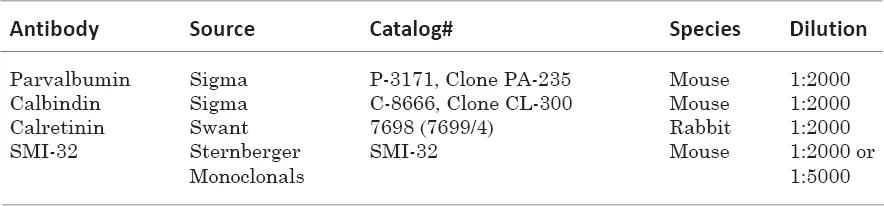
Free-floating sections were preincubated in the phosphate buffered saline (PBS), containing 0.5% Triton-X 100 and 5% normal serum (normal horse serum for parvalbumin, calbindin, and SMI-32, or normal goat serum for calretinin) and 2% bovine serum albumin (BSA) for 60 minutes at room temperature. The sections were then incubated in PBS containing 0.3% Triton-X 100, 3% normal serum, 1% BSA, and the primary antibody for 2 days at 4°C. After washing with PBS, sections were incubated in PBS containing 0.3% Triton-X 100, normal serum, 0.5% BSA, and the biotiny- lated horse antimouse IgG (for parvalbumin, calbindin, and SMI-32), or biotinylated goat antirab- bit IgG (calretinin) for 90 minutes at room temperature. After washing with PBS, sections were reacted and stained in a solution containing 0.05 M tris buffer (pH 7.2-7.4), 0.05% diaminobenzidine (DAB) and 0.003% hydrogen peroxide (H2O2). The sections were then mounted on the glass slides, air-dried, dehydrated, and coverslipped with Entellan.
Data analysis
The sections were observed with a light microscope under bright field illumination. To make the figures, the horizontal and coronal histology sections were scanned, using a Nikon Film Scanner (LS-4500AF), and the scanned images were processed in Adobe Photoshop. Although we blocked and cut the left and right hemispheres in the stereotaxic plane (corresponding to MRI planes), the processed histological sections did not match exactly with the corresponding MR images. To correct this mismatch, we rotated and digitally reslice...
Table of contents
- Cover image
- Title page
- Table of Contents
- Dedication
- Copyright
- Preface
- About the Authors
- Acknowledgments
- Comments
- Chapter 1. Introduction, Methods and Presentation of Data
- Chapter 2. Cytoarchitectonic and Chemoarchitectonic Organization of Cortical and Subcortical areas
- Chapter 3. Horizontal Series
- Chapter 4. Coronal Series
- Chapter 5. Sagittal Series
- Chapter 6. Selected Cortical and Subcortical Areas in three Different MRI Planes and Different Cases
- Index of Abbreviations
Frequently asked questions
Yes, you can cancel anytime from the Subscription tab in your account settings on the Perlego website. Your subscription will stay active until the end of your current billing period. Learn how to cancel your subscription
No, books cannot be downloaded as external files, such as PDFs, for use outside of Perlego. However, you can download books within the Perlego app for offline reading on mobile or tablet. Learn how to download books offline
Perlego offers two plans: Essential and Complete
- Essential is ideal for learners and professionals who enjoy exploring a wide range of subjects. Access the Essential Library with 800,000+ trusted titles and best-sellers across business, personal growth, and the humanities. Includes unlimited reading time and Standard Read Aloud voice.
- Complete: Perfect for advanced learners and researchers needing full, unrestricted access. Unlock 1.5M+ books across hundreds of subjects, including academic and specialized titles. The Complete Plan also includes advanced features like Premium Read Aloud and Research Assistant.
We are an online textbook subscription service, where you can get access to an entire online library for less than the price of a single book per month. With over 1.5 million books across 990+ topics, we’ve got you covered! Learn about our mission
Look out for the read-aloud symbol on your next book to see if you can listen to it. The read-aloud tool reads text aloud for you, highlighting the text as it is being read. You can pause it, speed it up and slow it down. Learn more about Read Aloud
Yes! You can use the Perlego app on both iOS and Android devices to read anytime, anywhere — even offline. Perfect for commutes or when you’re on the go.
Please note we cannot support devices running on iOS 13 and Android 7 or earlier. Learn more about using the app
Please note we cannot support devices running on iOS 13 and Android 7 or earlier. Learn more about using the app
Yes, you can access A Combined MRI and Histology Atlas of the Rhesus Monkey Brain in Stereotaxic Coordinates by Kadharbatcha S. Saleem,Nikos K. Logothetis in PDF and/or ePUB format, as well as other popular books in Biological Sciences & Neurology. We have over 1.5 million books available in our catalogue for you to explore.