- 672 pages
- English
- ePUB (mobile friendly)
- Available on iOS & Android
eBook - ePub
Veterinary Virology
About this book
Veterinary Virology deals with basic biomedical virology and the clinical discipline of infectious diseases. The book discusses the principles of virology as effecting future developments in the search for preventive and management of infectious diseases in animals, whether singly or as a whole herd or flock. Part I explains the principles of animal virology including the structure, composition, classification, nomenclature, cultivation, and assay of viruses. This part also discusses viral genetics, replication, and evolution (including mutation and genetic engineering). The book also reviews the pathogenesis of viruses, host resistance and susceptibility, as well as the mechanisms of persistent infections and tumor induction. Part II deals with viruses found in domestic animals; this part also explains in detail the properties, replication methods, pathogenesis, immunity, diagnosis, and control of some common viruses. The book discusses some other families of viruses of which no members are yet known as to have caused serious or important diseases in animals. Veterinarians, immunologists, virologists, molecular researchers, students, and academicians in the discipline of virology and cellular biology, as well as livestock owners will find this book helpful.
Trusted by 375,005 students
Access to over 1.5 million titles for a fair monthly price.
Study more efficiently using our study tools.
Information
Part I
PRINCIPLES OF ANIMAL VIROLOGY
CHAPTER 1
Structure and Composition of Viruses
Publisher Summary
Viruses are smaller and simpler in construction than unicellular microorganisms, and they contain only one type of nucleic acid—either DNA or RNA—never both. As viruses have no ribosomes, mitochondria, or other organelles, they are completely dependent on their cellular hosts for energy production and protein synthesis. They replicate only within cells of the host that they infect. Unlike any microorganism, many viruses can, in suitable cells, reproduce themselves from their genome, a single nucleic acid molecule, that is, their nucleic acid alone is infectious. Outside a susceptible cell, the virus particle like a bacterial spore is metabolically inert; on the other hand, when replicating in a cell, it exhibits all the characteristics of life. The new group of microorganisms are known as the filterable viruses. The filtration studies has shown that virus particles (virions) range from about the size of the smallest unicellular microorganisms (300 nm) down to objects little bigger than the largest protein molecules (20 nm). In the simpler viruses, the virion consists of a single molecule of nucleic acid surrounded by a protein coat, the capsid; the capsid and its enclosed nucleic acid together constitute the nucleocapsid.
The unicellular microorganisms can be arranged in order of decreasing size and complexity: protozoa, fungi, bacteria, mycoplasmas, rickettsiae, and chlamydiae. These microorganisms, however small and simple, are cells. They always contain DNA as the repository of their genetic information, they contain RNA, and they have their own machinery for producing energy and macromolecules. Microorganisms grow by synthesizing their own macromolecular constituents (nucleic acid, protein, carbohydrate, and lipid), and they multiply by binary fission.
Viruses, on the other hand, are smaller and simpler in construction than unicellular microorganisms, and they contain only one type of nucleic acid—either DNA or RNA, never both. Furthermore, since viruses have no ribosomes, mitochondria, or other organelles, they are completely dependent on their cellular hosts for energy production and protein synthesis. They replicate only within cells of the host that they infect. Indeed, unlike any microorganism, many viruses can, in suitable cells, reproduce themselves from their genome, a single nucleic acid molecule; i.e., their nucleic acid alone is infectious. Are viruses alive? The question is rhetorical. Outside a susceptible cell, the virus particle, like a bacterial spore, is metabolically inert; on the other hand, when replicating in a cell it exhibits all the characteristics of life. The key differences between viruses and microorganisms are listed in Table 1-1.
TABLE 1-1
Contrasting Properties of Unicellular Microorganisms and Viruses
| Property | Bacteria | Mycoplasmas | Rickettsiae | Chlamydiae | Viruses |
| <300 nm diameter | – | ± | – | ± | + |
| Growth on nonliving media | + | + | – | – | – |
| Binary fission | + | + | + | + | – |
| DNA and RNA | + | + | + | + | – |
| Nucleic acid infectious | – | – | – | – | +a |
| Ribosomes | + | + | + | + | – |
| Metabolism | + | + | + | ± | – |
| Sensitivity to antibiotics | + | + | + | + | – |
| Sensitivity to interferon | – | – | – | + | + |
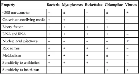
aSome, among both DNA and RNA viruses.
Several important practical consequences flow from these differences. For example, some viruses (but no microorganisms) may persist in cells by the integration of their DNA (or a DNA copy of their RNA) into the genome of the host cell, and they are not susceptible to antibiotics that act against specific steps in the metabolic pathways of bacteria.
MORPHOLOGY
For many years it has been known that viruses are smaller than unicellular microorganisms. Independently, in 1898–1899, the Dutch plant pathologist Beijerinck, working on tobacco mosaic disease, and the German veterinarians Loeffler and Frosch, working on foot-and-mouth disease of cattle, showed that these diseases could be transmitted by material which could pass through a filter with pores too small to allow passage of bacteria. The new group of “microorganisms” became known as the “filterable viruses.” Filtration studies showed that virus particles (virions) range from about the size of the smallest unicellular microorganisms (300 nm) down to objects little bigger than the largest protein molecules (20 nm). For a time they were also called “ultramicroscopic,” since they are too small to be seen with the ordinary light microscope. Only with the advent of the electron microscope did it become possible to study their morphology properly. In 1959, our knowledge of viral ultrastructure was transformed when Brenner and Horne applied negative staining to the electron microscopy of viruses. Potassium phosphotungstate, which is electron-dense, fills the interstices of the viral surface, giving the resulting electron micrograph a degree of detail not previously possible. Electron micrographs of negatively stained preparations of the virions of all fam...
Table of contents
- Cover image
- Title page
- Table of Contents
- Copyright
- Preface
- Peter A. Bachmann
- Part I: PRINCIPLES OF ANIMAL VIROLOGY
- Part II: VIRUSES OF DOMESTIC ANIMALS
- Glossary
- Index
Frequently asked questions
Yes, you can cancel anytime from the Subscription tab in your account settings on the Perlego website. Your subscription will stay active until the end of your current billing period. Learn how to cancel your subscription
No, books cannot be downloaded as external files, such as PDFs, for use outside of Perlego. However, you can download books within the Perlego app for offline reading on mobile or tablet. Learn how to download books offline
Perlego offers two plans: Essential and Complete
- Essential is ideal for learners and professionals who enjoy exploring a wide range of subjects. Access the Essential Library with 800,000+ trusted titles and best-sellers across business, personal growth, and the humanities. Includes unlimited reading time and Standard Read Aloud voice.
- Complete: Perfect for advanced learners and researchers needing full, unrestricted access. Unlock 1.5M+ books across hundreds of subjects, including academic and specialized titles. The Complete Plan also includes advanced features like Premium Read Aloud and Research Assistant.
We are an online textbook subscription service, where you can get access to an entire online library for less than the price of a single book per month. With over 1.5 million books across 990+ topics, we’ve got you covered! Learn about our mission
Look out for the read-aloud symbol on your next book to see if you can listen to it. The read-aloud tool reads text aloud for you, highlighting the text as it is being read. You can pause it, speed it up and slow it down. Learn more about Read Aloud
Yes! You can use the Perlego app on both iOS and Android devices to read anytime, anywhere — even offline. Perfect for commutes or when you’re on the go.
Please note we cannot support devices running on iOS 13 and Android 7 or earlier. Learn more about using the app
Please note we cannot support devices running on iOS 13 and Android 7 or earlier. Learn more about using the app
Yes, you can access Veterinary Virology by Frank J. Fenner,Peter A. Bachmann,E. Paul J. Gibbs in PDF and/or ePUB format, as well as other popular books in Technology & Engineering & Immunology. We have over 1.5 million books available in our catalogue for you to explore.