- 334 pages
- English
- ePUB (mobile friendly)
- Available on iOS & Android
eBook - ePub
Characterization of Biomaterials
About this book
Biomaterials and medical devices must be rigorously tested in the laboratory before they can be implanted. Testing requires the right analytical techniques. Characterization of biomaterials reviews the latest methods for analyzing the structure, properties and behaviour of biomaterials.Beginning with an introduction to microscopy techniques for analyzing the phase nature and morphology of biomaterials, Characterization of biomaterials goes on to discuss scattering techniques for structural analysis, quantitative assays for measuring cell adhesion, motility and differentiation, and the evaluation of cell infiltration and tissue formation using bioreactors. Further topics considered include studying molecular-scale protein-surface interactions in biomaterials, analysis of the cellular genome and abnormalities, and the use of microarrays to measure cellular changes induced by biomaterials. Finally, the book concludes by outlining standards and methods for assessing the safety and biocompatibility of biomaterials.With its distinguished editors and international team of expert contributors, Characterization of biomaterials is an authoritative reference tool for all those involved in the development, production and application of biomaterials.
- Reviews the latest methods for analyzing the structure, properties and behaviour of biomaterials
- Discusses scattering techniques for structural analysis, quantitative assays for measuring cell adhesion, and motility and differentiation
- Examines the evaluation of cell infiltration and tissue formation using bioreactors
Trusted by 375,005 students
Access to over 1.5 million titles for a fair monthly price.
Study more efficiently using our study tools.
Information
Subtopic
Biotechnology in Medicine1
Microscopy techniques for analyzing the phase nature and morphology of biomaterials
R.T. Dombrowski, Nanoview Associates,USA
Abstract:
This chapter provides a review of all the major microscopy imaging techniques that are available to the modern researcher for the characterization of biomaterials. Today, with the melding of both biology and materials science to produce both natural and man-made biomaterials, imaging has become a major characterization technique to carry out the further development of these materials that will be implanted in the human body to perform, augment or replace natural bodily functions. Microstructural imaging techniques utilizing light, electrons and molecular mechanical probes are covered. The various chapter sections for each of these major imaging modes contain a mix of useful foundational theory and practical application knowledge which is meant to allow the researcher to maximize the imaging data obtained using each technique.
Key words
microscopy
light microscopy
phase contrast microscopy
polarized light microscopy
differential interference contrast microscopy (DIC)
laser scanning confocal microscopy (LSCM)
scanning electron microscopy (SEM)
scanned probe microscopy (SPM)
atomic force microscopy (AFM)
biomaterials
characterization
imaging
phases
morphology
microstructure
1 Introduction: basic imaging concepts
From the seventeenth century onward, imaging has been an important tool that has allowed the advancement of knowledge in the biological sciences. We can say it all started with Antonie van Leeuwenhoek, the Dutch lens maker and scientist, when he produced his early forerunners of the modern microscope. With his handcrafted microscopes, van Leeuwenhoek was the first to observe and describe bacteria, muscle fibers, spermatozoa and blood flow in capillaries. His early studies also established imaging as a major research tool for the biomedical sciences. Today, with the melding of both biology and materials science to produce both natural and man-made bio-materials, imaging has become a major characterization technique to carry out the further development of these materials that will be implanted in the human body to perform, augment or replace natural bodily functions. Though there are many types of imaging used in the biomedical sciences, in this chapter imaging refers to observations and measurements carried out with microscopes using light, electrons or scanned molecular mechanical probes.
The following definitions for images, imaging and microscopy are presented in order to clarify imaging concepts that appear later in this chapter:
Image – the optical counterpart of a self-luminous or illuminated object formed by the light rays that traverse an optical system made up of a series of lenses; each point of the object has a corresponding point in the image from which rays diverge or appear to diverge. This definition holds true for optical systems using illuminating beams other than light, such as electrons.
Imaging – the formation of images of objects that can be created using light (optical microscopy), lasers (confocal microscopy), electrons (scanning and transmission electron microscopies) and scanned molecular mechanical probes (atomic force microscopy).
Microscopy – the interpretive application of magnification created by a microscope to the study of materials that cannot be seen properly by the unaided eye.
Microscopes do not only magnify objects. A more important measurable property of microscopes is their capability to resolve or clearly determine two separate points, or objects, as singular, distinguished entities. This property is known as microscope resolution. The lower resolution limit of a microscope system decreases from microns to nanometers and angstroms as an analyst goes from producing images with light rays to producing them with electrons and scanned molecular mechanical probes. These image techniques are shown in Table 1.1 together with the resolution relationships.
Table 1.1
Image techniques – resolution relationships
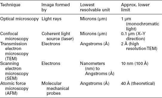
As the field of biomedical engineering moves toward the ability to more precisely engineer biomaterials, cells and tissue replacements there is an increased necessity to image sub-micron details in a wide variety of specimen types. The increased use of scanning electron microscopy (SEM), transmission electron microscopy (TEM) and atomic force microscopy (AFM) (each with ever lower resolution limits) as a multi-technique integrated imaging tool set has allowed the almost routine observation of important biological and chemical phenomena such as cell–cell and cell–extracellular matrix interactions, intracellular events and nano-scale changes in biomaterials.
Imaging using an integrated tool set of powerful microscopes is becoming a critical component of the science of biomaterials characterization. The almost routine use of these instruments allows both researchers and engineers to fabricate new materials, coatings and devices; study both their surface and bulk properties which effect cell adhesion and biocompatibility; and study the behavior of cells and tissues at the biomaterials interface. The micro and nano-structural information obtained can be used to formulate structure-property-performance models which allow fabrication processes and materials properties to be modified to produce the desired final bioma-terials performance properties. We have come a long way from the days of Antonie van Leeuwenhoek’s handcrafted microscopes and the future will hold even more opportunities for microscopy to play a critical role in the development of new cutting-edge biomaterials.
1.2 Image perception and interpretation
The prime optical instrument that we use to observe and interpret the world around us is the human eye. We are able to perceive colors within the visible portion of the spectrum and variations in the intensity of light being absorbed and reflected by the objects around us. Microscopes can be seen as tools that extend our sight, helping us magnify and resolve object details that we would not be able to detect with the unaided eye.
1.2.1 Image creation by the human eye
The human eye is capable of distinguishing color in the visible portion of the electromagnetic spectrum. This ‘white’ light portion of the spectrum extends from violet to green to yellow to orange and ends at red. This visible portion of the spectrum includes radiation varying in wavelength between approximately 400 and 750 nm. The eye cannot directly see ultra-violet or infra-red radiation. The eye is also able to detect differences in brightness or light intensity ranging from black to white and all the gray levels in between. In order for an image to be seen by the human eye, the image must be comprised of the colors of the visible spectrum and variations of light intensity or varying degrees of light intensity alone.
The eye with both its optical and structural components can be thought of as a bio-based optical instrument. For an image to be seen clearly, it must be spread on the retina of the eye at a sufficient visual angle. Unless the light falls on non-adjacent rows of retinal cells, due to magnification and spreading of the image, we are unable to see or ‘resolve’ details lying close together as being separate. There must also be sufficient variation in light intensity or contrast between adjacent details and the background to make the magnified, resolved image visible. All the conditions described above must be met for humans to see or adequately produce images of the world around us.
1.2.2 Image creation by lens-based imaging systems
In this section we will explore how lens-based imaging systems help the human eye visualize magnified images with increased resolution of detail. Due to the limited ability of the eye’s lens to change its shape, objects brought very close to the eye cannot have their images brought to focus on the retina. The object can be focused when a simple glass magnifier or convex lens, thicker in the center than the periphery, is placed between the object and the eye. These magnifiers or ‘simple microscopes’ aid the cornea and eye lens to spread the image of the object on the retina by magnification through increasing the visual angle on the retina.
Antonie van Leeuwenhoek’s handcrafted microscopes described earlier in this chapter operated in much the same manner as described above for a simple magnifier. The image produced by a van Leeuwenhoek ‘simple microscope’, when the microscope was held close to the observer’s eye, appeared as if it were on the same side of the lens as the object itself. Such an image, seen as if it were ten inches from the eye, is known as a virtual image and cannot be captured on film. During the seventeenth century, van Leeuwenhoek’s work was built upon by others to produce what we know today as the compound microscope. In its basic form, it consisted of two convex lenses aligned in series. An object glass or objective was placed closer to the specimen and an eyepiece or ocular was placed closer to the observer’s eye. There was also a way of adjusting the position of the specimen and the microscope lenses. The compound microscope achieves a two-stage magnification. In essence, the objective projects a magnified image into the body tube of the microscope and the eyepiece further magnifies the image projected by the ...
Table of contents
- Cover image
- Title page
- Table of Contents
- Copyright
- Contributor contact details
- Woodhead Publishing Series in Textiles
- Chapter 1: Microscopy techniques for analyzing the phase nature and morphology of biomaterials
- Chapter 2: Scattering techniques for structural analysis of biomaterials
- Chapter 3: Quantitative assays for measuring cell adhesion and motility in biomaterials
- Chapter 4: Assays for determining cell differentiation in biomaterials
- Chapter 5: Bioreactors for evaluating cell infiltration and tissue formation in biomaterials
- Chapter 6: Studying molecular-scale protein–surface interactions in biomaterials
- Chapter 7: Assessing the mutagenic effects of biomaterials: analyzing the cellular genome and abnormalities
- Chapter 8: Using microarrays to measure cellular changes induced by biomaterials
- Chapter 9: Standards and methods for assessing the safety and biocompatibility of biomaterials
- Index
Frequently asked questions
Yes, you can cancel anytime from the Subscription tab in your account settings on the Perlego website. Your subscription will stay active until the end of your current billing period. Learn how to cancel your subscription
No, books cannot be downloaded as external files, such as PDFs, for use outside of Perlego. However, you can download books within the Perlego app for offline reading on mobile or tablet. Learn how to download books offline
Perlego offers two plans: Essential and Complete
- Essential is ideal for learners and professionals who enjoy exploring a wide range of subjects. Access the Essential Library with 800,000+ trusted titles and best-sellers across business, personal growth, and the humanities. Includes unlimited reading time and Standard Read Aloud voice.
- Complete: Perfect for advanced learners and researchers needing full, unrestricted access. Unlock 1.5M+ books across hundreds of subjects, including academic and specialized titles. The Complete Plan also includes advanced features like Premium Read Aloud and Research Assistant.
We are an online textbook subscription service, where you can get access to an entire online library for less than the price of a single book per month. With over 1.5 million books across 990+ topics, we’ve got you covered! Learn about our mission
Look out for the read-aloud symbol on your next book to see if you can listen to it. The read-aloud tool reads text aloud for you, highlighting the text as it is being read. You can pause it, speed it up and slow it down. Learn more about Read Aloud
Yes! You can use the Perlego app on both iOS and Android devices to read anytime, anywhere — even offline. Perfect for commutes or when you’re on the go.
Please note we cannot support devices running on iOS 13 and Android 7 or earlier. Learn more about using the app
Please note we cannot support devices running on iOS 13 and Android 7 or earlier. Learn more about using the app
Yes, you can access Characterization of Biomaterials by M Jaffe,W. Hammond,P Tolias,T Arinzeh in PDF and/or ePUB format, as well as other popular books in Technology & Engineering & Biotechnology in Medicine. We have over 1.5 million books available in our catalogue for you to explore.