eBook - ePub
Toxoplasma Gondii
The Model Apicomplexan. Perspectives and Methods
- 800 pages
- English
- ePUB (mobile friendly)
- Available on iOS & Android
eBook - ePub
About this book
Toxoplasmosis is caused by a one-celled protozoan parasite known as Toxoplasma gondii. In the United States, it is estimated that approximately 30% of cats, the primary carriers, have been infected by T. gondii. Most humans contract toxoplasmosis by eating cyst-contaminated raw or undercooked meat, vegetables, or milk products or when they come into contact with the T. gondii eggs from cat feaces while cleaning a cat's litterbox, gardening, or playing in a sandbox. Approx 1 in 4 (more than 60 million) people in the USA are infected with the parasite, and in the UK between 0.5 and 1% of individuals become infected each year. By the age of 50, 40% of people test positive for the parasite. The predilection of this parasite is for the central nervous system (CNS) causing behavioral and personality alterations as well as fatal necrotizing encephalitis, and is especially dangerous for HIV infected patients.Though there have been tremendous strides in our understanding of the biology of Toxoplasma gondii in the last decade, there has been no systemic review of all of the information that has accumulated. Toxoplasma gondii provides the first comprehensive summary of literature on this organism by leading experts in the field who were responsible for organising the 7th International Congress on Toxoplasmosis in May 2003. It offeres systematic reviews of the biology of this pathogen as well as descriptions of the methods and resources used. Within the next year the T. gondii genome will be completed making this an indispensable research resource for biologists, physicians, parasitologists, and for all those contemplating experiments using T. gondii.* Serves as a model for understanding invasion of host cells by parasites, immune response, motility, differentiation, phylogenetics, evolution and organelle acquisition* Discusses the protocols related to genetic manipulation, cell biology and animal models while also providing reference material on available resources for working with this organism
Trusted by 375,005 students
Access to over 1.5 million titles for a fair monthly price.
Study more efficiently using our study tools.
Information
1
The History and Life Cycle of Toxoplasma gondii
J.P. Dubey
Publisher Summary
Infections by the protozoan parasite Toxoplasma gondii are widely prevalent in humans and other animals on all continents. This chapter provides a history of the milestones in the acquisition of knowledge of the biology of this parasite. Toxoplasmosis in sheep deserves special attention because of its economic impact. The identification of T. gondii abortion in ewes is considered a landmark discovery in veterinary medicine; prior to that, protozoa were not recognized as a cause of epidemic abortion in livestock. The ability to identify T. gondii infections based on a simple serological test opened the door for extensive epidemiological studies on the incidence of infection. It became clear that T. gondii infections are widely prevalent in humans in many countries. It also demonstrated that the so-called tetrad of clinical signs considered indicative of clinical congenital toxoplasmosis occurred in other diseases and assisted in the differential diagnosis. Vaccination of sheep with a live cystless strain of T. gondii reduces neonatal mortality in lambs, and the vaccine is available commercially.
1.1 INTRODUCTION
Infections by the protozoan parasite Toxoplasma gondii are widely prevalent in humans and other animals on all continents. There are many thousands of references to this parasite in the literature, and it is not possible to give equal treatment to all authors and discoveries. The objective of this chapter is, rather, to provide a history of the milestones in our acquisition of knowledge of the biology of this parasite.
1.2 THE ETIOLOGICAL AGENT
Nicolle and Manceaux (1908) found a protozoan in tissues of a hamster-like rodent, the gundi, Ctenodactylus gundi, which was being used for leishmaniasis research in the laboratory of Charles Nicolle at the Pasteur Institute in Tunis. They initially believed the parasite to be Leishmania, but soon realized that they had discovered a new organism and named it Toxoplasma gondii, based on the morphology (mod. L. toxo = arc or bow, plasma = life) and the host (Nicolle and Manceaux, 1909). Thus, its complete designation is Toxoplasma gondii. In retrospect, the correct name for the parasite should have been Toxoplasma gundii; Nicolle and Manceaux (1908) had incorrectly identified the host as Ctenodactylus gondi. Splendore (1908) discovered the same parasite in a rabbit in Brazil, also erroneously identifying it as Leishmania, but he did not name it.
1.3 PARASITE MORPHOLOGY AND LIFE CYCLE
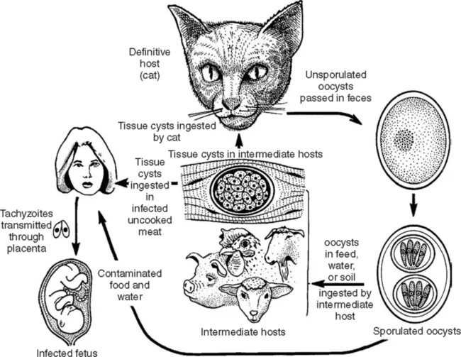
The life cycle of Toxoplasma gondii is illustrated in Figure 1.1.
1.3.1 Tachyzoites
The tachyzoite (Frenkel, 1973) is lunate (Figure 1.2A), and is the stage that Nicolle and Manceaux (1909) found in the gundi. This stage has also been called trophozoite, the proliferative form, the feeding form, and endozoite. It can infect virtually any cell in the body. It divides by a specialized process called endodyogeny, first described by Goldman et al. (1958). Gustafson et al. (1954) first studied the ultrastructure of the tachyzoite. Sheffield and Melton (1968) provided a complete description of endodyogeny when they fully described its ultrastructure.
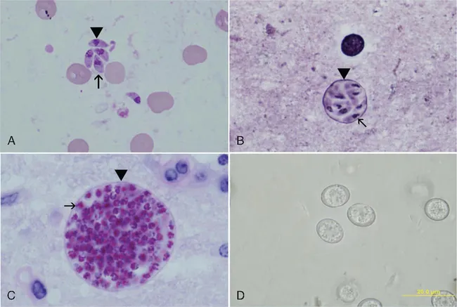
(A) Tachyzoites (arrowhead) in smear. Giemsa stain. Note nucleus dividing into two nuclei (arrow).
(B) A small tissue cyst in smear stained with Giemsa and a silver stain. Note the silver-positive tissue cyst wall (arrowhead) enclosing bradyzoites that have a terminal nucleus (arrow).
(C) Tissue cyst in section, PAS. Note PAS-positive bradyzoites (arrow) enclosed in a thin PAS-negative cyst wall (arrowhead).
(D) Unsporulated oocysts in cat feces, unstained.
1.3.2 Bradyzoite and tissue cysts
The term ‘bradyzoite’ (Gr. brady = slow) was proposed by Frenkel (1973) to describe the stage encysted in tissues. Bradyzoites are also called cystozoites. Dubey and Beattie (1988) proposed that cysts should be called tissue cysts (Figures 1.2B, 1.2C) to avoid confusion with oocysts. It is difficult to determine from the early literature who first identified the encysted stage of the parasite (Lainson, 1958). Levaditi et al. (1928) apparently were the first to report that T. gondii may persist in tissues for many months as ‘cysts’ however, considerable confusion between the terms ‘pseudocysts’ (group of tachyzoites) and ‘tissue cysts’ existed for many years. Frenkel and Friedlander (1951) and Frenkel (1956) characterized cysts cytologically as containing organisms with a subterminal nucleus and periodic acid Schiff (PAS)-positive granules (Figure 1.2C) surrounded by an argyrophilic cyst wall (Figure 1.2B). Wanko et al. (1962) first described the ultrastructure of the T. gondii cyst and its contents. Jacobs et al. (1960a) first provided a biological characterization of cysts when they found that the cyst wall was destroyed by pepsin or trypsin, but the cystic organisms were resistant to digestion by gastric juices (pepsin-HCl) whereas tachyzoites were destroyed immediately. Thus, tissue cysts were shown to be important in the life cycle of T. gondii because carnivorous hosts can become infected by ingesting infected meat. Jacobs et al. (1960b) used the pepsin digestion procedure to isolate viable T. gondii from tissues of chronically infected animals. When T. gondii oocysts were discovered in cat feces in 1970, oocyst shedding was added to the biological description of the cyst (Dubey and Frenkel, 1976).
Dubey and Frenkel (1976) performed the first in-depth study of the development of tissue cysts and bradyzoites, and described their ontogeny and morphology. They found that tissue cysts formed in mice as early as 3 days after their inoculation with tachyzoites. Cats shed oocysts (Figure 1.2D) with a short prepatent period (3–10 days) after ingesting tissue cys...
Table of contents
- Cover image
- Title page
- Table of Contents
- Contributors
- Preface
- Acknowledgements
- Chapter 1: The History and Life Cycle of Toxoplasma gondii
- Chapter 2: The Ultrastructure of Toxoplasma gondii
- Chapter 3: Population Structure and Epidemiology of Toxoplasma gondii
- Chapter 4: Clinical Disease and Diagnostics
- Chapter 5: Ocular Disease Due to Toxoplasma gondii
- Chapter 6: Toxoplasmosis in Wild and Domestic Animals
- Chapter 7: Toxoplasma Animal Models and Therapeutics
- Chapter 8: Biochemistry and Metabolism of Toxoplasma gondii
- Chapter 9: The Apicoplast and Mitochondrion of Toxoplasma gondii
- Chapter 10: Calcium Storage and Homeostasis in Toxoplasma gondii
- Chapter 11: Toxoplasma Secretory Proteins and their Roles in Cell Invasion and Intracellular Survival
- Chapter 12: Alterations in Host-Cell Biology due to Toxoplasma gondii
- Chapter 13: Bradyzoite Development
- Chapter 14: Development and Application of Classical Genetics in Toxoplasma gondii
- Chapter 15: Genetic Manipulation of Toxoplasma gondii
- Chapter 16: Gene Regulation
- Chapter 17: The Secretory Protein Repertoire and Expanded Gene Families of Toxoplasma gondii and Other Apicomplexa
- Chapter 18: Comparative Aspects of Nucleotide and Amino-acid Metabolism in Toxoplasma gondii and other Apicomplexa
- Chapter 19: Toxoplasma as a Model System for Apicomplexan Drug Discovery
- Chapter 20: Proteomics of Toxoplasma gondii
- Chapter 21: Cerebral Toxoplasmosis: Pathogenesis and Host Resistance
- Chapter 22: Innate Immunity in Toxoplasma gondii Infection
- Chapter 23: Adaptive Immunity and Genetics of the Host Immune Response
- Chapter 24: Vaccination Against Toxoplasmosis: Current Status and Future Prospects
- Epilogue
- INDEX
Frequently asked questions
Yes, you can cancel anytime from the Subscription tab in your account settings on the Perlego website. Your subscription will stay active until the end of your current billing period. Learn how to cancel your subscription
No, books cannot be downloaded as external files, such as PDFs, for use outside of Perlego. However, you can download books within the Perlego app for offline reading on mobile or tablet. Learn how to download books offline
Perlego offers two plans: Essential and Complete
- Essential is ideal for learners and professionals who enjoy exploring a wide range of subjects. Access the Essential Library with 800,000+ trusted titles and best-sellers across business, personal growth, and the humanities. Includes unlimited reading time and Standard Read Aloud voice.
- Complete: Perfect for advanced learners and researchers needing full, unrestricted access. Unlock 1.5M+ books across hundreds of subjects, including academic and specialized titles. The Complete Plan also includes advanced features like Premium Read Aloud and Research Assistant.
We are an online textbook subscription service, where you can get access to an entire online library for less than the price of a single book per month. With over 1.5 million books across 990+ topics, we’ve got you covered! Learn about our mission
Look out for the read-aloud symbol on your next book to see if you can listen to it. The read-aloud tool reads text aloud for you, highlighting the text as it is being read. You can pause it, speed it up and slow it down. Learn more about Read Aloud
Yes! You can use the Perlego app on both iOS and Android devices to read anytime, anywhere — even offline. Perfect for commutes or when you’re on the go.
Please note we cannot support devices running on iOS 13 and Android 7 or earlier. Learn more about using the app
Please note we cannot support devices running on iOS 13 and Android 7 or earlier. Learn more about using the app
Yes, you can access Toxoplasma Gondii by Louis M. Weiss,Kami Kim in PDF and/or ePUB format, as well as other popular books in Technology & Engineering & Public Health, Administration & Care. We have over 1.5 million books available in our catalogue for you to explore.