- 56 pages
- English
- ePUB (mobile friendly)
- Available on iOS & Android
eBook - ePub
Virus Structure
About this book
Virus Structure describes the physical characteristics of isolated viruses that represent typical structural groups, with particular reference to those features analyzed with the aid of the electron microscope. For descriptive purposes, the book has been divided into sections starting with the small icosahedral viruses and leading to the larger and more sophisticated structures, regardless of whether they are animal, plant, or bacterial viruses. These include double-stranded DNA icosahedral viruses, herpesvirus, viruses with helical symmetry, and viruses with complex or a combination of symmetries. Many common architectural features will be found in those viruses selected for discussion in each of the sections, and for these reasons the introduction places some emphasis on the symmetry elements rather than the shapes of viruses. The mechanism by which viruses enter host cells and the events that follow once the cell has been infected are only mentioned briefly as the virus-host interaction is a relatively complex one.
Trusted by 375,005 students
Access to over 1 million titles for a fair monthly price.
Study more efficiently using our study tools.
Information
INTRODUCTION
Nor do I doubt if the most formidable armies ever heere upon earth is a sort of soldiers who for their smallness are not visible. (52)
Petty, 1640
The modern concept of an infective virus particle is that it consists of a type of nucleic acid enclosed in a coat of protein or lipoprotein. The coat has the main function of protecting the infective nucleic acid or genome and in many instances may play some vital role in the initial attachment of the virus particle to the host cell and its subsequent penetration of viral material into the cytoplasm. Viruses can only multiply within a living cell and cannot be grown outside cells on an artificial medium. The techniques for maintaining animal cells in tissue culture are well established and they play an important role in the study and propagation of viruses. When viruses are isolated from the original host, they can be replicated in suitable tissue culture cells and relatively large numbers of progeny virus obtained. In the case of plant viruses, the techniques of cell culture and virus production are less advanced, but recent developments suggest that plant viruses may well be replicated in the near future in a similar way to animal viruses.
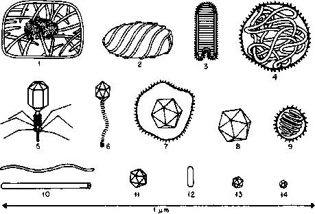
Viruses are incredibly small and, apart from one size group, they can only be visualized directly in the electron microscope. It is for this reason that most of the information concerning the size, shape, and symmetry of viruses has come from the application of electron microscopy to animal, plant, and bacterial viruses. Their size range together with a variety of morphological forms is shown in Fig. 1.
Units of Measurement
Because of the resolution and magnification required to visualize viruses and their components in the electron microscope and to some extent the overlap in the structural features of viruses which have been determined by X-ray diffraction methods, some unit of measurement is essential which will cover common structural dimensions within the range of macromolecules and atomic dimensions. The dimensions and distances measured from electron micrographs and X-ray diffraction patterns are expressed in angstrom units or nanometers. The current scale of measurement is shown in Table I.
TABLE I
CURRENT SCALE OF MEASUREMENTa
| Millimeter (mm) | Micrometer (µm) or micron (μ) | Millimicron (mµ) or nanometer (nm) | Angstrom units (Å) |
| 1 | 10−3 mm | 10−6 mm | 10−7 mm |
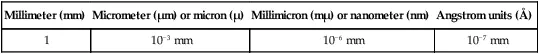
aExample: A virus particle seen on an electron micrograph possessing a diameter of 10 millimeters at a magnification of × 100,000 will be 1000 Å or 100 nanometers across.
In addition to the morphological features determined from electron micrographs, vital information about virus structure, symmetry, and chemical composition has resulted from biochemical, hydrodynamic, and X-ray diffraction studies carried out over a period of many years. Sufficient data from these techniques has enabled viruses to be grouped according to their various biological and structural characteristics, but for the purposes of this volume it is necessary to limit the details to those within the scope of ultrastructure. Although a voluminous published literature describes numbers of viruses that have been isolated from many different hosts, space is only available to describe typical examples.
It is not possible, nor is it within the scope of this monograph to discuss the details of diseases caused by viruses in man, animals, plants, and bacteria. For a more detailed account of the clinical and pathological aspects of virus diseases, the reader is referred to some of the literature cited at the end of the volume.
Terminology
The study of viruses and their components has involved the application of sophisticated technical methods within the fields of biochemistry, molecular biology, X-ray diffraction, and electron microscopy. It is not surprising to find, therefore, that different terms were used within the various disciplines to describe viral components and in many instances has led to some confusion in the published literature. In recent years, there has been some agreement about the terminology that can be applied to viruses generally (13, 54, 89). The diagram shown in Fig. 2 illustrates the terminology currently being applied to a wide range of viruses. For the larger and more complex viral structures included elsewhere in this volume, the terminology applied to these specific viruses will be discussed later in the relevant sections.
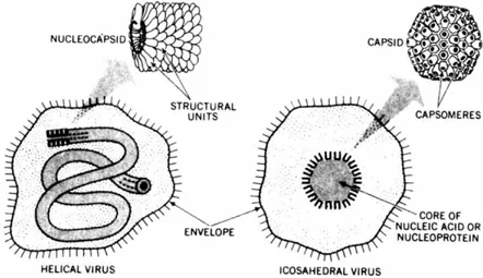
A large number of rodlike or filamentous simple viruses consist of a strand of nucleic acid enclosed by a protein shell. The helical rods of tobacco mosaic virus (TMV) serve as a good example (see Fig. 17). Part of another helical structure enclosed in an envelope is illustrated diagrammatically at the top left-hand side of Fig. 2. The intact and infective virus particle is known as a virion. The protein shell enclosing the coil of nulceic acid is composed of identical protein molecules known as structure units. The structure units are considered to be the same in this particular instance as the chemical units. When the structure or chemical units are combined or assembled with the nucleic acid to form a symmetrical or linear structure, it is referred to as the nucleocapsid.
The form taken by the helical nucleocapsid can be straight rods or flexible filaments. In the case of certain viruses, the nucleocapsid is enclosed in an envelope as illustrated in the particle shown in the lower left of Fig. 2.
The viruses of approximately spherical shape, as illustrated in the top right of Fig. 2, are composed of a protein shell or capsid that is assembled...
Table of contents
- Cover image
- Title page
- Table of Contents
- Copyright
- PREFACE
- ACKNOWLEDGMENTS
- Chapter 1: INTRODUCTION
- Chapter 2: SYMMETRY IN VIRIONS
- Chapter 3: SMALL DNA ICOSAHEDRAL VIRUSES
- Chapter 4: SMALL RNA ICOSAHEDRAL VIRUSES (NAPOVIRUSES AND PICORNAVIRUSES)
- Chapter 5: DOUBLE-STRANDED DNA ICOSAHEDRAL VIRUSES
- Chapter 6: HERPESVIRUS
- Chapter 7: VIRUSES WITH HELICAL SYMMETRY
- Chapter 8: VIRUSES WITH COMPLEX OR A COMBINATION OF SYMMETRIES
- SUMMARY
- REFERENCES
- INDEX
Frequently asked questions
Yes, you can cancel anytime from the Subscription tab in your account settings on the Perlego website. Your subscription will stay active until the end of your current billing period. Learn how to cancel your subscription
No, books cannot be downloaded as external files, such as PDFs, for use outside of Perlego. However, you can download books within the Perlego app for offline reading on mobile or tablet. Learn how to download books offline
Perlego offers two plans: Essential and Complete
- Essential is ideal for learners and professionals who enjoy exploring a wide range of subjects. Access the Essential Library with 800,000+ trusted titles and best-sellers across business, personal growth, and the humanities. Includes unlimited reading time and Standard Read Aloud voice.
- Complete: Perfect for advanced learners and researchers needing full, unrestricted access. Unlock 1.4M+ books across hundreds of subjects, including academic and specialized titles. The Complete Plan also includes advanced features like Premium Read Aloud and Research Assistant.
We are an online textbook subscription service, where you can get access to an entire online library for less than the price of a single book per month. With over 1 million books across 990+ topics, we’ve got you covered! Learn about our mission
Look out for the read-aloud symbol on your next book to see if you can listen to it. The read-aloud tool reads text aloud for you, highlighting the text as it is being read. You can pause it, speed it up and slow it down. Learn more about Read Aloud
Yes! You can use the Perlego app on both iOS and Android devices to read anytime, anywhere — even offline. Perfect for commutes or when you’re on the go.
Please note we cannot support devices running on iOS 13 and Android 7 or earlier. Learn more about using the app
Please note we cannot support devices running on iOS 13 and Android 7 or earlier. Learn more about using the app
Yes, you can access Virus Structure by Robert W. Horne in PDF and/or ePUB format, as well as other popular books in Biological Sciences & Biophysics. We have over one million books available in our catalogue for you to explore.