
- 584 pages
- English
- ePUB (mobile friendly)
- Available on iOS & Android
eBook - ePub
About this book
Nutritional cosmetics is an emerging area of intense research and marketing and encompasses the concept that orally consumed dietary products can support healthier and more beautiful skin. There are numerous dietary ingredients now being marketed for their potential skin health and beauty benefits and many of these are supported by growing scientific evidence. The purpose of this book is to compile the scientific evidence showing the potential benefits of some of the more extensively researched ingredients. As far as possible, information about the benefits of ingredients consumed orally for skin health is presented.
The information contained in this book will help provide insights into an emerging research area and provide scientific background for the potential clinical effectiveness for some of the better researched nutricosmetic ingredients.
ABOUT THE EDITORS
Aaron Tabor, M.D. is the CEO of Physicians Pharmaceuticals and author of The Revival Slim & Beautiful Diet. A graduate of the Johns Hopkins School of Medicine, Dr. Tabor oversees all clinical research on the Revival Slim & Beautiful Diet plan, conducting randomized, double-blinded, placebo-controlled studies at leading hospitals in the U.S. Areas of note include weight loss, skin/hair/nail appearance, energy, menopause, PMS, cholesterol, memory, and diabetic health. He is also responsible for directing new Revival product development based on clinical research results.
Robert M. Blair, Ph.D. is the Research Manager for Physicians Pharmaceuticals, Inc. and manages the daily activities of the Research and Nutrition departments. Dr. Blair received his Ph.D. from Oklahoma State University in the field of Reproductive Physiology. Before joining Physicians Pharmaceuticals, Inc., he worked as an Assistant Professor of Comparative Medicine at the Wake Forest University School of Medicine where he examined the effects of dietary soy on cardiovascular health and cognitive function.
- Reviews the most-popular and most-researched nutricosmetic ingredients
- Presents information specifically about the benefits of ingredients consumed orally for skin health
- Considers the benefits of whey protein, rosemary, soy – and green tea and milk thistle, specifically, for protection against sun damage and photocarcinogenesis
- Provides information on antioxidants, incl: potential benefits of botanical antioxidants; carotenoids; coenzyme Q10; healthy fruits; olive fruit; and natural enzymes
Trusted by 375,005 students
Access to over 1.5 million titles for a fair monthly price.
Study more efficiently using our study tools.
Information
Subtopic
Fashion & Textile IndustryIndex
BusinessTHE BIOLOGY OF HEALTHY
AND AGING SKIN
1
Structure and Function of the Skin
Department of Dermatology, Catholic University of Sacred Heart, Rome, Italy
1.1 Introduction
1.2 The Structure of the Skin
1.2.1 Macroscopic Characteristics
1.2.2 Microscopic Characteristics
1.2.3 The Epidermis
1.2.3.1 The Keratinocytes
1.2.3.2 The Melanocytes
1.2.3.3 Langerhans Cells
1.2.3.4 Merkel Cells
1.2.4 The Dennis
1.2.5 The Hypodermis
1.2.6 Vascularization
1.2.7 Innervation
1.2.8 Skin Annexes
1.2.8.1 The Hair System
1.2.8.2 Anatomy and Histology of Hair
1.2.8.3 The Hair Life Cycle
1.2.8.4 The Tricogram
1.2.8.5 Alopecias
1.2.8.6 Cutaneous Glands
1.2.8.7 Sebaceous Glands
1.2.8.8 Sweat Glands
1.2.8.9 The Nails
1.3 Skin Functions and Physiology
1.3.1 The Physiology of the Skin
1.3.1.1 Keratinization
1.3.1.2 Cellular Dynamics of Keratinization
1.3.1.3 Epidermal Lipids
1.3.1.4 Keratinosomes (Odland Bodies)
1.3.1.5 The Keratins
1.3.1.6 The Hydrolipid Film
1.3.1.7 Skin Hydration
1.3.1.8 SkinpH
1.3.1.9 Skin Flora
1.3.2 Functions of the Skin
1.3.2.1 Barrier Function
1.3.2.2 Protective Function
1.3.2.3 Immunological Function
1.3.2.4 Secretory Function
1.3.2.5 Thermoregulatory Function
1.3.2.6 Sensitivity Functions
1.3.2.7 Absorption Function
References
Aaron Tabor and Robert M. Blair (eds.), Nutritional Cosmetics: Beauty from Within, 1–45, © 2009 William Andrew Inc.
1.1 Introduction
“The skin draws the line between the end of the organism and the beginning of the world outside. Internally, the skin shelters and protects all the physiochemical phenomenon necessary for life, externally it is a barrier against mechanical forces, both physical and chemical, which can be hostile to life. The most important role of the skin both for man and for every other organism, vertebrate or invertebrate, unicellular or multicellular, is to create an obstacle for all those things outside the organism: the rest of the world.
“If life were hermetically sealed, like a pod, survival would be impossible for a being that depended on the outside world. An organism must, therefore, develop in relation to the environment in which it must live and with which it must communicate. So, the barrier which protects the organism from the outside, must at the same time inform the interior of all that is occurring outside itself. It is the perfect balance of these two barrier functions that determines survival.” Furthermore, human skin acts as an organ of attraction between individuals. The appearance of the skin and hair is the “first image” that others have of us. Personal expression changes with variations in the condition of our hair and skin, whose appearance is derived from their intrinsic well-being. Modern cosmetology has the task of interacting with physiology in maintaining its “good condition.”
1.2 The Structure of the Skin
1.2.1 Macroscopic Characteristics
The skin is the largest, most extensive organ of our body. In fact, the average adult has about 170–200 cm2 of skin with a weight that varies between 15 kg and 17 kg (obviously varying according to the subject’s height and dimensions).
The thickness of the epidermis, the outermost layer of the skin, can be from 0.5 mm in the thinnest areas (the eyelids, for example) to 4–6 mm at its thickest points (as on the palm of the hand and the sole of the foot). This thickness parameter becomes especially important when a substance is applied to the skin, be it a pharmaceutical or cosmetic product. In fact, once in contact with the skin any substance can penetrate the cutaneous barrier in a way directly proportional to the skin’s thickness at that point.
The skin, even if it appears smooth and compact to our eyes, is in reality marked over its entire surface by grooves, some shallow, others deeper, which by their layout mark many small polygons (Fig. 1.1). On the palm of the hand and the sole of the foot these grooves (dermatoglyphics) have become so evident as to characterize each individual and so unique that they are a distinct identification for each person. These apparently unimportant grooves are, however, necessary to accomplish an essential function—that of permitting the skin to stretch; if the skin were completely smooth, many of our movements would be impossible (Fig. 1.1).
The skin tissue houses within its structure other important constituents: hairs, nails, etc. (the skin’s annexes). Even with the naked eye one can see that (with the exception of the palm and sole) the whole of the skin is covered with hairs. In some areas the hairs are more developed and more coloured, as on the scalp, in the pubic region, and in the armpit. In other areas they are finer and much paler. These characteristics vary above all according to sex but also with individual biology and in the presence of certain pathologies.
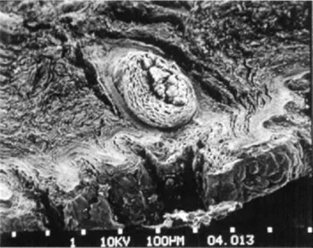
Figure 1.1 Human skin as seen with the scanning electron microscope.
Furthermore, tiny, invisible openings are found over the entire skin surface. These are the outlets of the eccrine sudoriparous glands, which, together with the apocrine sudoriparous glands and the sebaceous glands, will be handled in more detail later.
1.2.2 Microscopic Characteristics
When we consider the skin in its entirety, three different superimposed tissues can be identified (Fig. 1.2):
1. epidermis: the most external layer in contact with the environment
2. dermis: below the epidermis it is the structural component of the skin and the underlying organs
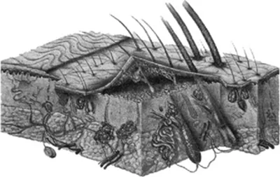
Figure 1.2 Diagram of a skin section (from W. Montagna).
3. hypodermis: immediately below the dermis, composed of a layer of adipose cells and representing a “cushion” of fat between the skin and the organs underneath
The boundary between epidermis and dermis, the “dermo-epidermal junction” (Fig. 1.2), is an “undulating” area resulting from many introflexions of the dermis and extroversions of the epidermis, dermal papillae, and epidermic crests, respectively. Along the entire dermo-epidermal junction there is a thin membrane, the “basal membrane.” The interconnection of the dermic papillae and epidermic crests is made functional by the presence of the basal membrane; this junction is a true structure fundamental for the relationship that exists between exchange and semipermeable barrier between the epidermis and the dermis and, consequently, also between the external environment and the internal organs. This boundary changes from zone to zone: it is flatter in the area of the forehead and highly accentuated on the back and on the soles of the feet.
The basal membrane (dermo-epidermal junction) is really a complex structure formed of many components: the cytoplasmic membrane of the basal keratinocytes, two thin layers (the lucid layer and basal lamina), and finally a fibrous structure in contact with the dermis. Toward the innermost side of the basal membrane there are special structures called “emidesmosomes,” which have an anchoring function. The fibrous structures below the basal lamina are of dermal origin and ensure the correct adhesion between epidermis and dermis.
We will now examine the epidermis, dermis, and hypodermis individually as each one contributes to the physiology of the cutaneous organ through its specific functions.
1.2.3 The Epidermis
The epidermis is composed of different types of cells that overlap, not randomly but in a well-defined manner. There are four different cell types:
1. keratinocytes
2. melanocytes
3. Langerhans cells
4. Merkel cells
1.2.3.1 The Keratinocytes
The “keratinocytes” are the predominant cell type and owe their name to the characteristic protein they produce in the course of their life, “keratin.” This protein is responsible for specific, important skin functions.
Keratinocytes are formed, grow, and die “rising” toward the surface of the epidermis. Gradually as they mature they gain particular morphological characteristics. This mechanism is defined as “epidermal cell turnover” and is the basis of the continuous and incessant renewal of the epidermis.
Under the microscope the epidermis is an obvious superimposition of cell layers, each clearly different from the others, these being the maturing phases of the keratinocytes (Fig. 1.3).
The basal layer is composed of a single line of more or less cylindrical cells that are densely packed and adherent to the basal membrane. These cells show intense metabolic activity due to their rapid division. They are in fact the “parent” cells of all the epidermal keratinocytes and thanks to their continuous division they are able to replace all the surface cells, which are continuously lost by exfoliation. Situated above the basal layer is the spinous layer (or Malpighian layer) formed by many layers of polygonal cells (moving upwards, the keratinocytes tend to flatten) that have already begun to produce keratin. Keratin, as previously mentioned, is the structural protein specific to the epidermis. The chemical structure of this protein makes possible certain fundamental functions of the skin: resistance against environmental attack and its impermeability to substances with which it comes into contact.
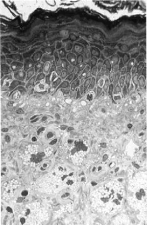
Figure 1.3 Image of a skin section seen under the light microscope, from the dermis to the upper epidermis (semi-thin section). The different layers of the epidermis are well visible. In the dermis istyocites and between the dermal fibres are other dermal cells.
Under the microscope the keratinocytes of the spinous layer are seen to bear thin “spines” that protrude from the cell membrane. It is from this characteristic that the spinous layer derives its name. The cellular spines are really firm points of contact between one cell and another.
Proceeding to the surface one finds the granular layer, where the various lines of largely polygonal, but by now flattened, keratinocytes contain “granules” in their cytoplasm. The granules are aggregates of keratin, produced in abundant quantities by the keratinocytic cells of the granular layer.
From this point on the keratinocytes mature, changing their function and also their morphology: from polygons they tend to become squashed and to lose their regular shape, and they become less active and accumulate intracellularly large quantities of keratin. So in the next layer, the lucid layer, is formed of one or two strata of keratinocytes. The cells are flat, without either a nucleus or other cytoplasmic organelles, and contain a characteristic homogenous substance called “eleidin.” This layer is found only in certain areas of the skin, namely the palms of the hand and soles of the feet.
This last formation of keratinocytes constitutes the horny layer. In this area the keratinocytes take the name of “corneocytes” and possess particular characteristics. They are completely without metabolic activity (due to the loss of the nucleus and cytoplasmic organelles), contain large amounts of keratin, and possess a peculiar cell structure, resembling “leaves” tiled one on top of the other.
In the most external layers the corneocytes gradually lose their cohesion...
Table of contents
- Cover
- Halftitle
- Personal Care and Cosmetic Technology
- Title
- Copyright
- Dedication
- Contents
- Contributors
- Foreword
- Preface
- Introduction
- Part 1: The Biology of Healthy and Aging Skin
- Part 2: Beauty from the Inside and the Outside
- Part 3: Micronutrient Support for Beautiful Hair and Skin
- Part 4: Protect Your Skin with Natural Antioxidants
- Part 5: Supporting a Solid Foundation for Firmer Skin
- Part 6: Natural Moisturizers for Smoother Skin
- Part 7: Natural Support for a Healthier Complexion
- Part 8: Natural Protection from Photocarcinogenesis
- Index
Frequently asked questions
Yes, you can cancel anytime from the Subscription tab in your account settings on the Perlego website. Your subscription will stay active until the end of your current billing period. Learn how to cancel your subscription
No, books cannot be downloaded as external files, such as PDFs, for use outside of Perlego. However, you can download books within the Perlego app for offline reading on mobile or tablet. Learn how to download books offline
Perlego offers two plans: Essential and Complete
- Essential is ideal for learners and professionals who enjoy exploring a wide range of subjects. Access the Essential Library with 800,000+ trusted titles and best-sellers across business, personal growth, and the humanities. Includes unlimited reading time and Standard Read Aloud voice.
- Complete: Perfect for advanced learners and researchers needing full, unrestricted access. Unlock 1.5M+ books across hundreds of subjects, including academic and specialized titles. The Complete Plan also includes advanced features like Premium Read Aloud and Research Assistant.
We are an online textbook subscription service, where you can get access to an entire online library for less than the price of a single book per month. With over 1.5 million books across 990+ topics, we’ve got you covered! Learn about our mission
Look out for the read-aloud symbol on your next book to see if you can listen to it. The read-aloud tool reads text aloud for you, highlighting the text as it is being read. You can pause it, speed it up and slow it down. Learn more about Read Aloud
Yes! You can use the Perlego app on both iOS and Android devices to read anytime, anywhere — even offline. Perfect for commutes or when you’re on the go.
Please note we cannot support devices running on iOS 13 and Android 7 or earlier. Learn more about using the app
Please note we cannot support devices running on iOS 13 and Android 7 or earlier. Learn more about using the app
Yes, you can access Nutritional Cosmetics by Aaron Tabor,Robert M. Blair in PDF and/or ePUB format, as well as other popular books in Business & Fashion & Textile Industry. We have over 1.5 million books available in our catalogue for you to explore.