eBook - ePub
Basic Neurochemistry
Molecular, Cellular and Medical Aspects
- 1,016 pages
- English
- ePUB (mobile friendly)
- Available on iOS & Android
eBook - ePub
Basic Neurochemistry
Molecular, Cellular and Medical Aspects
About this book
Basic Neurochemistry: Molecular, Cellular and Medical Aspects, a comprehensive text on neurochemistry, is now updated and revised in its Seventh Edition. This well-established text has been recognized worldwide as a resource for postgraduate trainees and teachers in neurology, psychiatry, and basic neuroscience, as well as for graduate and postgraduate students and instructors in the neurosciences. It is an excellent source of information on basic biochemical processes in brain function and disease for qualifying examinations and continuing medical education.
- Completely updated with 60% new authors and material, and entirely new chapters
- Over 400 fully revised figures in splendid color
Trusted by 375,005 students
Access to over 1.5 million titles for a fair monthly price.
Study more efficiently using our study tools.
Information
PART I
Cellular Neurochemistry and Neural Membranes
CHAPTER 1 Neurocellular Anatomy
UNDERSTANDING NEUROANATOMY IS NECESSARY TO STUDY NEUROCHEMISTRY
Diverse cell types are organized into assemblies and patterns such that specialized components are integrated into a physiology of the whole organ
CHARACTERISTICS OF THE NEURON
General structural features of neurons are the perikarya, dendrites and axons
Neurons contain the same intracellular components as do other cells
Molecular motors move organelles and particles along axonal microtubules
Molecular markers can be used to identify neurons
CHARACTERISTICS OF NEUROGLIA
Virtually nothing can enter or leave the central nervous system parenchyma without passing through an astrocytic interphase
Oligodendrocytes are myelin-producing cells in the central nervous system
The microglial cell plays a role in phagocytosis and inflammatory responses
Ependymal cells line the brain ventricles and the spinal cord central canal
The Schwann cell is the myelin-producing cell of the peripheral nervous system
The extracellular space between peripheral nerve fibers is occupied by bundles of collagen fibrils, blood vessels and endoneurial cells
UNDERSTANDING NEUROANATOMY IS NECESSARY TO STUDY NEUROCHEMISTRY
Despite the advent of molecular genetics in neurobiology, our understanding of the functional relationships of the components of the CNS remains in its infancy, particularly in the areas of cellular interaction and synaptic modulation. Nevertheless, the fine structural relationships of most elements of nervous system tissue have been described well [1–5]. The excellent neuroanatomical atlases of Peters et al. [3] and Palay and Chan-Palay [1] should be consulted for detailed ultrastructural analyses of specific cell types, particularly of neurons with their diverse forms and connections. This chapter provides a concise description of the major cytoarchitectural features of the nervous system, an introduction to the historical literature and a structural roadmap for the neurochemist. Although the fine structure of the organelles of the CNS and PNS is not peculiar to these tissues, the interactions between cell types, such as synaptic contacts between neurons and myelin sheaths around axons, are unique. These specializations and those that allow for the sequestration of the CNS from the outside world, namely, the blood–brain barrier (BBB) and the absence of lymphatics, become major issues in considerations of normal and disease processes in the nervous system. For the sake of simplicity, the present section is subdivided into a section on general organization and a section regarding major cell types.
Diverse cell types are organized into assemblies and patterns such that specialized components are integrated into a physiology of the whole organ.
The CNS parenchyma is made up of nerve cells and their afferent and efferent extensions, dendrites and axons, all closely enveloped by glial cells. Coronal section of the cerebral hemispheres of the brain reveals an outer convoluted rim of gray matter overlying the white matter (Fig. 1-1). Gray matter, which also exists as islands within the white matter, contains mainly nerve cell bodies and glia and lacks significant amounts of myelin, the lipid component responsible for the whiteness of white matter. More distally along the neuraxis in the spinal cord, the cerebral situation is reversed: white matter surrounds gray matter, which is arranged in a characteristic H formation (Fig. 1-2).
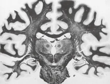
FIGURE 1-1 Coronal section of the human brain at the thalamic level stained by the Heidenhain technique for myelin. Gray matter stains faintly; all myelinated regions are black. The thalamus (*) lies beneath the lateral ventricles and is separated at this level by the beginning of the third ventricle. The roof of the lateral ventricles is formed by the corpus callosum (small arrows). Ammon’s horns are shown by the large arrows. Note the outline of gyri and sulci at the surface of the cerebral hemispheres, sectioned here near the junction of the frontal and parietal cortices.
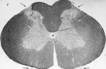
FIGURE 1-2 Transverse section of a rabbit lumbar spinal cord at L-1. Gray matter is seen as a paler-staining area in an H configuration formed by the dorsal and ventral horns with the central canal in the center (*). The dorsal horns would meet the incoming dorsal spine nerve roots at the upper arrows. The anterior roots can be seen below (lower arrows), opposite the ventral horns, from which they received their fibers. The white matter occupies a major part of the spinal cord and stains darker. Epon section, 1 μm, stained with toluidine blue.
A highly diagrammatic representation of the major CNS elements is shown in Figure 1-3. The entire CNS is bathed both internally and externally by cerebrospinal fluid (CSF), which circulates throughout the ventricular and leptomeningeal spaces. This fluid, a type of plasma ultrafiltrate, plays a significant role in protecting the CNS from mechanical trauma, balancing electrolytes and protein and maintaining ventricular pressure (see Ch. 32). The outer surface of the CNS is invested by the triple-membrane system of the meninges. The outermost is the dura mater, derived from the mesoderm, which is tightly adherent to the inner surfaces of the calvaria. The arachnoid membrane is closely applied to the inner surface of the dura mater. The innermost of the meninges, the pia mater, loosely covers the CNS surface. The pia and arachnoid together, derived from the ectoderm, are called the leptomeninges. CSF occupies the subarachnoid space, between the arachnoid and the pia, and the ventricles. The CNS parenchyma is overlaid by a layer of subpial astrocytes, which in turn is covered on its leptomeningeal aspect by a basal lamina (Fig. 1-3). On the inner, or ventricular, surface, the CNS parenchyma is separated from the CSF by a layer of ciliated ependymal cells, which are thought to facilitate the movement of CSF. The production and circulation of CSF are maintained by the choroid plexus, grape-like collections of specialized vascular elements and cells that protrude into the ventricles. Resorption of CSF is effected by vascular structures known as arachnoid villi, located within the leptomeninges over the surface of the brain (see Ch. 32).
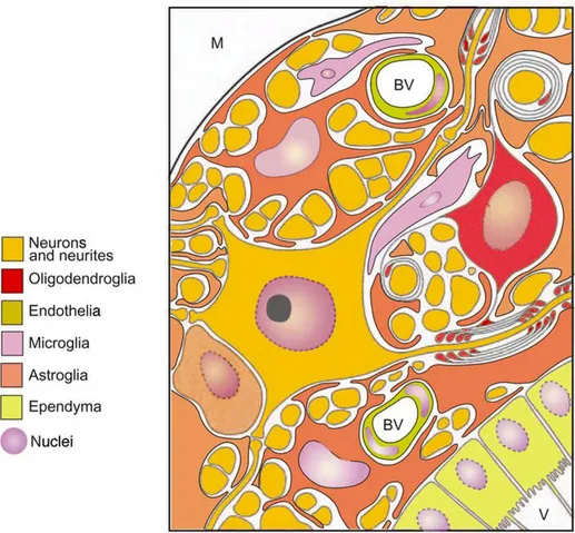
FIGURE 1-3 The major components of the CNS and their interrelationships. Microglia are depicted in light purple. In this simplified schema, the CNS extends from its meningeal surface (M) through the basal lamina (solid black line) overlying the subpial astrocyte layer of the CNS parenchyma and across the CNS parenchyma proper (containing neurons and glia) and subependymal astrocytes, to ciliated ependymal cells lining the ventricular space (V). Note how the astrocyte also invests blood vessels (BV), neurons and cell processes. The pia-astroglia (glia limitans) provides the barrier between the exterior (dura and blood vessels) and the CNS parenchyma. One neuron is seen (center), with synaptic contacts on its soma and dendrites. Its axon emerges to the right and is myelinated by an oligodendrocyte (above). Other axons are shown in transverse section, some of which are myelinated. The oligodendrocyte to the lower left of the neuron is of the nonmyelinating satellite type. The ventricles (V) and the subarachnoid space of the meninges (M) contain cerebrospinal fluid.
Ependymal cells abut layers of astrocytes, which in turn envelop neurons, neurites and vascular components. In addition to neurons and glial cells, such as astrocytes and oligodendrocytes, the normal CNS parenchyma contains blood vessels and microglial cells, the resident macrophages of the CNS.
The PNS and the autonomic nervous system consist of bundles of myelinated and nonmyelinated axons, extending to and from the CNS, which are intimately enveloped by Schwann cells, the PNS counterpart of oligodendrocytes. Nerve bundles are enclosed by the perineurium and the epineurium, which are tough, fibrous, elastic sheaths. Between individual nerve fibers are isolated connective tissue, or endoneurial cells, and blood vessels...
Table of contents
- Cover image
- Title page
- Table of Contents
- Copyright
- Section Editors
- Contributors
- Acknowledgments and History
- Preface to the Seventh Edition
- Pierre Morell, Ph.D. 1941–2003
- PART I: Cellular Neurochemistry and Neural Membranes
- PART II: Intercellular Signaling
- PART III: Intracellular Signaling
- PART IV: Growth, Development and Differentiation
- PART V: Metabolism
- PART VI: Inherited and Neurodegenerative Diseases
- PART VII: Sensory Transduction
- PART VIII: Neural Processing and Behavior
- Glossary
- Index
Frequently asked questions
Yes, you can cancel anytime from the Subscription tab in your account settings on the Perlego website. Your subscription will stay active until the end of your current billing period. Learn how to cancel your subscription
No, books cannot be downloaded as external files, such as PDFs, for use outside of Perlego. However, you can download books within the Perlego app for offline reading on mobile or tablet. Learn how to download books offline
We are an online textbook subscription service, where you can get access to an entire online library for less than the price of a single book per month. With over 1.5 million books across 990+ topics, we’ve got you covered! Learn about our mission
Look out for the read-aloud symbol on your next book to see if you can listen to it. The read-aloud tool reads text aloud for you, highlighting the text as it is being read. You can pause it, speed it up and slow it down. Learn more about Read Aloud
Yes! You can use the Perlego app on both iOS and Android devices to read anytime, anywhere — even offline. Perfect for commutes or when you’re on the go.
Please note we cannot support devices running on iOS 13 and Android 7 or earlier. Learn more about using the app
Please note we cannot support devices running on iOS 13 and Android 7 or earlier. Learn more about using the app
Yes, you can access Basic Neurochemistry by R. Wayne Albers,Donald L. Price,Scott Brady,George Siegel,Donald Price in PDF and/or ePUB format, as well as other popular books in Biological Sciences & Neuroscience. We have over 1.5 million books available in our catalogue for you to explore.